4678
MRI for the assessment of pre-treatment regional cardiac dysfunction in thoracic cancer patients
El-Sayed H. Ibrahim1, Antonio Sosa1, Jadranka Stojanovska2, Lindsay Puckett1, Elizabeth Gore1, and Carmen Bergom3
1Medical College of Wisconsin, Milwaukee, WI, United States, 2New York University, New York, NY, United States, 3Washington University, St Louis, MO, United States
1Medical College of Wisconsin, Milwaukee, WI, United States, 2New York University, New York, NY, United States, 3Washington University, St Louis, MO, United States
Synopsis
Keywords: Heart, Heart
Radiation therapy (RT) plays a key role in treating thoracic cancer, although the incidence of RT-induced cardiac complications could be as high as 33%. Nevertheless, characterization of baseline heart function, myocardial tissue characteristics and hemodynamic parameters in this patient population is not well elucidated, which was investigated in this study. The results revealed suboptimal cardiac function (both systolic and diastolic) and associations between different MRI cardiovascular parameters at baseline. Therefore, the condition of the cardiovascular system at baseline should be taken into consideration as a contributing factor in the development of RT-induced cardiotoxicity in thoracic cancer patients.Introduction
Thoracic cancer is the most frequently diagnosed cancer worldwide and the leading cause of cancer-related deaths, where patients present with locally advanced cancer and are treated with definitive radiation therapy (RT). As part of RT treatment, most patients receive incidental radiation exposure to the heart, where the incidence of cardiac complications post-RT is as high as 33%. Recent trials have not been able to correlate early deaths with clinically evident cardiotoxicity, suggesting that early non-cancer deaths are at least partially due to subclinical cardiac dysfunction caused by RT exposure to the heart and/or borderline baseline cardiac function before RT treatment, where there are limited data about the latter. In this study, we used a comprehensive cardiac MRI exam to study baseline cardiac function, myocardial tissue characteristics, and hemodynamic parameters in this patient population.Methods
A total of eight thoracic cancer patients (males; age = 65±4 y.o.) scheduled for RT underwent a comprehensive cardiac MRI exam on a GE 3T MRI scanner. The exam included cine, tagging, T1 and T2 mappings, and 4D flow sequences. The images were analyzed using the Circle cvi42 software. The cine images were analyzed to generate measures of global cardiac function, including ejection fraction (EF), end-diastolic volume (EDV), end-systolic volume (ESV), stroke volume (SV), and LV mass. Volume and mass measurements were indexed by the patients’ body surface area (BSA). The tagged images were analyzed using the SinMod technique to measure global longitudinal (GLS), circumferential (GCS), and radial (GRS) strains, as well as regional strain measurements at the basal, mid-ventricular, and apical levels. The T1 (both pre-contrast and post-contrast) and T2 images were processed to generate T1, T2, and extracellular volume (ECV) maps. Regional T1 and T2 measurements were measured at the basal, mid-ventricular, and apical levels. Blood flow through the mitral and tricuspid valves was measured to obtain LV and RV early-to-atrial (E/A) filling ratios, respectively, which are measures of diastolic cardiac function. The results were compared to normal ranges from the literature. Statistical analysis was conducted to study the correlations among different MRI parameters. P < 0.05 was considered statistically significant.Results
The study population characteristics are shown in Table 1. Table 2 shows the resulting global cardiac measurements. Ejection fraction was 58±6%, reflecting normal systolic function. However, the E/A ratios measured through the mitral and tricuspid valves were < 1 (0.84±0.1 and 0.72±0.3, respectively), reflecting diastolic cardiac dysfunction. Table 3 shows global and regional myocardial strains. GLS, GCS, and GRS were -14±2%, -10±2%, and 15±9%, respectively. All strain values were smaller than the normal threshold value of 17%. In general, circumferential strains were less than longitudinal strains, which in turn were less than radial strains. There existed slight differences in strain measurements between different regions (basal, mid-ventricular, apical). Table 4 shows myocardial tissue characterization parameters. ECV (38%) exceeded the normal threshold value of 30%, while T1 and T2 (1282±84 ms and 52±5 ms, respectively) were close the threshold normal values (1300 ms and 50 ms for T1 and T2, respectively). There existed small differences between T1 and T2 values at the basal, mid-ventricular, and apical levels. Figure 1 shows the correlation heat map between different MRI parameters. Significant correlations are represented by BOLD face font. There existed significant high positive correlations between: EDV vs ESV (r=0.86), mass (r=0.90) and T2 (r=0.77); ESV vs mass (r=0.76); T2 vs ECV (r=0.88); and GRS vs tricuspid E/A (r=0.93). There existed a significant high negative correlation between EF vs ESV (r=-0.76).Conclusions
Cardiac MRI is a valuable modality for comprehensive assessment of baseline heart health in thoracic cancer patients undergoing RT, which showed borderline cardiac function in these patients. Correlation analysis demonstrated associations between different aspects of the cardiovascular system: cardiac function (myocardial strain), myocardial tissue characterization (T2, ECV), and hemodynamics (E/A, volumes). Therefore, the condition of the cardiovascular system at baseline should be taken into consideration as a contributing factor in the development of RT-induced cardiotoxicity in thoracic cancer patients, which would help with treatment management.Acknowledgements
Funding supports from Radiation Oncology Institute (ROI) and GE Healthcare.References
1. W Haque et al. Int J Radiat Oncol Biol Phys. 2018;100:470-477.
2. J Bradley et al. Lancet Oncol. 2015;16:187-199.
3. D Hardy et al. Ann Oncol 2010;21:1825-33.
4. C Johnson et al. Curr Opin Cardiol. 2015;30:197-204.
5. RT Dess et al. J Clin Oncol 2017;35:1395-402.
6. EH Ibrahim. Heart Mechanics. MRI. CRC Press 2017.
Figures
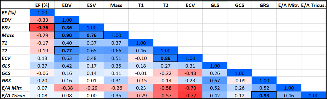
Figure 1. Heat map showing
correlations between different MRI parameters. Significant correlations are
represented by BOLD face font inside boxes. EF: ejection fraction. EDV:
end-diastolic volume. ESV: end-systolic volume. GLS, GCS, and GRS: global
longitudinal, circumferential, and radial strains, respectively. E/A: early to
atrial filling ratio.
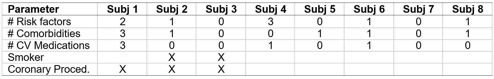
Table 1.
Study population characteristics. CV = cardiovascular.
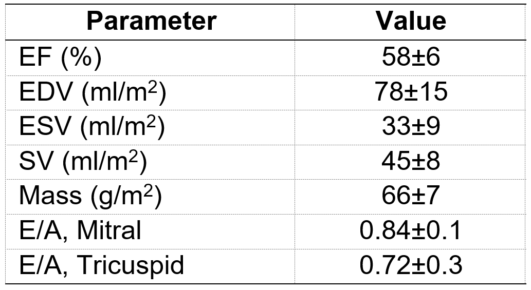
Table
2. Baseline global cardiac function measurements (mean±SD).
EF (systolic function) is normal, while E/A ratios are <1 (diastolic
dysfunction). Abbreviations: EF:
ejection fraction. EDV: end-diastolic volume. ESV: end-systolic volume. SV:
stroke volume. E/A: early-to-atrial filling ratio.
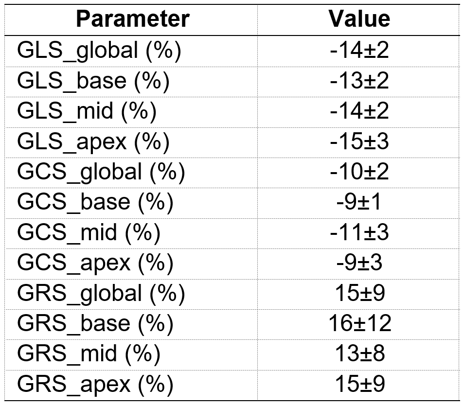
Table
3. Baseline regional cardiac function measurements (mean±SD).
Global and regional strain measurements are below normal threshold value
of 17%. Abbreviations: GLS:
longitudinal strain. GCS: circumferential strain. GRS: radial strain.
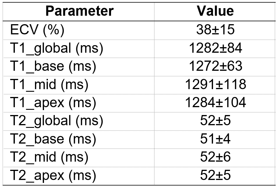
Table
4. Baseline tissue characteristics measurements (mean±SD).
Extracellular volume (ECV) exceeds the normal threshold value of 30%, while T1
and T2 are close to threshold values of 1300 ms and 50 ms, respectively.
DOI: https://doi.org/10.58530/2023/4678