4656
Spiral-GRASP-UTE: 4D Real-Time MRI of the Lung with Sub-Second Temporal Resolution1Biomedical Engineering and Imaging Institute and Department of Radiology, Icahn School of Medicine at Mount Sinai, New York, NY, United States, 2Ming Hsieh Department of Electrical and Computer Engineering, University of Southern California, Los Angeles, CA, United States, 3MR Applications Predevelopment, Siemens Healthcare GmbH, Erlangen, Germany
Synopsis
Keywords: Lung, Lung
The purpose of this work was to develop a dynamic lung MRI framework, called Spiral-GRASP-UTE, for 4D real-time imaging of the lung with sub-second temporal resolution. Spiral-GRASP-UTE combines continuous ultra-short echo time (UTE) variable-density stack-of-spirals acquisition with a recently developed GRASP-Pro reconstruction technique that is based on a low-rank subspace model. Compared to other state-of-the-art lung MRI methods using 3D radial Kooshball sampling, Spiral-GRASP-UTE enables fast free-breathing dynamic lung imaging (2-3 minutes) and a high temporal resolution of less than one second per volume. This eliminates the need for respiratory motion detection and compensation, which is often challenging in lung MRI.Introduction
Recent advances of MRI acquisition and reconstruction have enabled high-quality free-breathing lung MRI with high spatial resolution [1–4]. State-of-the-art lung MRI methods typically use a 3D radial Kooshball trajectory with center-out radial sampling to achieve ultra-short echo time (UTE). However, this sampling trajectory suffers from relatively low imaging efficiency and thus long scan time even with iterative reconstruction [2, 3]. Furthermore, due to half-spoke radial sampling, respiratory motion detection from acquired data becomes more challenging. Another way to acquire lung MRI data, which has recently attracted great attention, is to use a stack-of-spirals UTE sequence that is known as Spiral-UTE. Spiral-UTE employs center-out spiral sampling with varying echo times along the slice dimension to achieve ultra-short echo times [5–7]. In addition, Spiral-UTE offers more efficient sampling to reduce scan time and flexible selection of spatial resolution in three spatial dimensions. However, respiratory motion remains a challenge for Spiral-UTE in free-breathing lung imaging.The purpose of this work was to develop a novel 4D dynamic MRI framework, called Spiral-GRASP-UTE, for free-breathing dynamic imaging of the lung. Spiral-GRASP-UTE combines Spiral-UTE with GRASP-Pro (Golden-angle RAdial Sparse Parallel imaging with imProved performance), a recently developed dynamic MRI method based on a low-rank subspace model [8, 9]. In this work, GRASP-Pro has been extended to spiral sampling for highly-accelerated 4D lung MRI with sub-second temporal resolution, so that respiratory motion can be intrinsically resolved without the need for explicit motion compensation. From the hundreds of reconstructed dynamic image volumes, lung images at a similar motion phase can be combined to further improve image quality.
Methods
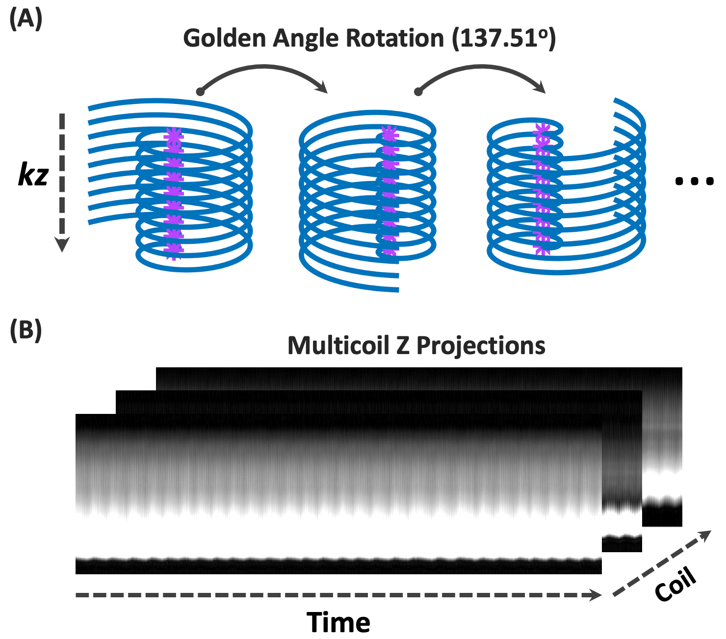
Spiral-GRASP-UTE acquisition employs variable-density spiral sampling in the kx-ky plane and fully sampled Cartesian sampling along the kz dimension. The echo time for the central slice is minimized to achieve UTE sampling, and it increases gradually when sampling outer slices [7]. As shown in Figure 1A, data acquisition is segmented into multiple spiral stacks rotating by a golden angle (~137.51°) from the previous one. For each spiral stack, the centers of k-space (the first k-space sample in each spiral readout) from all the slices generated a slice-oriented Z projection after 1D FFT (Figure 1B). The projections from all spiral stacks are used to estimate a temporal basis that is needed for GRASP-Pro reconstruction [9].Spiral-GRASP-UTE aims to reconstruct 4D real-time images of the lung with high temporal resolution by exploiting the low-rank property of dynamic images. Specifically, image reconstruction is performed as follows:
$$\tilde{\mathbf{V}}_{\mathbf{k}}=\underset{\mathbf{V_k}}{\mathrm{argmin}}\Vert{\mathbf{E}\mathbf{U_k}\mathbf{V_k} - \mathbf{y}}\Vert{^2_2}+\lambda _{1}R_{1}(\mathbf{U_k}\mathbf{V_k})+\lambda_{2}R_{2}(\mathbf{V_k})$$
Here, $$$\mathbf{E}$$$ denotes a multicoil encoding operator, $$$\mathbf{y}$$$ represents undersampled dynamic k-space and $$$R$$$ is a regularizer that can be enforced on spatial and/or temporal dimensions. The dynamic images to be reconstructed $$$(\mathbf{x})$$$ can be decomposed as $$$\mathbf{x}=\mathbf{UV}$$$, where $$$\mathbf{U}$$$ denotes the temporal basis that can be estimated from the multicoil Z projections using principal component analysis (PCA) [10], and $$$\mathbf{V}$$$ denotes the spatial basis, which provides spatial characteristics for the dynamic images under $$$\mathbf{U}$$$. Due to the low-rank condition of dynamic MR images, only a few dominant components in $$$\mathbf{U}$$$ are sufficient to represent $$$\mathbf{x}$$$, which leads to $$$\mathbf{x}\approx\mathbf{U_k}\mathbf{V_k}$$$, where $$$\mathbf{k}$$$ represents the first $$$\mathbf{k}$$$ major basis components in $$$\mathbf{U}$$$. Solving the equation obtains $$$\mathbf{V_k}$$$, and dynamic images are given as $$$\tilde{\mathbf{x}}=\mathbf{U_k}\mathbf{V_k}$$$.
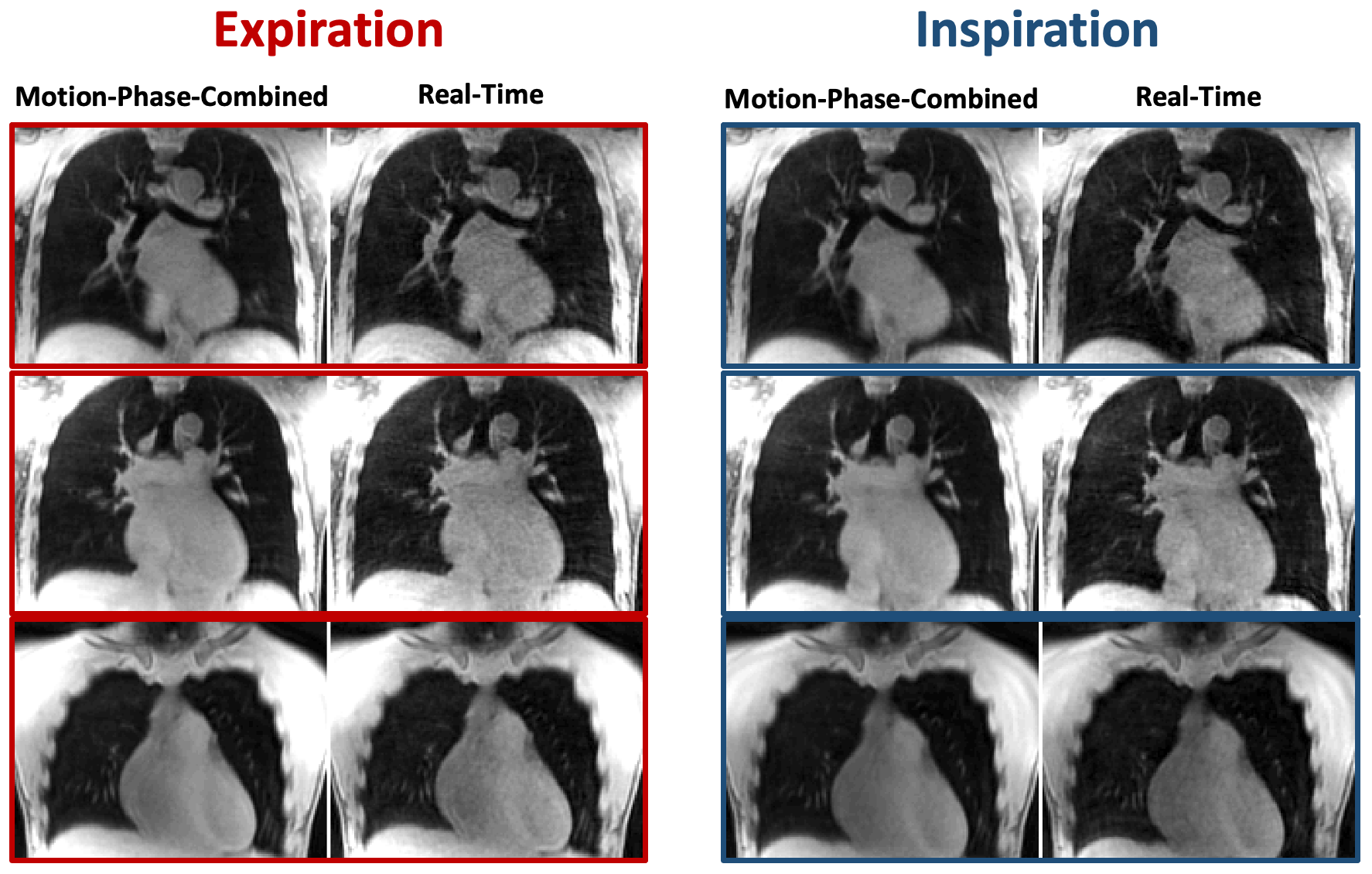
The new Spiral-GRASP-UTE technique was tested in a healthy volunteer for free-breathing 4D MRI of the lung on a 3T MR scanner (MAGNETOM Skyra, Siemens Healthcare, Erlangen, Germany). Relevant imaging parameters included: FOV=480x480mm2, matrix size=256x256, slice thickness=5mm, number of slices=48, TR/TE=4.18/0.05ms, flip angle=5°, and spiral readout duration=1.26ms. A total of 700 spiral interleaves were acquired for each partition, and fat saturation was enabled. The total acquisition time was 160 seconds. 4D real-time images (without motion sorting) were reconstructed to generate a total of 344 dynamic volumes by combining only three spiral stacks for each dynamic volume. This resulted in a temporal resolution of 0.69ms per 3D volume to intrinsically resolve respiratory motion without the need for motion detection and explicit motion compensation. The abundant dynamic information provided by the real-time images also permits visualization of lung ventilation. Furthermore, after real-time image reconstruction, similar motion phases can be easily identified (e.g., based on the structure similarity index) and combined to further improve image quality for assessment of lung structure.
Results
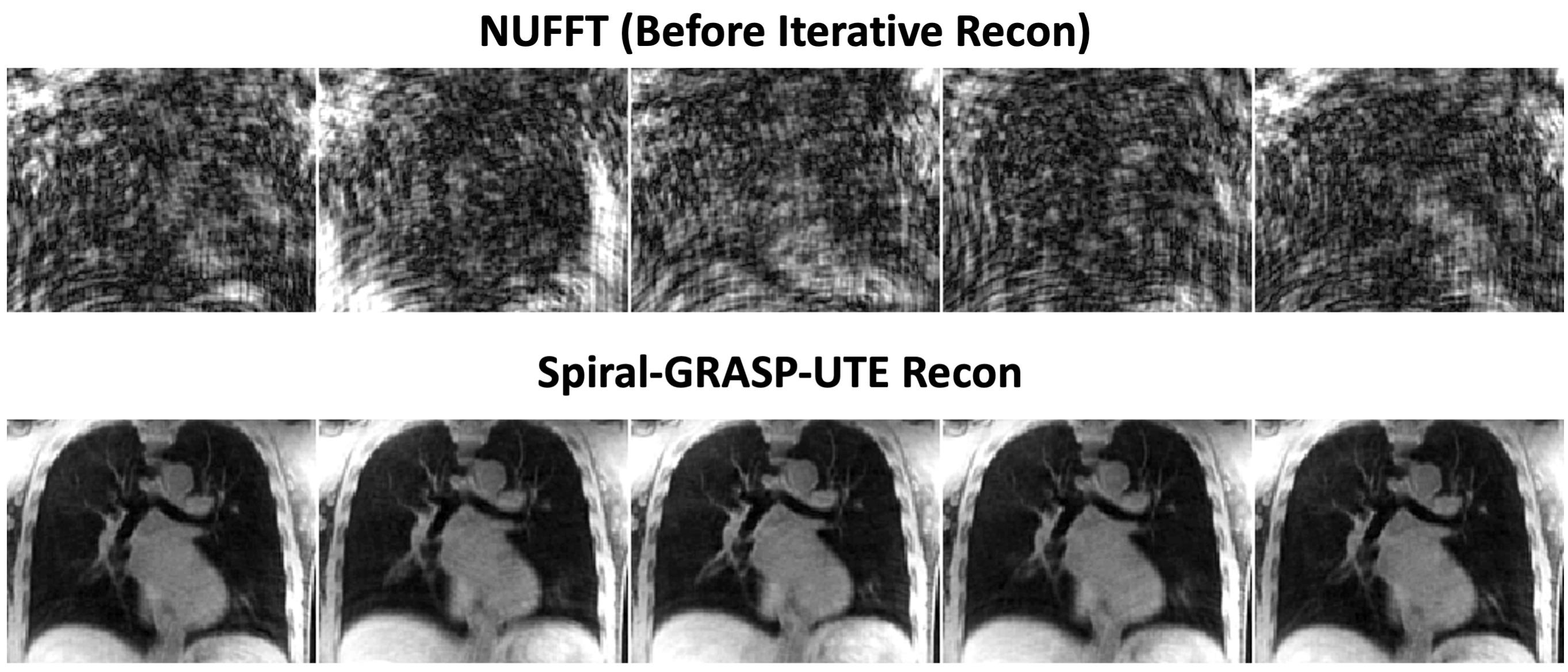
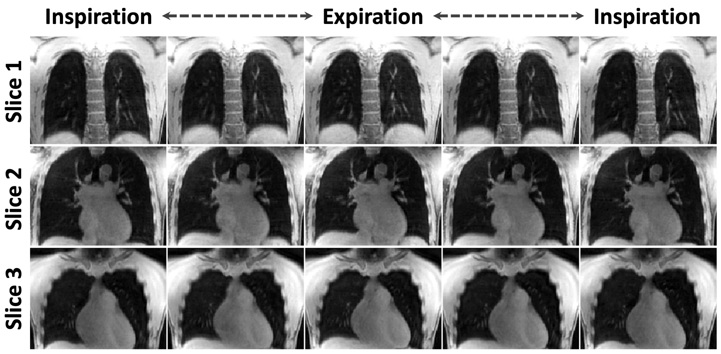
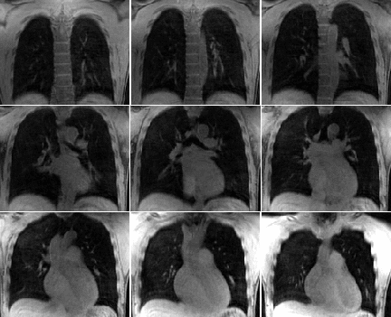
Figure 2 shows one representative slice of lung images from standard NUFFT reconstruction and corresponding results from Spiral-GRASP-UTE. With only 3 spiral stacks for each dynamic volume, NUFFT reconstruction shows substantial undersampling artifacts, while Spiral-GRASP-UTE reconstruction was able to remove them and recover most of the structure in the lung. Figure 3 shows three slices of lung images spanning a respiratory cycle reconstructed using Spiral-GRASP-UTE, indicating good delineation of lung structure and boundaries. By combining image volumes at a similar motion phase, image quality can be further improved for better assessment of lung structure, as shown in Figure 4. Finally, Figure 5 shows 9 slices of dynamic lung images in real time.Conclusion
This work proposes Spiral-GRASP-UTE, a novel 4D real-time lung MRI framework combining the Spiral-UTE sequence with GRASP-Pro reconstruction. It provides abundant dynamic information that may potentially be useful for simultaneous assessment of lung structure and ventilation.Acknowledgements
This work was supported in part by the NIH R01EB031083. The authors thank Dr. Michael Bush and Dr. Xiaoying Cai for help with modification of the Spiral-UTE sequence.References
[1] Delacoste J, Chaptinel J, Beigelman-Aubry C, Piccini D, Sauty A, Stuber M: A double echo ultra short echo time (UTE) acquisition for respiratory motion-suppressed high resolution imaging of the lung. Magn Reson Med 2018; 79:2297–2305.
[2] Jiang W, Ong F, Johnson KM, et al.: Motion robust high resolution 3D free-breathing pulmonary MRI using dynamic 3D image self-navigator. Magn Reson Med 2018; 79:2954–2967.
[3] Feng L, Delacoste J, Smith D, et al.: Simultaneous Evaluation of Lung Anatomy and Ventilation Using 4D Respiratory-Motion-Resolved Ultrashort Echo Time Sparse MRI. J Magn Reson Imaging 2019; 49:411–422.
[4] Zhu X, Chan M, Lustig M, Johnson KM, Larson PEZ: Iterative motion-compensation reconstruction ultra-short TE (iMoCo UTE) for high-resolution free-breathing pulmonary MRI. Magn Reson Med 2020; 83:1208–1221.
[5] Mugler III JP, Fielden S, Meyer C, Altes T, Miller GW, Stemmer A. Breath-hold UTE lung imaging using a stack-of-spirals acquisition. InProc Intl Soc Mag Reson Med 2015 (Vol. 23, p. 1476).
[6] Mugler III JP, Meyer CH, Pfeuffer J, Stemmer A, Kiefer B. Accelerated stack-of-spirals breath-hold UTE lung imaging. InProc Intl Soc Mag Reson Med 2017 (Vol. 25, p. 4904).
[7] Qian Y, Boada FE: Acquisition-weighted stack of spirals for fast high-resolution three-dimensional ultra-short echo time MR imaging. Magn Reson Med 2008; 60:135–145.
[8] Feng L, Wen Q, Huang C, Tong A, Liu F, Chandarana H: GRASP-Pro: imProving GRASP DCE-MRI through self-calibrating subspace-modeling and contrast phase automation. Magn Reson Med 2020; 83:94–108.
[9] Feng L: 4D GRASP MRI at Sub-Second Temporal Resolution. NMR Biomed 2022:e4844.
[10] Liang ZP: Spatiotemporal imaging with partially separable functions. 2007 4th IEEE Int Symp Biomed Imaging From Nano to Macro - Proc 2007:988–991.
Figures