4653
Improved Fast Whole-Brain High Resolution Diffusion Imaging on 7 Tesla MRI
Merry Mani1, Xinzeng Wang2, and Baolian Yang3
1University of Iowa, Iowa City, IA, United States, 2GE Healthcare, Houston, TX, United States, 3GE Healthcare, Waukesha, WI, United States
1University of Iowa, Iowa City, IA, United States, 2GE Healthcare, Houston, TX, United States, 3GE Healthcare, Waukesha, WI, United States
Synopsis
Keywords: Brain Connectivity, High-Field MRI
Achieving high resolution in diffusion MRI is challenging due to its inherently low SNR, vulnerability to motion and other EPI-related artifacts. On higher field-strengths, the SNR advantage can be exploited to push the resolution if the echo-time can be effectively reduced and the increased number of slices can be efficiently acquired. Combining acceleration techniques such as multi-band and parallel imaging is critical for this approach. Here we combine two deep-learned reconstruction priors, one pertaining to the q-space and another pertaining to image artifact removal, into a model-based iterative reconstruction framework to improve the quality of highly accelerated high-resolution 7T DWIs.Introduction
Diffusion MRI (dMRI) is a well-established tool for studying brain microstructure and connectivity. However, the spatial resolution of the dMRI technique is inherently limited. This is because of the long echo-time (TE) associated with the single-shot EPI acquisition typically used for dMRI studies. To keep the geometric distortions low while maximizing the spatial resolution, the use of parallel imaging (PI) and partial Fourier (pF) acceleration are critical, which helps to keep the TE low. However, the high under-sampling can lead to SNR loss.Imaging on the 7T MRI is an alternative solution for high spatial resolution imaging while addressing the SNR issue. The higher field strength allows to achieve isotropic voxel dimensions by reducing the slice thickness also. In this scenario, the acceleration capability of the multi-band (MB) technique will become crucial to collect the increased number of slices in a reasonable scan time. However, the combination of MB and high in-plane acceleration makes the reconstruction problem ill-posed. In this work, we present an improved reconstruction method for this case.
Methods
Previously, we introduced the qModeL method1 for the joint recovery of MB and PI accelerated dMRI. Here, we further improve the reconstruction by integrating another deep-learned prior into the qModeL framework that is pre-trained to de-noise generic MRI images2-3.The qModeL reconstruction is a model-based reconstruction which makes use of pre-learned deep q-space priors, that enables the joint recovery of the DWIs from highly under-sampled data while exploiting the SNR available from all q-space samples. It makes use of a SENSE-based forward-model to solve the multi-band un-aliasing in a model-based iterative framework. AIR Recon DL is a deep learning-based MR reconstruction pipeline which removes both noise and ringing artifacts from generic MR images2-3. In this work, we show that the AIR Recon DL can be effectively integrated into the qModeL framework which can then reconstruct the multi-band and PI accelerated data collected on the 7T MRI at very high resolution, without SNR loss. The success of this combination comes from the plug-and-play framework that is in-built in the qModeL formulation. This approach enables us to replace pre-learned priors effectively into the model-based reconstruction framework.
The combined qModeL + AIR Recon DL can be formulated as:$$\mathbf {S^{^*}} = argmin_{\tilde{ \mathbf S}} ||\mathcal {A}({\mathbf S})-\widehat{\mathbf Y }||_2^2 + \lambda_1 ~ ||\mathcal P_{\Theta}(\mathbf S) ||_2^2 + \lambda_2 ~|| \mathcal P_{I}(\mathbf S)||_2^2, $$
where $$$\mathcal P_{\Theta}(\mathbf S)$$$ is the q-space denoiser originally used with qModeL,
$$$ \mathcal P_{I}(\mathbf S)$$$ is the AIR Recon DL denoiser, that has replaced the TV denoiser originally used in qModeL, and S is the image to be reconstructed. For the reconstruction of the b0 images, only the AIR Recon DL denoiser $$$ \mathcal P_{I}(\mathbf S)$$$ is utilized.
To test the above reconstruction, in-vivo diffusion data were collected on the GE 7T SIGNA MRI scanner (Gmax=100mT/m, Gslewmax=200T/m/s) at the University of Iowa following IRB approval. A volunteer was scanned with a 32-channel receive coil . The images were acquired with a spatial resolution of 1x1x1mm3, TE=63.2ms, TR=5.4sec, b=1000s/mm2 with 60 diffusion directions. A MB factor of 3 was used with an in-plane parallel imaging factor of 2 and pF of 60%. The FOV was 240mm, with slice thickness of 1mm. 120 slices were collected with no gap to cover the whole brain in 6mins.
Results and Discussion
Figures 1-2 show the reconstruction of the b0 and DWI images reconstructed using standard MB reconstruction and its comparison to the AIR Recon DL, respectively. Here, the AIR Recon DL shows good performance on the b0 images because of the relatively higher SNR. The denoising property of the AIR Recon DL is evident and the image features appear sharer compared to the multi-band reconstruction. In the case of the DWIs, however, the performance of the AIR Recon DL is not impressive due to the very low SNR. Figure 2 also compares the performance of the qModeL reconstruction for this case, which provides significant gain in SNR due to the joint reconstruction. Here, a TV-denoiser was used as per the original implementation of the method1. Many of the structures that were previously under noise levels are now clearly delineated as can be seen in figure 2(c). However, qModeL is unable to compensate for the other systemic imperfections in the reconstruction, which carries over to the parameter maps. The AIR Recon DL is trained to correct for such artifacts, including the Gibbs ringing artifact removal. Figure 2(d) shows the improved performance of the proposed combined qModeL and AIR Recon DL approach for the reconstruction of the DWIs.Figure 3 shows the fractional anisotropy maps computed from the various reconstructions. The improvement in parameter maps can be seen from figure 3(d), where the proposed integrated iterative framework is noted to further remove the residual artifacts present in the qModeL-only, and AIR Recon DL-only parameter maps. Finally, figure 4 shows a comparison of fiber tracking from the various reconstruction methods for the same tracking parameters.
Conclusion
The integrated framework of combining AIR Recon DL into the qModeL reconstruction can provide improved results for highly accelerated high resolution DWIs. It removes systemic artifacts and preserves the SNR gains of the joint reconstruction scheme.Acknowledgements
This work was conducted on an MRI instrument funded by 1S10RR028821-01References
1. M. Mani, V. A. Magnotta, and M. Jacob, “qModeL: A plug‐and‐play model‐based reconstruction for highly accelerated multi‐shot diffusion MRI using learned priors,” Magn. Reson. Med., vol. 86, no. 2, pp. 835–851, Aug. 2021, doi: 10.1002/mrm.28756.2.
2 Lebel, R.M. "Performance characterization of a novel deep learning-based MR image reconstruction pipeline". August 2020. http://arxiv.org/abs/2008.065593.
3. Xinzeng Wang, Baolian Yang, Marc Lebel, Steen Moeller and Suchandrima Banerjee, "High Resoultion DiffusionTensor Imaging at 7T with multi-band multi-shot EPI acquisition and Deep Learning Reconstruction", ISMRM 2022, vol 3966
Figures
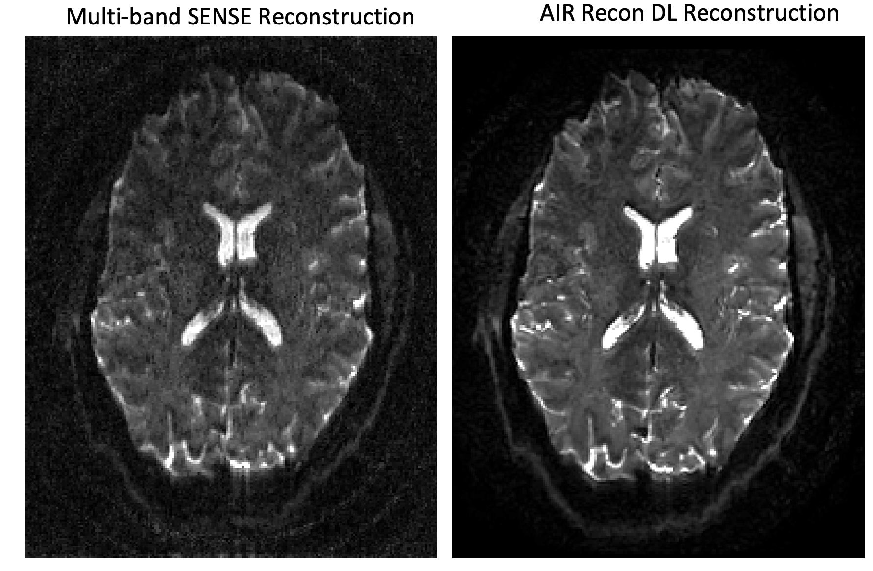
Figure 1: Comparison of standard multi-band (left) and AIR Recon DL reconstruction (right) for the b0 volume of a highly accelerated (MB=3, PI=2, pF=.6), high-resolution (1x1x1 mm) dataset.
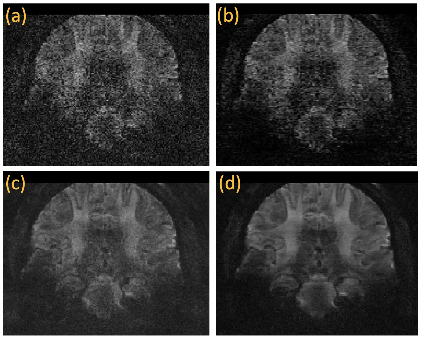
Figure 2: Comparison of the various reconstruction methods from a diffusion weighted volume. (a) Standard multi-band reconstruction, (b) AIR Recon DL only, (c) qModeL only, (d) qModeL combined with AIR Recon DL.
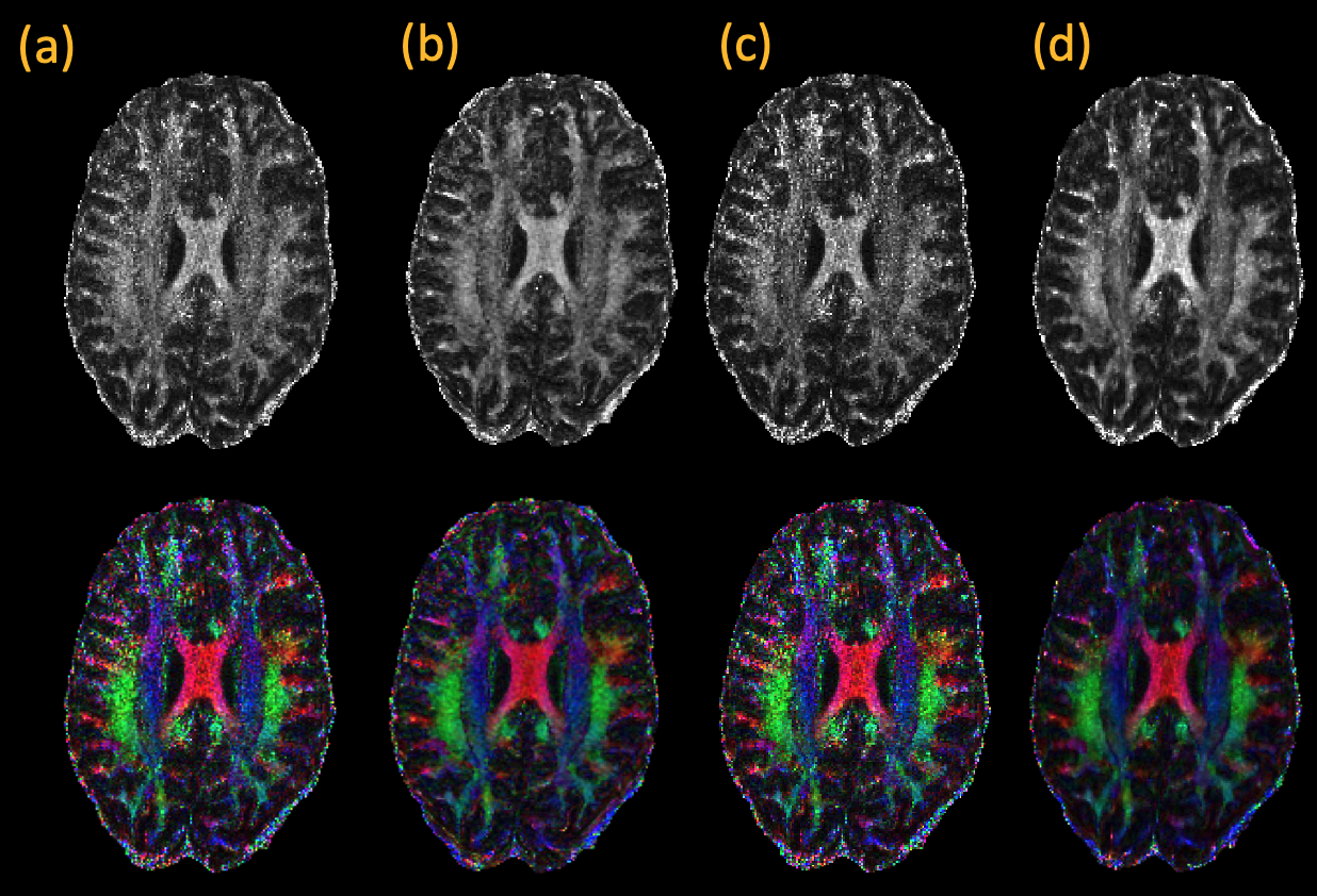
Figure 3: Comparison of parameter maps computed using the various methods: (a) multi-band reconstruction, (b) AIR Recon DL only, (c) qModeL only, (d) qModeL combined with AIR Recon DL.
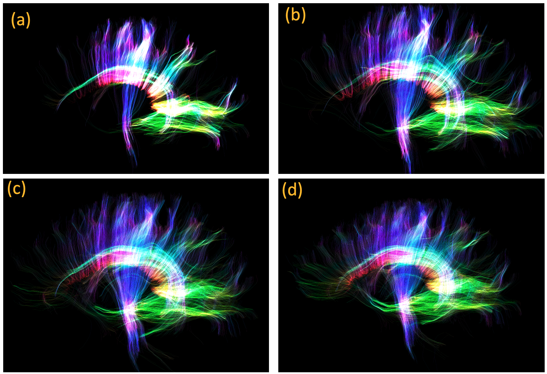
Figure 4: Comparison of fiber tracking from data reconstructed using (a) multi-band reconstruction, (b) AIR Recon DL only, (c) qModeL only and (d) qModeL combined with AIR Recon DL. Only 100000 tracks with a minimum length of 60mm are displayed for all the methods. The improvement in the frontal regions are visible in (d) compared to (b) and (c) as the systemic imperfections are better denoised by the combined approach.
DOI: https://doi.org/10.58530/2023/4653