4641
The effect of acceleration factor on brain magnetic resonance imaging based on artificial intelligence compressed sensing technology
shuai hu1, haonan zhang1, nan wang1, qingwei song1, and ailian liu1
1the First Affiliated Hospital of Dalian Medical University, dalian, China
1the First Affiliated Hospital of Dalian Medical University, dalian, China
Synopsis
Keywords: New Devices, Brain
ACS based cranial MRI has good feasibility in clinical use. Scanning time is reduced by 37%, 40%, and 71% for recommended AF in T1-FLAIR, T2WI, and T2-FLAIR, while the image quality meets the requirement of diagnosis.Objective
To investigate the effect of acceleration factor (AF) on the quality of craniocerebral magnetic resonance images based on AI-assisted compressed sensing (ACS).Methods
Twenty-seven volunteers (13 males and 14 females, average 56.2±13.4 years old) were recruited and randomly divided into 3 groups with 9 subjects in each group. MRI scans were performed on a 3.0 T MR scanner (Omega, UIH), including axial T1-FLAIR, T2WI and T2-FLAIR sequences. ACS with different AF (2, 2.5, 3, 3.5 and 4) was enabled for all contrasts (FIG.1), and conventional parallel imaging (PI) with AF =2 was involved for comparison. Detailed MRI parameters are listed in Table 1. Regions of interest (ROIs) with controlled size of 15-16 mm2 (FIG.2) were manually determined at corpus callosum, cerebellum, pons, and white matter on images of all the sequences. Signal to noise ratio (SNR) and contrast to noise ratio (CNR) were calculated from the signal intensity (SI) of each ROI. The images were scored by two observers (five and four years of MRI experience) using a quartet-scale method based on visibility of anatomical structures, diagnostic certainty, and image noise (Table 2). Kappa test was used to evaluate the consistency of subjective scores between observers. Friedman test was used to evaluate the differences of SNR, CNR and subjective scores among different accelerating settings. Further, pairwise comparison using Wilcoxon test was performed in subjects with significant difference found in Friedman test.Results
Supervisor scores were consistent between the two observers (Kappa: 0.831, 0.835, 0.822). There were no significant differences in SNR and CNR among different accelerating settings in all ROIs of each contrast. Statistically significant differences were found in T1-FLAIR and T2WI subjective scores (p < 0.001) (Table 3). High AF (3.5 and 4) ACS images of T1-FLAIR and T2WI showed significant lower scores compared to conventional PI (p < 0.05), while no significant were found between low AF ACS (2.0 and 2.5) and PI (Table 4). Surprisingly, no significant difference was found in T2-FLAIR scores.Conclusions
ACS based craniocerebral MRI could save abundant scanning time without loss of SNR or CNR compared to conventional PI method. Reduction of imaging quality should be considered as costs when using high AF > 3, with the gain of saving extra 30% time. On the premise of ensuring general image quality (subjective score > 3), it is clinically recommended to perform AF 2.5, 3 and 4 respectively for T1-FLAIR, T2WI, and T2-FLAIR.Acknowledgements
No acknowledgement found.References
No reference found.Figures
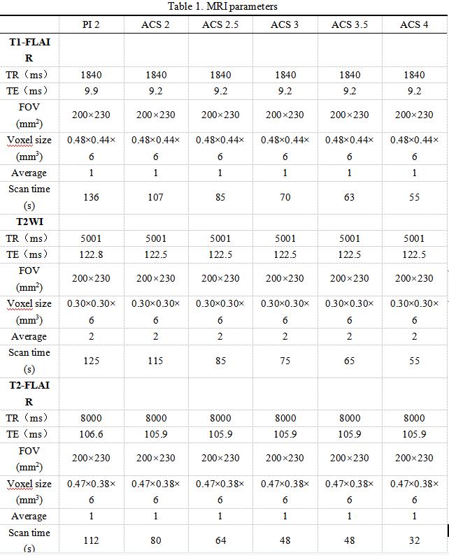
Table 1. MRI parameters
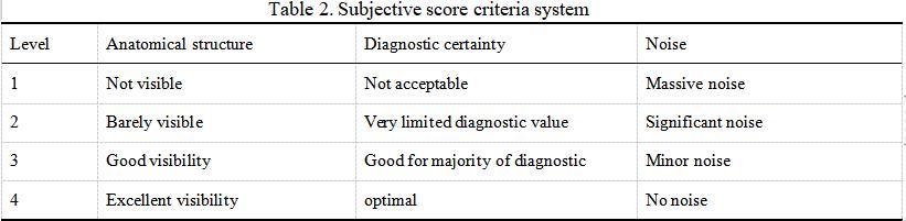
Table 2. Subjective score criteria system
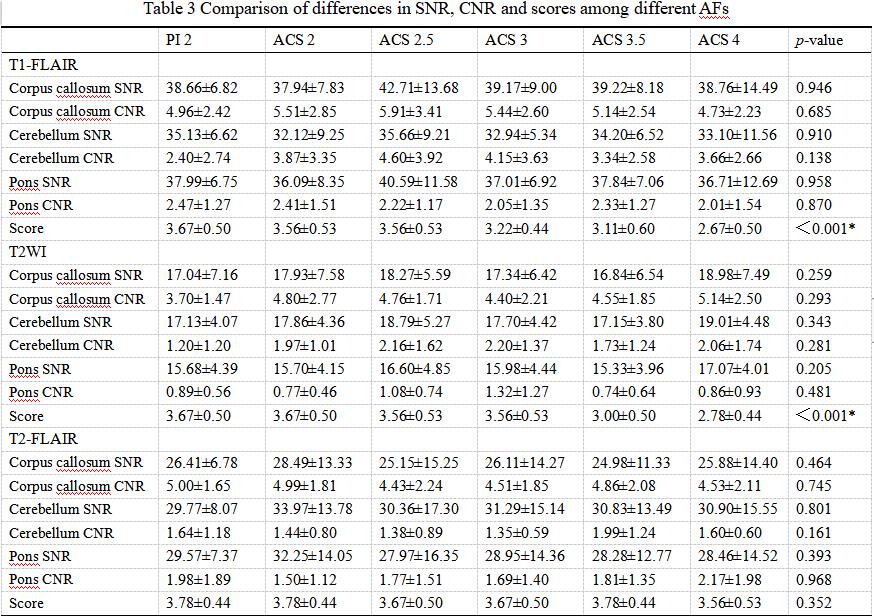
Table 3 Comparison of differences in SNR, CNR and scores among different AFs
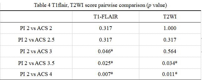
Table 4 T1flair, T2WI score pairwise comparison (p value)
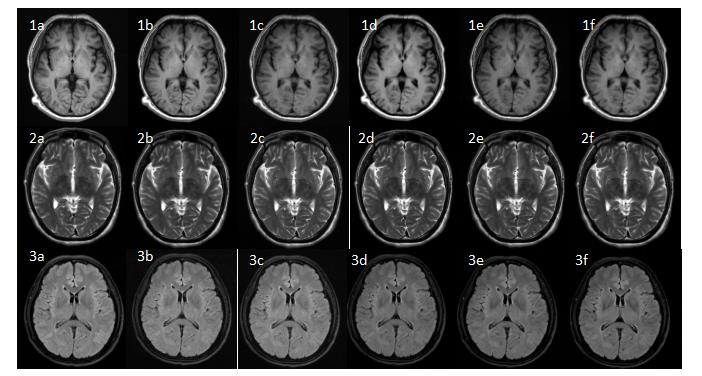
Figure 1 Male, 54-year-old. Panels 1a-1f are the axial T1-FLAIR images of the brain with PI AF=2, ACS AF=2, 2.5, 3, 3.5, and 4, respectively; Panels 2a-2f are the axial T2WI images of the brain with PI AF=2, ACS AF=2, 2.5, 3, 3.5, and 4, respectively. Panels 3a-3f are the axial T2-FLAIR images of the brain with PI AF=2, ACS AF=2, 2.5, 3, 3.5, and 4, respectively.
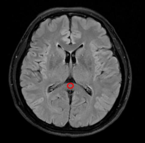
Figure 2 T2-FLAIR image of a 55 years old female. This figure shows an example of ROI at corpus callosum which was used to calculate SNR and CNR.
DOI: https://doi.org/10.58530/2023/4641