4595
Metasurface for passive RF shimming to improve brain imaging at 7T
Vladislav Koloskov1, Alena Shchelokova1, and Andrew Webb2
1School of Physics and Engineering, ITMO University, St. Petersburg, Russian Federation, 2Department of Radiology, C.J. Gorter Center for High Field MRI, Leiden University Medical Center, Leiden, Netherlands
1School of Physics and Engineering, ITMO University, St. Petersburg, Russian Federation, 2Department of Radiology, C.J. Gorter Center for High Field MRI, Leiden University Medical Center, Leiden, Netherlands
Synopsis
Keywords: Hybrid & Novel Systems Technology, Hybrid & Novel Systems Technology, metasurface, passive shimming
We propose a new passive radiofrequency (RF) shimming approach using a flexible, compact, low-cost, and long-term stability metasurface to improve brain imaging at 7T. The metasurface can be manufactured as a periodic structure based on a set of metal crosses connected by the capacitors, thus having a thickness of less than 1 mm. Numerical studies with the human voxel model showed up to 6% homogeneity improvement at the regions of interest (ROI) in the presence of the metasurface in comparison with the conventional case, which also outperforms the effect of the dielectric pad’s placement.Introduction
Ultra-high field MR scanners substantially improve MR image quality due to a higher signal-to-noise ratio (SNR) compared to clinical 1.5 and 3T systems. However, challenges related to the standing wave occur, severely degrading image quality and diagnostic value. High permittivity dielectric pads1,2 have proven themselves as an effective and inexpensive method for passive radiofrequency (RF) shimming. However, they have some drawbacks as fairly large thickness (around 5 mm) and deterioration of properties in time, that limit their application. Recently, the design of a thin and flexible metasurface (MS) was proposed to improve abdominal imaging at 3T3. Here, we propose and demonstrate via electromagnetic (EM) simulations with a human voxel model an optimized MS design to outperform the effect of B1+ homogenization in brain area at 7T achieved with high permittivity dielectric pads.Methods
All EM simulations were performed in CST Studio Suite 2021. The MS consists of a set of thin metal crosses (1x1 cm2) connected by capacitors (Fig. 1A). Such a design allows obtaining a secondary magnetic field (due to the conducting currents flowing along the structure) similar to one due to the displacement currents inside conventional dielectric pads. Note that the MS should be a non-resonant on an operating frequency of 298 MHz to act as a passive pad rather than a resonator. Thus, we obtained simulations to detect the MS’s resonant frequency dependent on the capacitance value with fixed geometrical properties such as a period (d=1.1cm) and a number of unit cells (N=15). In addition, for the reference case, we included in our simulations dielectric pads with ε=120, tan δ=0.05, and dimensions of 180×180×5 mm3 as an optimal one for head imaging1. A head birdcage-type coil (BC) loaded with the homogenous phantom and tuned and matched at 298 MHz was used (Fig. 2A). Dielectric pads and MS were positioned parallel to the axis of the BC (nearby temporal lobes) (Fig. 2B,C) in the aim of homogenizing the B1+-field in regions where minima can be observed. Based on the field distribution in the reference case with dielectric pads, we evaluated the optimization of capacitance to obtain a similar transmit efficiency along the x-axis in the central axial slice. Furthermore, a similar optimization was held with the voxel model Hugo placed inside the same BC. An optimization was based on achieving a gain in transmit efficiency and reducing B1+-field inhomogeneity (coefficient of variation (Cv)) in the entire brain area. For this purpose, the setups with one, two, and three MSs were considered. Cv was estimated as the ratio of the standard deviation of the |B1+|RMS (RMS=root mean square) and its mean value and multiplied by 100%. The B1+-field and SAR averaged over 10 grams of tissues (SARav.10g) distributions were calculated for 1W of the total accepted power. Additionally, we evaluated the RF safety without and with the MSs as |B1+|RMS divided by max SARav.10g.Results
Fig. 1B shows in blue the correlation between capacitance and the resonant frequency of the metasurface, and in yellow, the resonant frequency of the dielectric pad with ε=120, meaning that the capacitance gap for optimization is between 4 and 11 pF. Fig. 2 D-F shows correspondingly the |B1+|RMS maps for the reference case, with dielectric pads and two MSs. Dielectric pads increase the homogeneity in ROI by ~12.5%, whereas MS with 8pF capacitors can push that improvement further, up to 17.8%. |B1+|RMS maps in Fig. 3A-E show the same impact of MS even with a larger object. One MS (Fig. 3C) will improve Tx efficiency in the occipital lobe (ROI3) with a slight decrease of homogeneity in other ROI due to redistribution of the B1+-field. In the case of two MSs (Fig. 3D), we can redistribute the magnetic field that way to enhance both the B1+ field and its homogeneity. Since MS is flexible enough, more than two can be placed around the object, combining results from cases with one MS and two MSs leading to max efficiency of the proposed approach. Fig. 3F shows the cumulative table with all evaluated quantitative results. One can observe in Fig. 4 no significant increase in local SAR with RF safety values of 0.62 uT/√W/kg for the reference case, 0.59 uT/√W/kg with dielectric pads, and 0.59 uT/√W/kg, 0.58 uT/√W/kg, 0.57 uT/√W/kg with one, two and three MSs, respectively, with 5pF found after optimization.Discussion and conclusions
The metasurface design was optimized to improve the homogeneity of the B1+-field in the entire brain area at 7T. Due to its ultra-thin and flexible structure, metasurface can easily outperform conventional dielectric pads commonly used for passive RF shimming and improve patient comfort during the MR procedure. The proposed approach can be modified further depending on the specific task. By changing MS’s properties, such as the number of unit cells, periodicity, and capacitance, or changing the position of the structure relative to the brain, one can create different secondary magnetic field distributions, improving the transmit efficiency and field homogeneity in subjects with different shapes. Further studies will be provided with experimental approbation on volunteers.Acknowledgements
This work was supported by the Russian Science Foundation (Project 21-19-00707).References
- Teeuwisse WM, et al. Simulations of high permittivity materials for 7 T neuroimaging and evaluation of a new barium titanate‐based dielectric. Magnetic resonance in medicine 67.4 (2012): 912-918.
- Brink WM, et al. High permittivity dielectric pads improve high spatial resolution magnetic resonance imaging of the inner ear at 7 T. Investigative radiology 49.5 (2014): 271-277.
- Vorobyev V, et al. Improving homogeneity in abdominal imaging at 3 T with light, flexible, and compact metasurface. Magnetic Resonance in Medicine 87.1 (2022): 496-508.
Figures
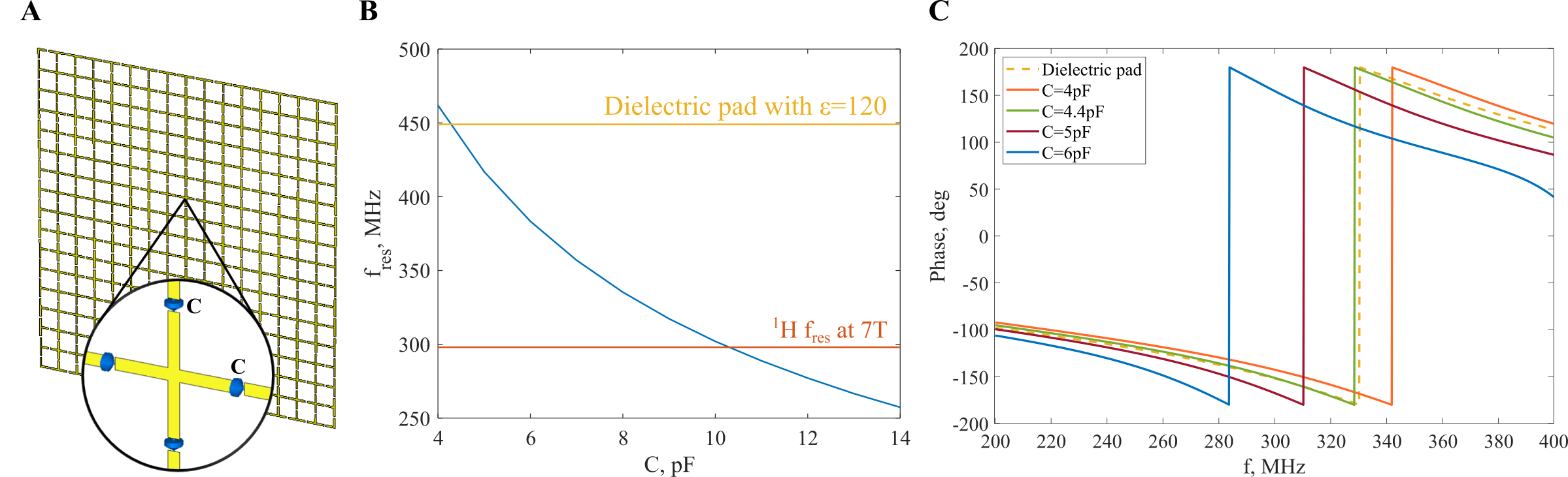
Figure 1: Metasurface modeling and its comparison to a dielectric pad: A, schematic representation of the MS based on unit cells with crosses connected with capacitors; B, Numerically calculated resonant frequency of MS as a function of capacity value; C, Numerically calculated phases of S21 (transmission) as a function of frequency values for the dielectric pad as a reference and MS with different capacitor values.

Figure 2: General view of the proposed setup: A, A reference case with a phantom placed in the head birdcage coil; B, The setup with dielectric pads; C, The setup with proposed metasurfaces. D-F, |B1+|RMS distribution for the reference case, with dielectric pads and metasurfaces (8pF). G, Numerically calculated normalized |B1+|RMS along black dashed lines in D-F. B1+ was normalized to 1 W of total accepted power. White dashed lines in D stand for ROI in temporal lobes.
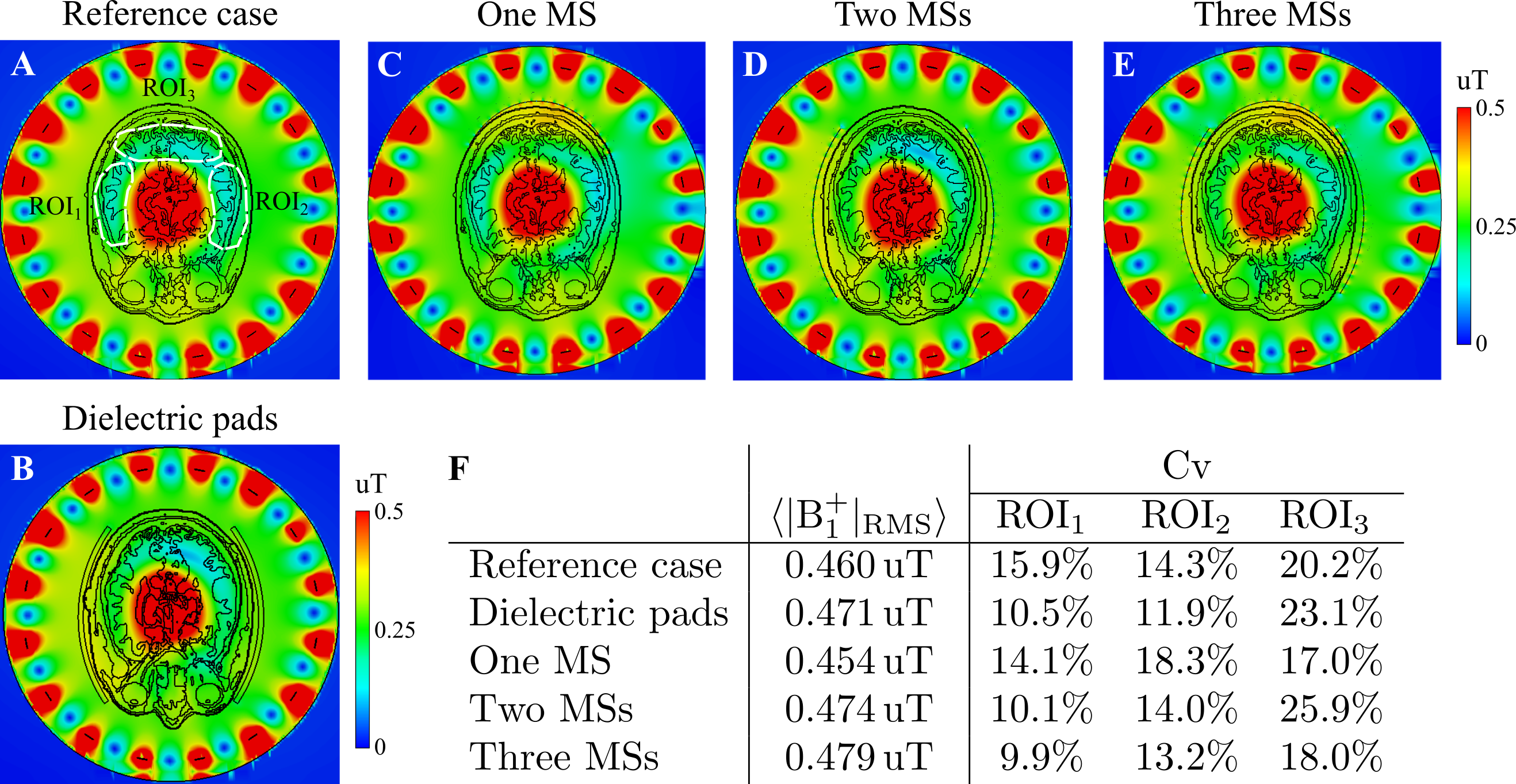
Figure 3: Numerically calculated |B1+|RMS maps for the voxel model Hugo inside head birdcage coil A, for a reference case, B, with dielectric pads, C-E, with one, two, and three metasurfaces (5pF), respectively. F, The cumulative table with mean B1+ values calculated in the slice crossing the center with z=0 and Cv calculated for all cases in all three ROI outlined with white dashed lines. B1+ maps were normalized to 1 W of total accepted power.
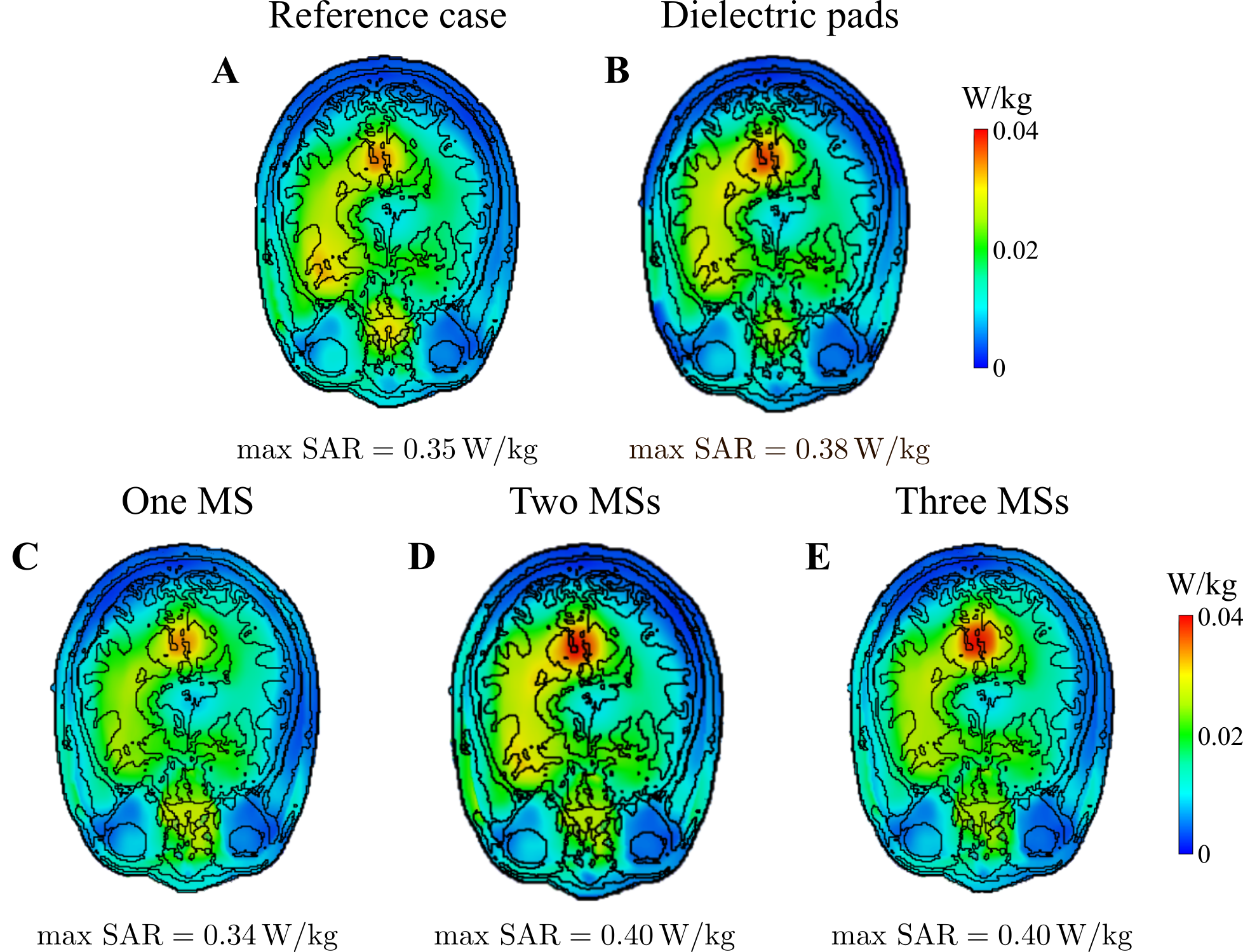
Figure 4: Numerically calculated SARav.10g maps for the voxel model Hugo inside head birdcage coil A, for a reference case, B, with dielectric pads, C-E, with one, two, and three metasurfaces (5pF), respectively. SAR maps were normalized to 1 W of total accepted power.
DOI: https://doi.org/10.58530/2023/4595