4585
Parallel transmission of cardiac imaging using statistical initial phase optimization at 5T1Paul C. Lauterbur Imaging Research Center, Shenzhen Institutes of Advanced Technology, Chinese Academy of Sciences, Shenzhen, China, 2School of Pharmacy and Bioengineering, Chongqing University of Technology, Chongqing, China, 3Shanghai United Imaging Healthcare, Shanghai, China, 4Key Laboratory for Magnetic Resonance and Multimodality Imaging of Guangdong Province, Shenzhen, China, 5Department of Biomedical Engineering, State University of New York at Buffalo, Buffalo, NY, United States
Synopsis
Keywords: Parallel Transmit & Multiband, Shims
High-field magnetic resonance imaging is one of the main development directions of magnetic resonance cardiac imaging because of its high signal-to-noise ratio and more anatomical details. However, with the increase of field strength, the homogeneity of B1 field is a serious challenge, and parallel transmission is one of the important methods to solve this problem.In this work, we proposed a clinically applicable radiofrequency shimming method to improve the imaging quality of cardiac imaging at 5T MRI.Statistical methods were employed to find out a better initial phase of an eight-channel coil and enhance the stability of cardiac imaging.Introduction
High‐field (≥3 T) cardiac magnetic resonance imaging (CMR) offers considerable gains in signal-to-noise ratio and improved blood–tissue contrast,but these gains bring many challenges1 such as inhomogeneity of the radiofrequency (RF) transmission field (B1+).This problem can be improved by independently adjusting the relative complex weight applied to each channel in the parallel transmission system using the RF shimming approach, which is widely used in CMR with its simplicity and easy implementation2.In addition, the general RF shimming is regarded as an optimization problem of magnitude least squares3. It is difficult to guarantee that all objects can converge to good results by using the general conjugate gradient method starting from the circularly polarized phase(CP-Shimming), because it is a nonconvex overdetermined equation that affects the inhomogeneity of B1+ in the ROI4.In this study, in order to improve the stability of RF shimming, we counted the transmit phases from 19 volunteers with good results of CP-Shimming and did statistical analysis.By changing the initial point to statistical phase and applying it to subjects with poor CP-Shimming, the B1+field uniformity and image quality of the two RF shimming methods were compared on 5T MRI.Method
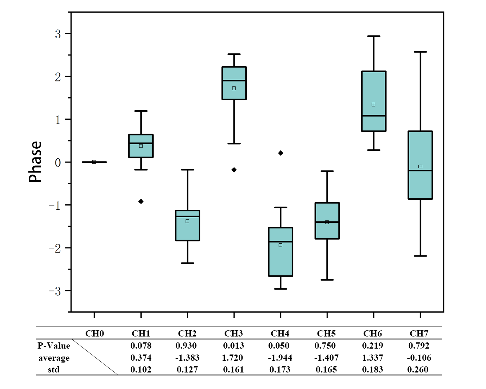
The goal of RF shimming was to make the B1+ field in the ROI uniform by adjusting the complex weight of each channel,so we adopted an eight-channel coil for the work.The overall workflow was shown in Figure 1.The statistical phases were obtained by 19 volunteers who had high image quality CMR and homogeneous field results after CP-Shimming.The Shapiro-Wilk test was used to determine whether they conformed to a normal distribution and whether it was statistically significant. It should be noted that we specify the first transmit channel as phase 0, which was regarded as the reference 0 for the other seven channels.Then, 4 new subjects with poor effect after RF shimming with CP-Shimming were selected.Started RF shimming from the phase obtained by statistics (STATS-Shimming), and compared the B1+ field uniformity and image quality between them.The coefficient of variation (CV) was used as the evaluation index of B1+ field uniformity. It was necessary to check the ROI manually, so the heart was selected five times independently to reduce the human subjective influence.RF shimming was performed for a gradient recalled echo cine(GRE-CINE) sequence with the following parameters:flip angle=10degree,TR=4.5ms,TE=2.445ms,FOV = 320×360mm2,slice thickness = 6 mm.Single-slice images were acquired in the four-chamber and short-axis view of the heart at a whole body 5T MRI system (United Imaging Healthcare, Shanghai, China).Result
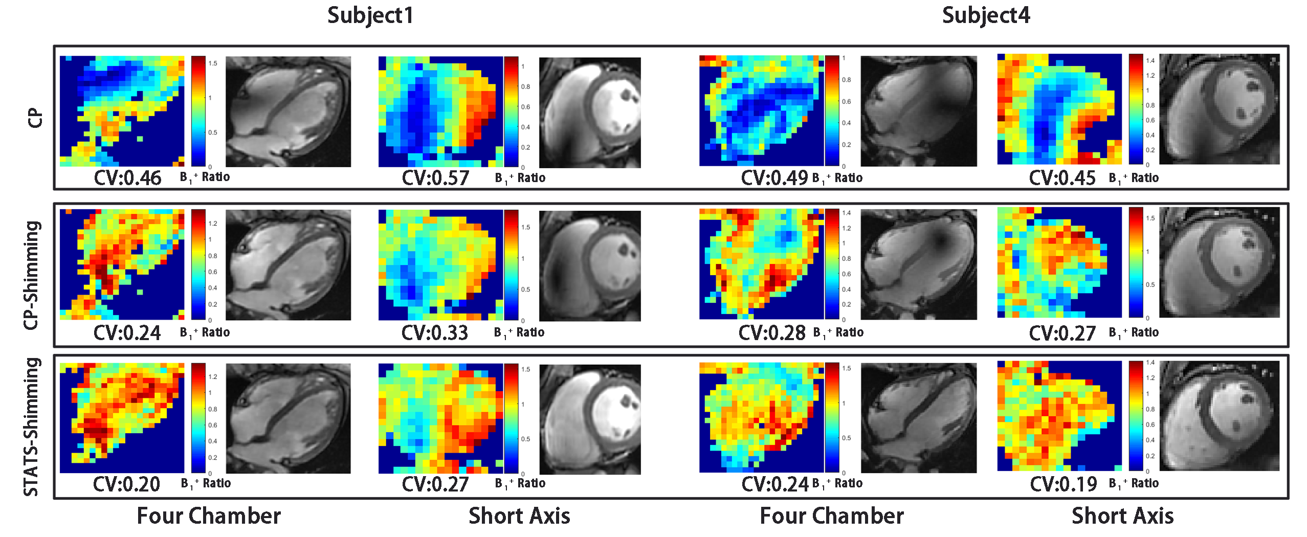
Figure 2 showed the phase distribution of shimming results of 19 volunteers in the direction of four chamber. Through Shapiro-Wilk test, P-Values of all channels except channel 3 were greater than 0.05, that is to say, for subjects with good CP-Shimming, the phase of each channel basically follows normal distribution.This revealed that there were some commonalities among different individuals,so a more general RF shimming method could be formulated.B1+ field and GRE-CINE results under CP-Shimming and STATS-Shimming were exhibited in Figure 3. It was observed that RF shimming can improve the large-scale under-excitation region in CP mode. Furthermore, compared with CP-Shimming, the results obtained by STATS-Shimming were still enhanced, the overall contrast distribution of the image was more uniform,and the under-excitation or over-excitation areas were amended.In addition, we tried the short-axis position with the same method, and some subjects also achieved good results.
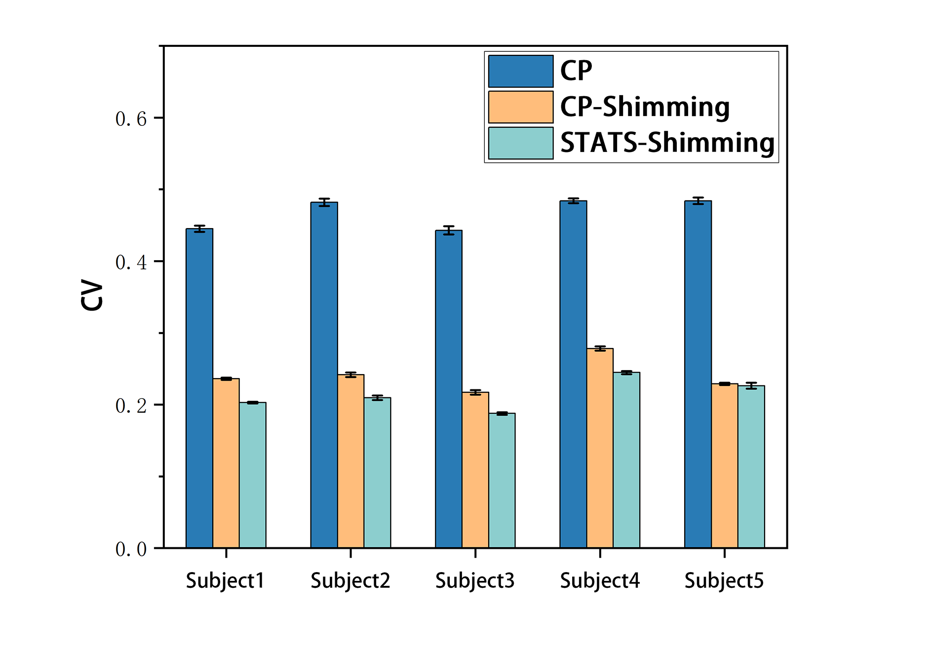
Figure 4 displayed the CV statistical results of B1+ field of all five subjects, including 4 subjects with poor performance in CP-Shimming and one good heart (Subject5). It could be observed that when the STATS-Shimming is applied, the CV values in ROI of all subjects reach the minimum value.Compared with CP shimming, the first four subjects increased by 13.24% on average, and kept good results for the better CP-shimming object subject5. That is, this method can image different hearts stably.
Conclusion and Discussion
This study demonstrated a method of cardiac RF shimming, which can improve the 'Invalid Shimming' that may appear in some subjects by changing the initial value.We used the statistical results as the initial point in RF shimming optimization to raise the uniformity of B1+ in the heart region, improve the under-excitation region in ROI, and thus enhance the quality of CMR. This method was verified at 5T MRI,which revealed that there may be some commonalities among individual differences of the cardiac shimming.Future research will focus on verifying more anatomical surfaces or using other methods to find the global optimal solution to further improve the stability of CMR.Acknowledgements
This work is supported by National Key Research and Development Program of China, 2021YFE0204400; the Strategic Priority Research Program of Chinese Academy of Sciences, XDB25000000; National Natural Science Foundation of China, U22A20344; Youth Innovation Promotion Association of CAS No. Y2021098; Key Laboratory Project of Guangdong Province, 2020B1212060051; Shenzhen city grant, RCYX20200714114735123.References
1. Noeske R, Seifert F, Rhein KH, Rinneberg H. Human cardiac imaging at 3T using phased array coils. Magn Reson Med. 2000;44:978-982.2.
2. Mueller A, Kouwenhoven M, Naehle C, et al. Dual-source radiofrequency transmission with patient-adaptive local radiofrequency shimming for 3.0-T cardiac MR imaging: initial experience.Radiology.2012;263:77–86.3.
3. Arian Beqiri,Anthony N. Price,et al.Extended RF shimming: Sequence-level parallel transmission optimization applied to steady-state free precession MRI of the heart.NMR in Biomedicine. 2017;e37014.
4. He X, Schmidt S, et al. Improved TSE imaging at ultrahigh field using nonlocalized efficiency RF shimming and acquisition modes optimized for refocused echoes (AMORE). Magn Reson Med. 2022;88(4):1702-1719.
Figures
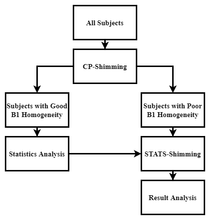
Figure 1:The overall workflow