4582
Slice-wise z-shimming for spinal cord fMRI with Shimming Toolbox1Neuropoly Lab, Institute of Biomedical Engineering, Polytechnique Montreal, Montreal, QC, Canada, 2Max Planck Institute for Human Cognitive and Brain Sciences, Leipzig, Germany, 3Department of Systems Neuroscience, University Medical Center Hamburg-Eppendorf, Hamburg, Germany, 4Mila - Quebec AI Institute, Montreal, QC, Canada, 5Functional Neuroimaging Unit, Centre de recherche de l'Institut universitaire de gériatrie de Montréal, Montreal, QC, Canada, 6Centre de recherche du CHU Sainte-Justine, Université de Montréal, Montreal, QC, Canada
Synopsis
Keywords: Shims, Spinal Cord
Spinal cord fMRI is challenging, in part due to B0 inhomogeneities. An EPI, z-shimming signal intensity based approach was implemented to automatically and dynamically shim the spinal cord. The technique was implemented in the open-source Shimming Toolbox. Z-shimming was tested on a healthy participant and showed a 14% increase in signal intensity as well as 19% increase in tSNR. The technique was generalized to allow for the use of any shim coil geometry as well as non-EPI data.Introduction
Spinal cord fMRI suffers from poor B0 shimming due to field inhomogeneities caused by its proximity to the lungs [1], as well as smaller scale variations due to connective tissue/vertebrae interfaces [2,3]. A complementary approach to the traditional volume-wise active shimming using field maps consists of applying different z-shim gradients to different EPI volumes and selecting the gradient that results in the highest signal intensity in the region of interest (ROI) for each slice [3]. This approach was automated in a recent paper [4] to reduce processing time, reduce user error and provide a better user experience. Here, we implement the automated EPI intensity-based z-shimming technique in Shimming Toolbox [5], a centralized, open-source software package to perform and prototype B0 and B1+ shimming experiments. Shimming Toolbox provides tools to convert DICOM files, create masks and compute shim coefficients (static, dynamic and real-time) using field map based methods, allowing for easy integration of the technique within the ecosystem.Methods
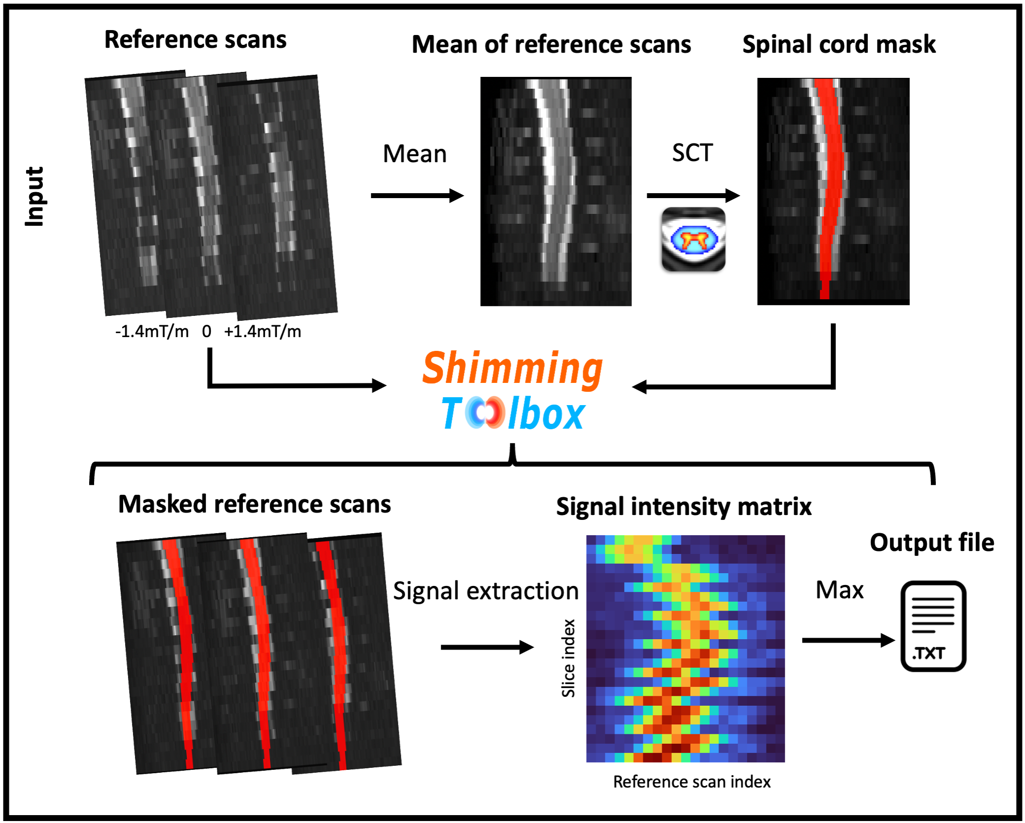
EPI intensity-based z-shimming in Shimming ToolboxThe intensity-based z-shimming technique involves the acquisition of multiple EPI volumes with different z-shimming gradients for each volume (i.e., referred to as the z-shim “reference” scans). A mask (ROI) of the spinal cord is generated from the reference scans and used to calculate the average signal in the ROI for each slice, and for each z-shim setting/EPI volume. The z-shim gradient yielding the maximum signal intensity for each slice is identified and output to a text file. The processing pipeline is shown in Figure 1.
Acquisitions
EPI scans of the spinal cord (C2/3 to T1/2) were acquired on a single healthy participant with proper ethics compliance to establish a baseline which was shimmed using the standard 2nd order shim procedure (Global 2SH) of the scanner (3 T Siemens Prisma). The acquisition parameters were: 100 volumes, 24 slices (5mm slices), matrix: 128 × 128, in-plane resolution: 1 × 1 mm2, TE: 40 ms, TR: 2338 ms, partial fourier: 7/8, phase encode: anterior-posterior, GRAPPA acceleration factor: 2, bandwidth per pixel: 1220 Hz/Pixel and fat saturation.
Z-shim reference scans were acquired using a custom EPI sequence using the same acquisition parameters as the baseline scan. However, 21 equidistant z-shim gradients were used, compensating field inhomogeneities between +0.21 and −0.21 mT/m (in steps of 0.021 mT/m), to acquire 21 EPI volumes (one z-shim gradient per volume).
Shimming Toolbox was installed on a standalone computer able to retrieve the acquisitions of the scanner and automatically processed the following pipeline (~15 s). It converted the DICOM into NIfTI files (~5 s). The 4D EPI was averaged along the 4th dimension (<1 s), and was processed by the Spinal Cord Toolbox (SCT) [6] to obtain a mask of the spinal cord (~6 s). Shimming Toolbox then calculated the z-shim gradients yielding the maximum signal intensity for each slice (~1 s).
The text file output by Shimming Toolbox was read by a custom EPI sequence and another set of 100 EPI volumes was acquired using the same acquisition parameters as the baseline scans, but with dynamic (slice-wise) z-shim settings that maximize the signal intensity in the spinal cord.
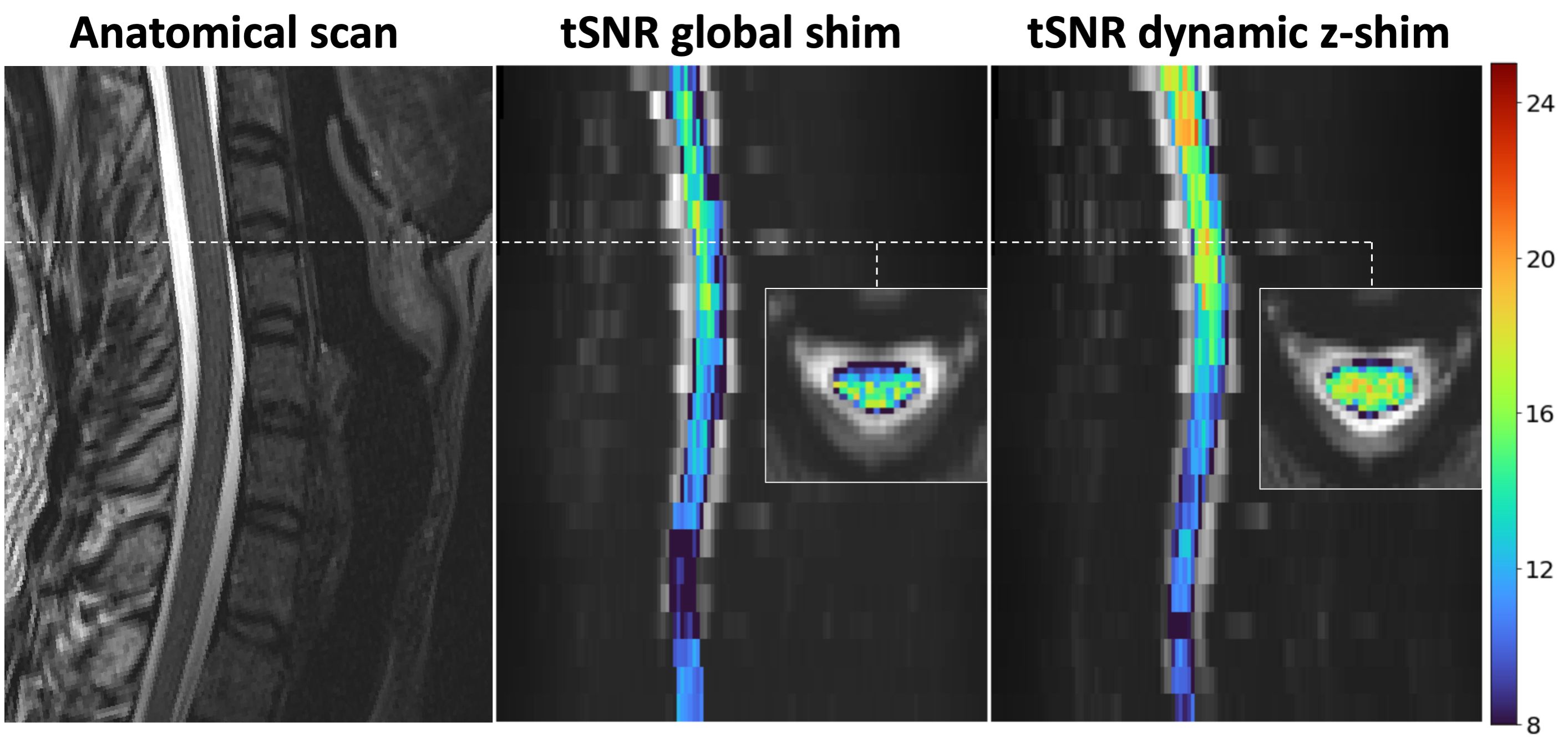
A T2-weighted acquisition was also acquired to provide anatomical landmarks.
Analysis
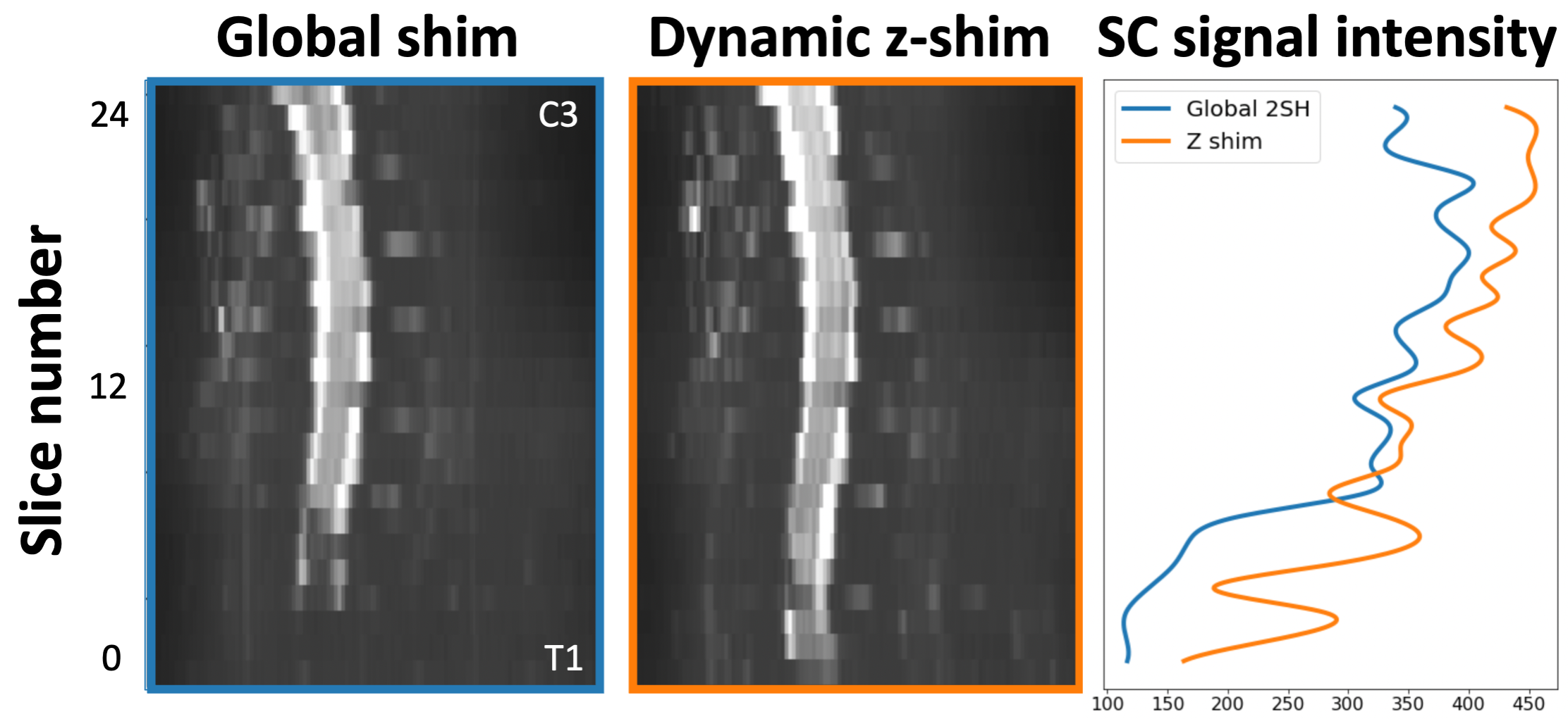
The baseline and z-shimmed EPI acquisitions were processed separately by extracting the spinal cord using SCT on the mean of the 100 EPI measurements. Both masks were manually assessed and no manual corrections were necessary. Both masks were also similar and no registration to a template was performed to avoid non-linear effects. The average signal intensity in the spinal cord for each slice was extracted using their respective masks. Temporal SNR (tSNR) maps were also calculated from the 100 EPI volumes of the baseline and z-shim scans.
Results
The signal intensity in the spinal cord for the baseline and z-shimmed scan is shown in Figure 2. Z-shimming resulted in a 14% increase in signal intensity across all slices and EPI volumes. tSNR was also computed in the spinal cord and is shown in Figure 3. tSNR increased by 19% across all EPI measurements and slices.Discussion
The main contribution of this work is a fully automated pipeline integrated in an open-source software package to perform EPI intensity-based z-shimming for the spinal cord. As previously demonstrated, z-shimming can increase signal intensity as well as tSNR in spinal cord fMRI time series. The intensity-based shimming technique is now offered alongside other field map based shimming tools in Shimming Toolbox. This shimming approach was also generalized to non-EPI data and can accommodate the computation of shim settings using coils of any geometry (1st, 2nd, 3rd order spherical harmonics, custom shim coils and others) in any target region.Conclusion
The EPI intensity-based z-shimming technique, which showed improvements in image quality for spinal cord fMRI, is now part of Shimming Toolbox. We hope this open-source platform will help researchers carry on advanced shimming experiments in a more convenient and reproducible manner. Future efforts will involve the expansion of this work to other body parts and the use of custom shim coil arrays.Acknowledgements
Funded by the Canada Research Chair in Quantitative Magnetic Resonance Imaging [950-230815], the Canadian Institute of Health Research [CIHR FDN-143263], the Canada Foundation for Innovation [32454, 34824], the Fonds de Recherche du Québec - Santé [322736], the Natural Sciences and Engineering Research Council of Canada [RGPIN-2019-07244], the Canada First Research Excellence Fund (IVADO and TransMedTech), the Courtois NeuroMod project, the Quebec BioImaging Network [5886, 35450], INSPIRED (Spinal Research, UK; Wings for Life, Austria; Craig H. Neilsen Foundation, USA), Mila - Tech Transfer Funding Program. Also funded by the Max Planck Society and the European Research Council (under the European Union’s Horizon 2020 research and innovation programme; grant agreement No 758974).References
1. Verma T, Cohen-Adad J. Effect of respiration on the B0 field in the human spinal cord at 3T. Magn Reson Med. 2014;72: 1629–1636.
2. Cooke FJ, Blamire AM, Manners DN, Styles P, Rajagopalan B. Quantitative proton magnetic resonance spectroscopy of the cervical spinal cord. Magn. Reson. Med. 2004. pp. 1122–1128. doi:10.1002/mrm.20084
3. Finsterbusch J, Eippert F, Büchel C. Single, slice-specific z-shim gradient pulses improve T2*-weighted imaging of the spinal cord. Neuroimage. 2012;59: 2307–2315.
4. Kaptan M, Vannesjo SJ, Mildner T, Horn U, Hartley-Davies R, Oliva V, et al. Automated slice-specific z-shimming for functional magnetic resonance imaging of the human spinal cord. Hum Brain Mapp. 2022. doi:10.1002/hbm.26018
5. D’Astous A, Cereza G, Papp D, Gilbert K, Stockmann J, Alonso-Ortiz E, Cohen-Adad J Shimming Toolbox: An open-source software toolbox for B0 and B1 shimming in Magnetic Resonance Imaging, Magn Reson. Med. 2022 DOI: 10.1002/mrm.29528, https://shimming-toolbox.org/ [in prod]
6. De Leener B, Lévy S, Dupont SM, Fonov VS, Stikov N, Louis Collins D, et al. SCT: Spinal Cord Toolbox, an open-source software for processing spinal cord MRI data. Neuroimage. 2017;145: 24–43.
Figures