4556
Silicon Carbide (SiC) Based Flexible and Stretchable Dielectric Pad for B1 Field Enhancement1School of Biological and Health Systems Engineering, Arizona State University, Tempe, AZ, United States, 2Department of Biomedical engineering, Yonsei University, Wonju, Korea, Republic of
Synopsis
Keywords: New Devices, RF Arrays & Systems, Invisible Dielectric pad, Silicon Carbide, SNR enhancement
We propose a stretchable, flexible, and MRI invisible dielectric pad to enhance the quality of acquired MR images. A silicon-based elastomer is mixed with Silicon Carbide (SiC) to develop this proposed dielectric pad. Preclinical in-vivo imaging of a mouse head was conducted at 9.4T to validate the performance of the dielectric pad. 2.54% (in dB) increase in the central SNR of the brain was observed after using the dielectric pad and peripheral brain locations have shown improved contrast. The flexible and stretchable dielectric pad could be used as a substitute for conventional dielectric pads, especially in irregular sample shapes.Introduction
Dielectric materials can increase the image quality by improving the transmit efficiency and the Signal-to-Noise Ratio (SNR) with aide of secondary magnetic fields across different magnetic field strengths.1,2 Different types such as slurries and rigid blocks have been widely studied and evaluated with MR imaging. These conventional forms of dielectric materials are bulky and present safety challenges. Rigid blocks are mostly sintered dielectric powder and cannot fit the subject shapes. Although solution or slurry based pads are flexible, they are susceptible to leakage which is a safety concern. Another important issue with conventional dielectric pads is being visible in MR images. A common solution to this issue is adding heavy water (D2O), however the effect is temporary, and the pad loses its invisibility over time.3,4 Silicon carbide (SiC) based dielectric materials are biocompatible, hemocompatible, and MRI invisible.5,6 The SiC pads also have a similar impact on the average and local Specific Absorption Rate (SAR) as Barium Titanate (BaTiO3) dielectric pads as well as the improved B1 field distribution when compared to the reference (without dielectric pad).6 In this study, we propose a new stretchable, flexible, and MRI invisible dielectric pad and evaluate its performance with the preclinical in-vivo imaging at 9.4T.Method
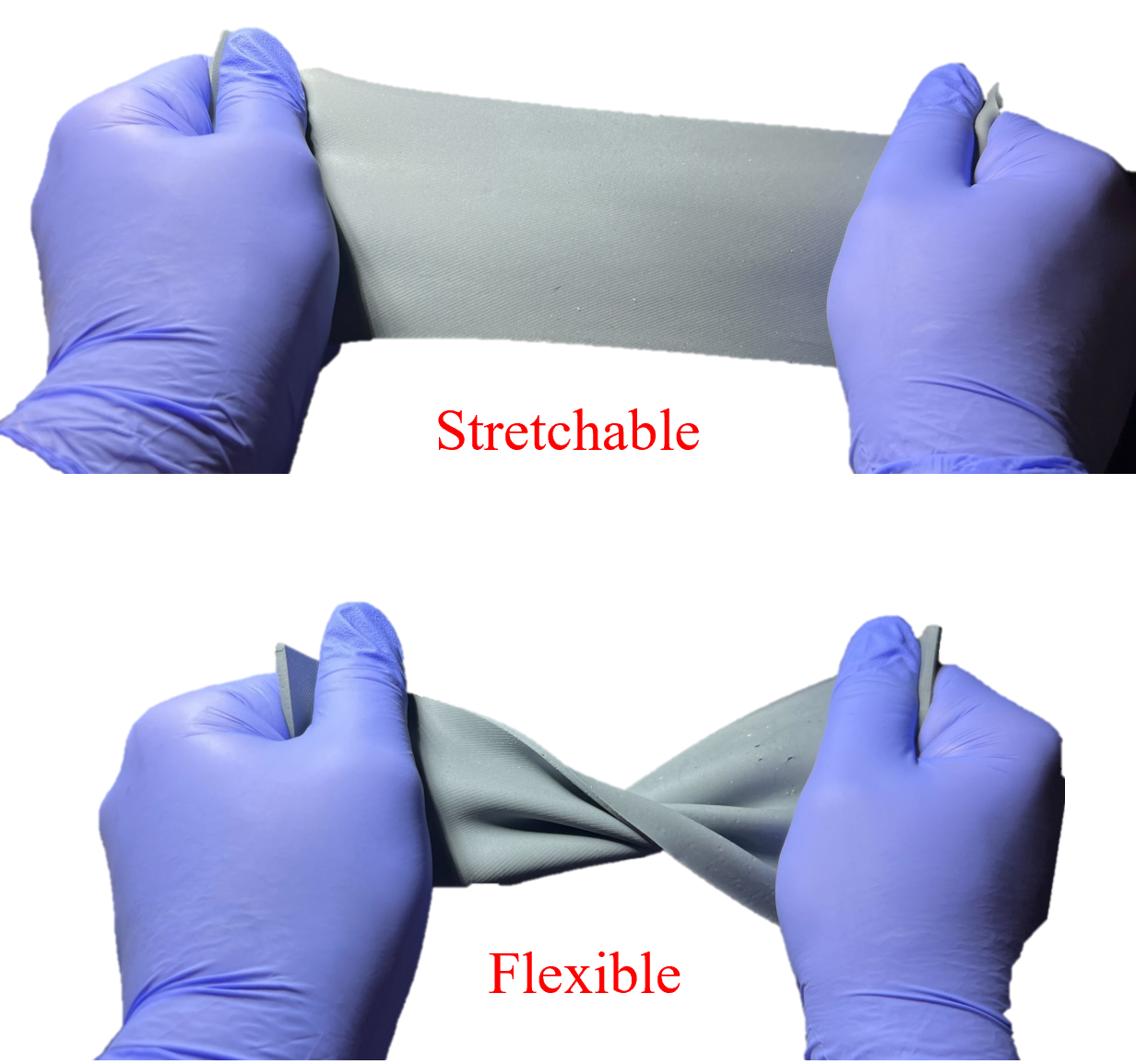
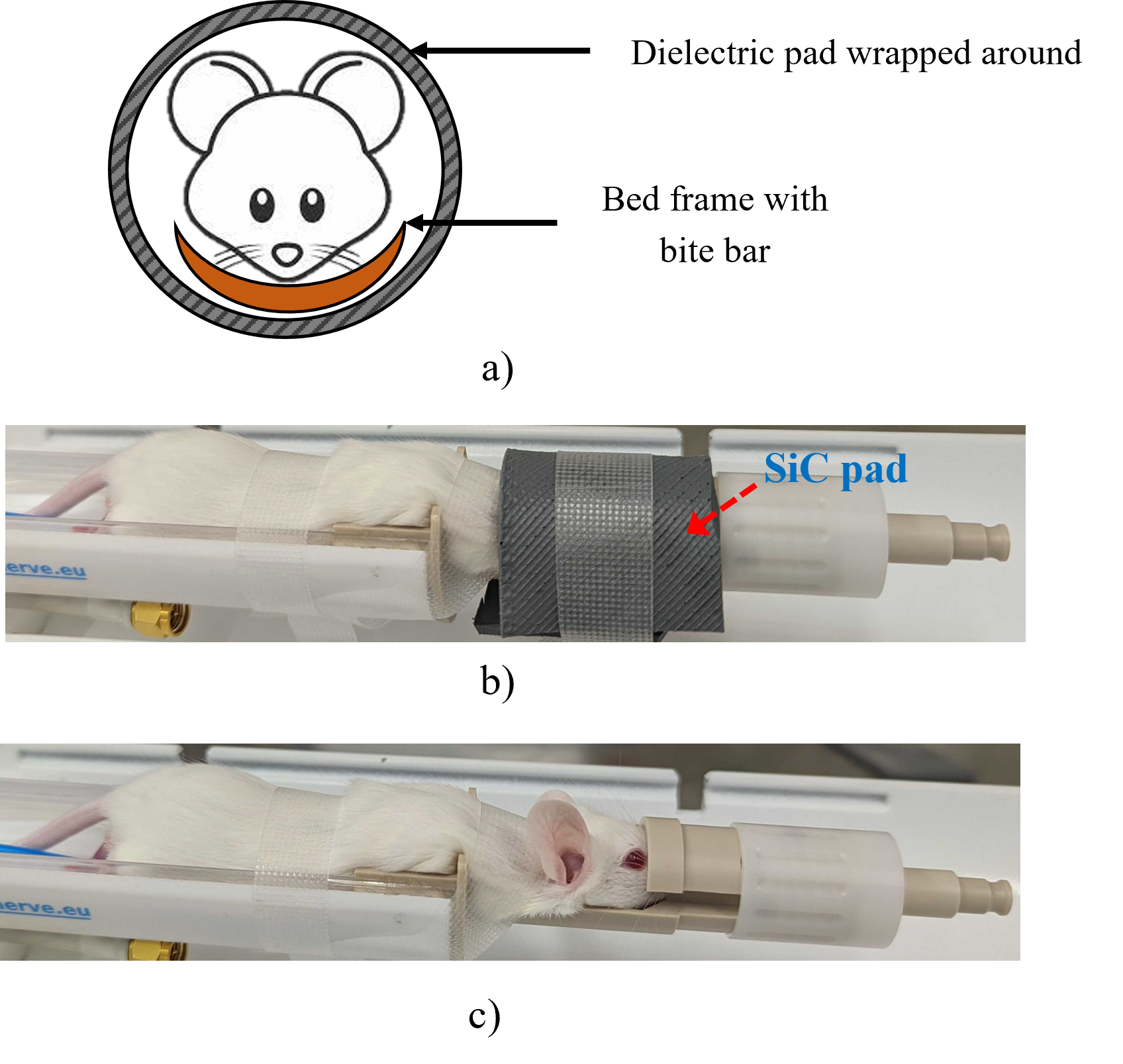
SiC powder (Poly Plastics, USA) was mixed with a biocompatible silicon elastomer (Ecoflex-Series, Smooth-On, USA) to create a 58% (weight to weight ratio) SiC dielectric pad. The solution was cured in vacuum chamber for 5 minutes to eliminate the air bubbles and was later transferred to a 3D print mold with the thickness of 3 mm. The mold was placed at room temperature (23℃) for 10 minutes and placed inside an oven to accelerate the curing process. Fig.1 demonstrates the stretchability and flexibility of the final pads. In order to assess the performance of the fabricated SiC pad, a preclinical 9.4T mouse head in-vivo imaging experiment was conducted at Magnetic Resonance Research Center - Arizona State University (MRRC-ASU) facility using a variable field strength scanner (MR Solutions, USA).Results and Discussion
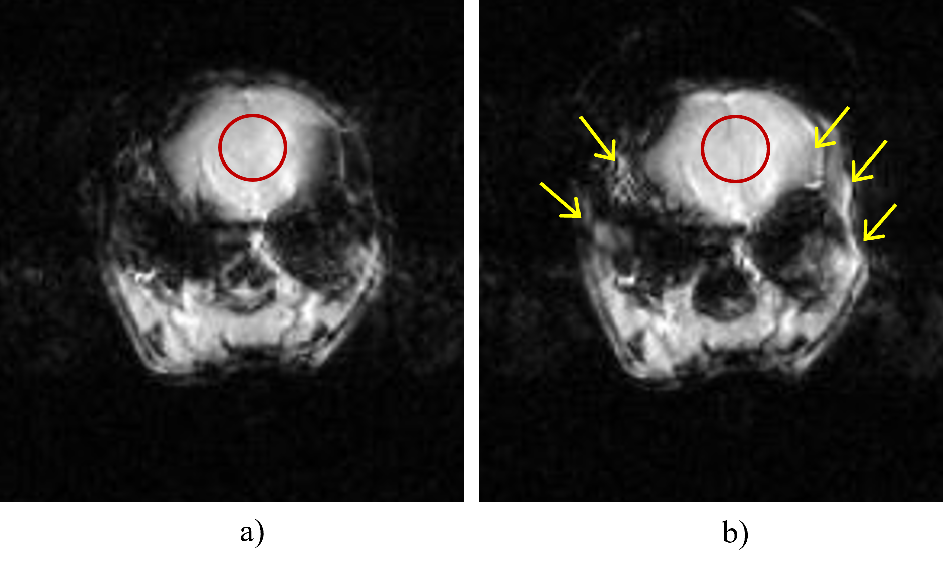
In the imaging experiment, Fast-Low-Angle-Shot (FLASH) sequence was employed with 200 ms repetition time (TR), 4 ms echo time (TE), 25 degrees flip angle (α), and the field of view (FOV) of 35 × 35 mm. In addition, 10 slices with a 1 mm slice thickness were acquired along the sample. In this experiment, a quadrature volume coil (MR Solutions, USA) was used for acquiring the images. We performed the in-vivo mouse brain imaging to fully characterize the impact of the proposed flexible dielectric pad in preclinical imaging at 9.4T as shown in Fig.2a, 2b, and 2c. Region of interest (ROI) is indicated with a red circle and the SNR of that area improved by 2.54% (from 36.78 dB to 37.73 dB) after wrapping the material around the mouse head, as shown in Fig.3a and 3b. Furthermore, the marked areas with red arrows in Fig.3b demonstrated a significant contrast enhancement in peripheral brain locations with the wrapped SiC dielectric pad around the mouse head. The elastomer used in this work demonstrates over 100% stretchability without a powder mix. However, the stretch ratio was reduced to 50% of its original dimensions when the SiC powder was mixed because of the addition of silicon carbide molecules to the silicon-based elastomer. This increases the strength of the bonds between the layers and degrades stretchability. Further material studies are in progress to better understand the structural limits of the elastomer, chemical interactions, change of dielectric properties over time with various dielectric materials, and performance of different materials across various field strengths.Conclusion
An MRI invisible, stretchable, and flexible dielectric pad was introduced and evaluated with in-vivo imaging. The SNR improvement of 2.54% was obtained using a single layer of pad in the mouse head image. Invisible peripheral structures in mouse brain were visible when SiC dielectric pad was applied.Acknowledgements
No acknowledgement found.References
1. Gandji NP, Sica CT, Lanagan MT, et al. Displacement current distribution on a high dielectric constant helmet and its effect on RF field at 10.5 T (447 MHz). Magn Reson Med. 2021;86(6):3292-3303. doi:10.1002/mrm.28923
2. Webb AG. Dielectric materials in magnetic resonance. Concepts Magn Reson Part A Bridg Educ Res. 2011;38 A(4):148-184. doi:10.1002/cmr.a.20219
3. Yang QX, Wang J, Zhang X, et al. Analysis of wave behavior in lossy dielectric samples at high field. Magn Reson Med. 2002;47(5):982-989. doi:10.1002/mrm.10137
4. O’Reilly TPA, Webb AG, Brink WM. Practical improvements in the design of high permittivity pads for dielectric shimming in neuroimaging at 7 T. J Magn Reson. 2016;270:108-114. doi:10.1016/j.jmr.2016.07.003
5. Saddow SE. Silicon Carbide Technology for Advanced Human Healthcare Applications. Micromachines. 2022;13(3). doi:10.3390/mi13030346
6. Raolison Z, Dubois M, Luong M, et al. Evaluation of new MR invisible silicon carbide based dielectric pads for 7 T MRI. Magn Reson Imaging. 2022;90(April):37-43. doi:10.1016/j.mri.2022.04.002
Figures