4554
Ultra-thin metasurface pads for spine MRI
Marc Dubois1, Amira Trabelsi1,2,3, Tania Vergara Gomez1,2,3, Pierre Jomin3, Djamel Berrahou1, David Bendahan2,4, Frank Kober2,4, Virginie Callot2,4, Stefan Enoch3, and Redha Abdeddaim3
1Multiwave Imaging, Marseille, France, 2Aix Marseille Univ, CNRS, CRMBM, Marseille, France, 3Aix Marseille Univ, CNRS, Centrale Marseille, Institut Fresnel, Marseille, France, 4APHM, Hopital Universitaire Timone, CEMEREM, Marseille, France
1Multiwave Imaging, Marseille, France, 2Aix Marseille Univ, CNRS, CRMBM, Marseille, France, 3Aix Marseille Univ, CNRS, Centrale Marseille, Institut Fresnel, Marseille, France, 4APHM, Hopital Universitaire Timone, CEMEREM, Marseille, France
Synopsis
Keywords: New Devices, Spinal Cord, Metamaterial, Metasurface
Spine imaging is usually conducted at 3T given the need of high spatial resolution. Unfortunately, the lumbar and thoracic spine regions of interest will lie in areas of weak transmit efficiency. Recent studies showed that high permittivity ceramic blocks can improve image quality. Here we present a new approach based on ultra-thin, lightweight, flexible metasurface pads yielding improved efficiency and homogeneity of the B1+ field along the spine in clinical 3T MRI settings. Our design offers seamless integration to regular equipment leading to more efficient and safer spine imaging protocols.Introduction
3T MR scanners are popular in a clinical context, offering higher SNR compared to the widely deployed 1.5T. Higher field strength leads to shorter scanning times or higher resolution images especially sought for spine imaging [1]. However, when imaging large cross-sectional areas such as the abdomen or the thorax, inhomogeneities of the radiofrequency field transmitted by the body coil generates a skewed flip angle distribution affecting image contrast [2]. High dielectric constant pads have been used as an efficient solution to mitigate radiofrequency inhomogeneities as presented in a recent review article [4]. Progress in MRI metamaterials has gained significant traction allowing more degrees of freedom in their design compared to traditional dielectric pads [5-7]. Focusing on 3T spine imaging, Koolstra et al. demonstrated that the use lead zirconate titanate blocks with relative permittivity reaching over a thousand can simultaneously improve image quality and lower power deposition [3]. However, the production of such materials is very limited, even in terms of dimensions. Moreover, the seven blocks used in the study were 3-cm thick, weighed more than a kilogram each and had to be placed carefully on the patient table. Here, we present an alternative approach based on ultra-thin, lightweight, and flexible metasurface pads. Our design offers a seamless integration into standard MRI equipment, as well as a robust and cost-effective solution leading to more efficient and safer 3T spine imaging protocols.Materials and Methods
The proposed metasurface works under the resonance hybridization principle [6,8]. A series of parallel conductors were printed on a thin polyimide substrate (200mm) and sealed in a polymer-coated fabric pouch. The conductors are inductively coupled to the body birdcage coil and redistribute the B1+ field [6]. Figure 1 presents photographs of the WearMe metasurface. A single pad was placed on top of the patient table beneath the volunteer. Experiments were performed on a single volunteer (Male, 193cm, BMI 29 kg/m2) in a 3T Vida MRI scanner (Siemens Healthineers, Erlangen, Germany). Twelve channels of the integrated table coils were activated during the experiments. Flip Angle (FA) maps were acquired at the thoraco-lumbar levels with a turbo-flash (TFL) MRI sequence [9] in sagittal orientations with a 650 V reference voltage, TR/TE= 5000/1.8ms, FA= 8°, FOV= 250x400 mm, matrix= 80x128 and 13 slices of 15-mm thickness. Raw images were used to delineate the position of the spinal cord and to report the region of interest in the FA maps. T1-weighted and T2-weighted images were also acquired with recalibrated reference voltage based on the FA maps (T1-w 2D-TSE: TR/TE= 641/9.6ms, ETL= 3, Acceleration factor= 3, FOV= 238x400 mm, matrix= 210x352, 30 slices of 5-mm thickness; T2-w 2D-TSE HASTE (80% partial Fourier) TR/TE= 2000/97ms, ETL= 109, FOV= 325x400 mm, matrix= 208x256, 30 slices of 5-mm thickness.Results
FA maps and profiles are shown in Figure 2. Along the spinal cord, a 36% increase in FA was observed with the WearMe configuration for a given reference voltage (Fig 2b). The homogeneity of the FA distribution was evaluated along the cord using the relative standard deviation (RSD)(Fig.2c). We observed an RSD of 17% in the reference case and a reduction to 9% with WearMe. FA maps were used to normalize the reference voltage to an 80° target angle in the lumbar spine: 747V for the reference and 548V with WearMe. Consequently, the input power was reduced by 47% in the presence of WearMe. SAR simulations (data not shown) were performed before the in vivo studies and showed that global and max 10g local SAR were marginally affected by the presence of WearMe. Furthermore, our results have shown that using WearMe, a similar FA can be obtained with half the input power thereby limiting power absorbed. High-resolution T2w and T1w images obtained with and without WearMe are reported in Figure 3 and 4. Each acquisition was performed with the specific reference voltage given above. A more homogeneous contrast was observed along the whole field of view, especially going toward the thoracic spine.Discussion
Our experimental results indicate that the initial FA inhomogeneity along the spine was reduced by 50% in the presence of WearMe leading to a 9% RSD along a 350mm profile following the spinal cord. High-resolution images in Figures 3 and 4 confirmed that the improved FA homogeneity resulted in a better contrast along the spinal cord with a single acquisition which could be helpful for an improved detection of signal abnormalities across such a large field of view. This can also be of interest for a scan time reduction as the imaging protocol may require multi-stage MRI acquisitions to cover all spine sections. The performance of the WearMe pad is currently assessed in a larger cohort of volunteers and in different MRI scanners with various birdcage coil designs.Conclusion
WearMe, the ultra-thin, lightweight, flexible metasurface pad, has been shown to dramatically improve transmit efficiency and contrast homogeneity over a 40cm-long field of view in the case of 3T spine MRI. Altogether, our design is seamlessly integrated with standard MRI equipment and offers a robust and cost-effective solution to improve and accelerate 3T spine MRI protocols.Acknowledgements
The project leading to this publication has received funding from the Excellence Initiative of Aix-Marseille University - A*MIDEX, a french "Investissements d'Avenir" programme.References
[1] Vertinsky, AT, et al. Neuroimaging Clin N Am 2007; 17:117–136
[2] Dietrich, O, et al. Eur J Radiol 2008; 65:29–35. 3
[3] Koolstra, K, et al., Magnetic Resonance in Medicine 2018; 79: 1192-1199
[4] Webb, AG, et al. Magnetic Resonance
Materials in Physics, Biology and Medicine 2022; 1-20
[5]
Dubois, M, et al.
Physical Review X 2018; 8.3: 031083
[6] Vergara, T, et al., Proc. Intl. Soc. Mag. Reson. Med. 2021;
29.1405
[7] Vorobyev, V, et al.
Magnetic Resonance in Medicine 2022; 87.1:496-508
[8] Jouvaud, C, et
al. Applied Physics Letters 2016; 108(2):023503.13
[9] Chung, S, et al. Magnetic Resonance in Medicine 2010;64(2):439–446
Figures

Figure 1. Photographs of the
WearMe metasurface pad, presenting ultra-thin (<1mm), lightweight (~200g),
and flexible design. Size: 65x40 cm.
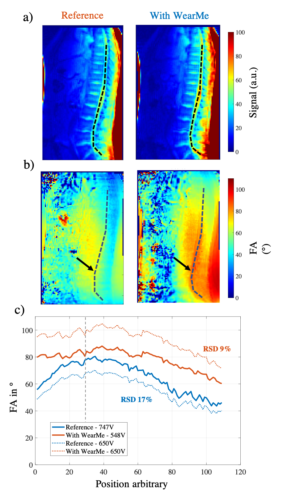
Figure 2. FA maps and FA profiles
with and without WearMe pads. (a) Raw magnitude image used to delineate the
thoraco-lumbar spinal cord with a black dotted line. (b) FA maps with an
identical reference voltage (650V). The dotted lines were reported in the FA
maps to extract FA profiles along the spinal cord. (c) Original and normalized
FA profiles with a normalized reference voltage for each experiment to reach
the target FA (80°) at the black arrow location. The relative standard
deviation (RSD) of FA across the z direction (dotted line) was reported for
each case.
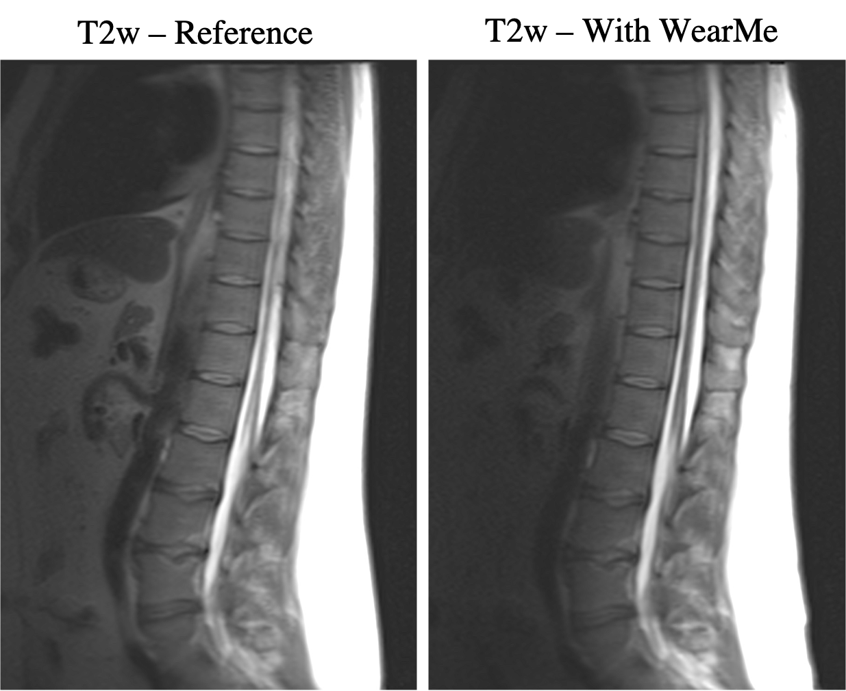
Figure 3. T2-weigthed sagittal
image acquired on the same volunteer. Experiments with and without the WearMe
pads were performed with a normalized reference voltage (747V for the reference
and 548V in presence of WearMe). The homogeneity of the FA distribution with
WearMe allowed a homogeneous contrast along the whole field of view.
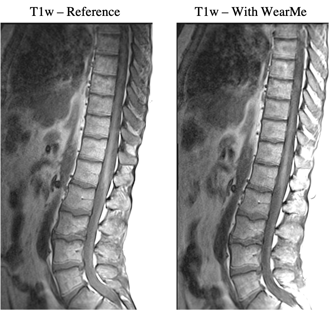
Figure 4. T1-weigthed sagittal
image acquired on the same volunteer. Experiments with and without the WearMe
pads were performed with a normalized reference voltage (747V for the reference
and 548V in presence of WearMe). The homogeneity of the FA distribution with
WearMe allowed a homogeneous contrast along the whole field of view.
DOI: https://doi.org/10.58530/2023/4554