4549
MRI IN RADIATION THERAPY: VALIDATION OF THE EFFECT FROM BLANKET MRI-COIL ON THE PELVIS REGION OF HEALTHY VOLUNTEERS
Yosef Al-Abasse1, Tova Roman Rung1, Peter Larsson1, Dan Josefsson1, Lena Lundkvist2, Kristina Redelius2, Kristina Hultman2, Maria Nilsson2, Jan Rzepecki2, and Peter Lundberg1,3
1Department of Health, Medicine and Care, Linköping University, Linköping, Sweden, 2Department of Oncology, and Department of Biomedical and Clinical Sciences, Linköping University, Linköping, Sweden, 3Center for Medical Imaging and Visualization (CMIV), Linköping University, Linköping, Sweden
1Department of Health, Medicine and Care, Linköping University, Linköping, Sweden, 2Department of Oncology, and Department of Biomedical and Clinical Sciences, Linköping University, Linköping, Sweden, 3Center for Medical Imaging and Visualization (CMIV), Linköping University, Linköping, Sweden
Synopsis
Keywords: Multimodal, Radiotherapy, Synthetic CT, T2-w, software
Conventional coils for dose planning purposes cannot be placed on the patient as the outer body contour then will be deformed. Coils are therefore placed on a special holder which creates a distance between coil and patient, but this leads to less SNR. The blanket-coils are significantly lighter and can be placed directly on the patient. Three similarity comparison methods were used here, Hausdorff distance, Dice similarity coefficient (DSC) and Surface DSC. Eight of eleven comparisons were within 4 mm difference for Hausdorff distance; Surface DSC was >99%, at a 3 mm tolerance, and DSC >98.5%.
Introduction
MRI-images are used in radiotherapy to delineate the target and organs at risk because of the superior image quality compared to CT-images [1]. Since the MRI scan is done with conventional array coils that are heavy, “coil spacers” are used leading to less signal-to-noise ratio (SNR), and thus worse image quality due to the distance between the coil and the body. The aim of this project was to examine if a super-light blanket MR-coil (‘AIR™-coil’) positioned directly on the skin surface of the pelvis would affect the body contours. In order to achieve this goal, we aimed to validate a software tool in MICE (NONPIMedical AB, Umeå, Sweden) using different methods for comparison. To compare two contours some established methods are widely used, such as the Hausdorff distance and Dice similarity coefficient (DSC). These are good measures for geometric similarity, but do not always correlate with clinical applicability of the contours (or time needed to adjust them [2]). In 2018 Nikolov, et al., [3] presented a new similarity measurement method, ‘Surface DSC’.Material and Methods
Thirteen healthy volunteers gave informed consent and were imaged in a GE Signa Architect 3.0 T MRI-scanner using “coil spacers” and then without; see Fig. 1 where the blanket MR-coil is placed directly on the skin contour. The pulse sequence that was used in this study was a T2-weighted sequence with a 44 cm FOV in the plane, 2.5 mm slice thickness and a total time of 6 min and 33 sec. For the evaluation of the blanket coil (‘AIR™ coil’; GE Healthcare), three different similarity comparison methods were used, Hausdorff Distance (HD), Dice Similarity Coefficient (DSC) and Surface Dice similarity Coefficient (SDSC). In Fig.2 the flow chart for the implementation in MICE of the image registration, segmentation and similarity measure methods are shown. After the images had been imported to MICE, only slice #40 – #60 were extracted, as this made the algorithm faster. Then a mask was created from the dorsal area, with the spacer. A mask for the ventral area was also created from the same MRI scan. Both masks were created using the centre position and size of the input image. The image registration was done using the node Elastix2 (in MICE), which registers images using specific parameters. Rigid translation registration was then used. The dorsal mask was also used as a fixed mask, this makes the sampler in MICE draw the required number of samples from within the selected image region. During segmentation the ventral mask was applied to the MRI-images, with and without the spacer, after the pre-processing (extraction of slices and registration). The two masks were then analysed using Overlap Measurement (in MICE) to get DSC and Hausdorff Distances. Both algorithms were already implemented in MICE as nodes. In addition, the Hausdorff distance node also calculated the average Hausdorff distance defined as the average of all minimum distances.Results
DSC and maximum HD are shown in Fig.3. The SDSC values for the skin contour using blanket coil are shown in Fig. 4. The maximum Hausdorff distances were all within 5 mm. For the patient with the largest deviation, which is Pat.# 5 in Fig.4, the maximum Hausdorff distances were less than 1%. Eight out of eleven image sets had a difference of less than 4 mm (HD), and a SDSC > 99% (3 mm tolerance). Nine out of eleven image sets had a DSC > 98.5%. At 3 mm tolerance subjects #4, #11 and #13 had all an SDSC of 100%. DSC measures gave above 98.5% for 9 out of 11 of the comparisons. Some comparison images are shown in Fig.5.Conclusion
The main purpose of this project was to develop a software tool in MICE to evaluate the blanket coil effect on the skin contour for the pelvis region. The results from the similarity comparison methods showed that the effect was negligible. The project itself could be further elaborated; it would be valuable to include more subjects, (including patients) in the evaluation of the blanket coil. Another future task could be to use a deep learning approach for segmentation.Acknowledgements
No acknowledgement found.References
1. Schmidt MA, Payne GS. Radiotherapy planning using MRI. Phys Med Biol. 2015;60(22):R323–61.
2. Vaassen , F, o.a. Evaluation of measures for assessing time-saving of automatic organ-at- risk segmentation in radiotherapy. Physics and imaging in radiation oncology. 2019, Vol. 13, ss. 1-6. https://doi.org/10.1016/j.phro.2019.12.001
3. Nikolov, S, o.a. Clinically Applicable Segmentation of Head and Neck Anatomy for Radiotherpy: Deep Learning Algorithm Development and Validation Study. Journal of medical Internet Research. e26151, 2021, Vol. 23, 7. https://doi.org/10.2196/26151.
Figures
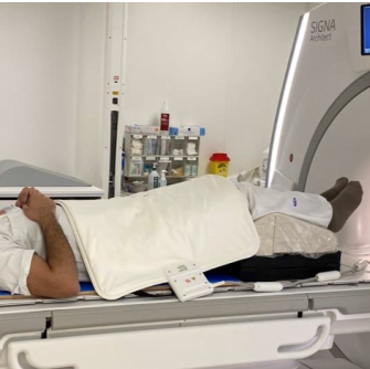
Figure 1. An MRI scan using the blanket coil directly on the pelvis, without the use of coil spacers.
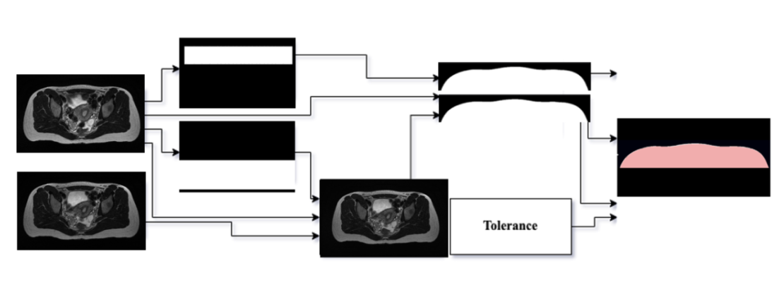
Figure 2. Visual flowchart over the segmentation algorithm, implemented in MICE.
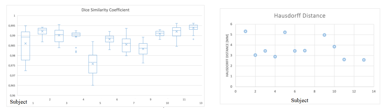
Figure 3. Left: Dice similarity coefficients for slice #40 – #60 for each subject. Right: The maximum Hausdorff distance [mm] for all subjects.
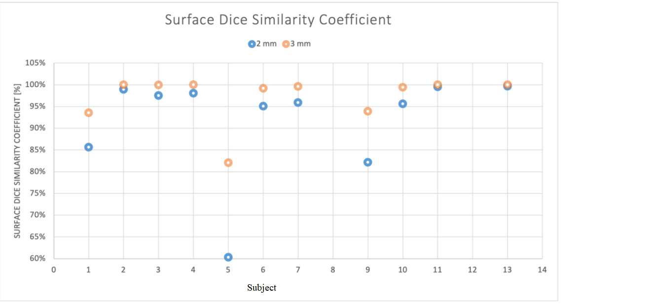
Figure 4. Surface Dice Similarity Coefficient,SDSC, [%] for tolerance criterion 2 mm and 3 mm, respectively, for the skin contour.
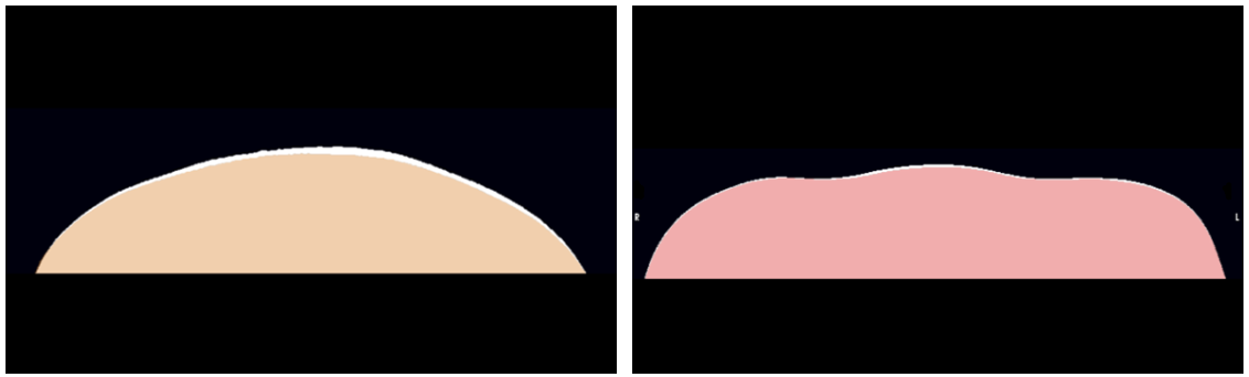
Figure 5. Left: Worst case: Subject #5, the difference between the segmentation using spacer, and without spacer. Right: Best case: Subject #13.
DOI: https://doi.org/10.58530/2023/4549