4546
Feasibility of high-frequency shear wave MR elastography of the parotid glands1Department of Biophysics, Institute of Physics, College of Natural Sciences, University of Rzeszow, Rzeszow, Poland, 2Department of Radiology, Mayo Clinic, Rochester, MN, United States, 3Center for Diagnostic Medical Sonography, Rzeszow, Poland, 4Department of Obstetrics and Gynecology, Institute of Medical Sciences, Medical College, University of Rzeszow, Rzeszow, Poland, 5Department of Otorhinolaryngology, Institute of Medical Sciences, Medical College, University of Rzeszow, Rzeszow, Poland, 6Institute of Medical Sciences, Medical College, University of Rzeszow, Rzeszow, Poland
Synopsis
Keywords: New Devices, Elastography
A specialized driver was developed to enable magnetic resonance elastography of the parotid and other salivary glands using high shear wave frequencies to provide improved spatial resolution and optimized ergonomics. The performance of this new device was tested in volunteer studies. Preliminary results showed that suitable shear wave illumination of the parotid gland is possible at frequencies as high as 120 Hz and that high-frequency shear wave MR elastography of the parotid gland is feasible.Introduction
Tumors of the salivary glands are most often first detected by noticing a palpable mass. This provides motivation to explore the potential of MR elastography for characterizing benign and malignant salivary gland tumors and to address the current limitations of conventional imaging techniques used to evaluate these lesions. Parotid glands are the most common site for the presentation of salivary gland tumors, accounting for approximately 86% of cases [1]. Previous studies have demonstrated the feasibility and promise of applying MRE technology to assess the mechanical properties of parotid tissues [2,3]. The goal of this work described here was to further advance MRE technology for this application by designing and testing an ergonomic driver device capable of generating usable shear wave fields in the glands at higher frequencies than the 60 Hz and 80 Hz values used in previous works. Higher shear wave frequencies provide shorter shear wavelengths and improve stiffness measurement accuracy in small lesions.Methods
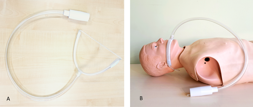
The design of the novel passive shear wave driver developed in this project was guided by considerations of patient comfort, ergonomics, convenience of use, and ability to localize the applied vibration to the anatomy of interest. The final design of the driver, fabricated from polyvinyl chloride tubing, is illustrated in figure 1. Volunteer subject testing of the performance of the novel driver was conducted using 3D vector MRE acquisition at vibration frequencies of 60, 90, and 120 Hz on a 1.5T whole-body Signa HDxt scanner (GE Healthcare, Milwaukee, WI, USA). The imaging parameters were as follows: field of view = 24 cm, repetition time = 3117.6 ms, echo time = 40.2 ms, spacing between slices = 3 mm, image matrix = 80 × 80, slice thickness = 3 mm, flip angle = 90°, time steps = 4. A full 3D direct inversion algorithm was used to process the MRE data.Results
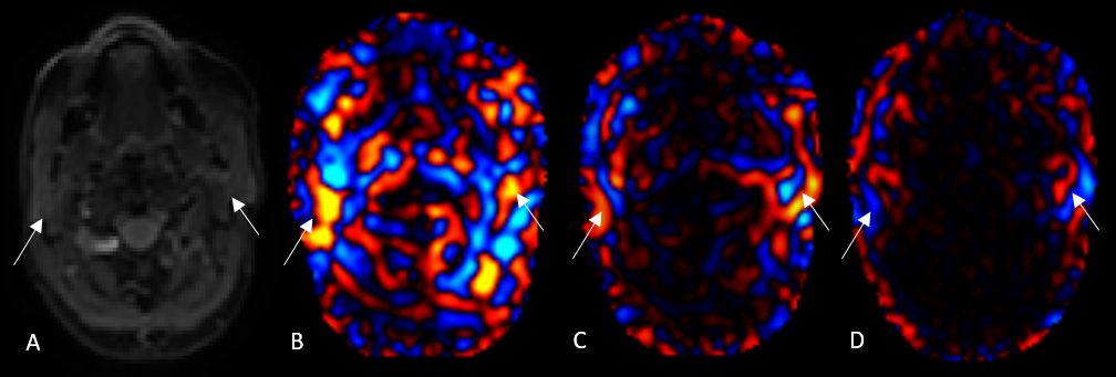
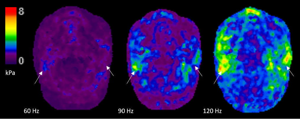
Testing demonstrated that the novel driver provided excellent shear wave illumination in the parotid gland at all tested frequencies, including 120 Hz, as demonstrated in figure 2. The calculated mean stiffness +/- SD in kPa of the parotid glands was 1.28 +/- 0.28 at 60 Hz, 2.31 +/- 0.36 at 90 Hz, and 3.39 +/- 0.51 at 120 Hz (Fig. 3).Discussion
In order to obtain reliable measurements of the stiffness of small lesions it is necessary for the spatial resolution of the acquisition to be sufficient to account for the spatial footprint of the inversion algorithm and for the displacement-induced phase shift across that necessarily small spatial footprint to be sufficiently high. The latter requirement is helped if the shear wavelength is short as possible across the footprint, which provides motivation for using higher shear wave frequencies. A further consideration is that shear wave attenuation in tissue increases with frequency. Designing the driver to be placed as close as possible to the region of interest addresses this factor.Conclusion
MR elastography at shear wave frequencies as high as 120 Hz is feasible with the use of the specialized shear wave driver developed in this project. The results provide the basis for planning studies aimed at assessing the potential role of MRE in characterizing benign and malignant parotid neoplasms.Acknowledgements
No acknowledgement found.References
1. Alvi S, Chudek D, Limaiem F. Parotid Cancer. [Updated 2022 Aug 8]. In: StatPearls [Internet]. Treasure Island (FL): StatPearls Publishing; 2022 Jan. Available from: https://www.ncbi.nlm.nih.gov/books/NBK538340/
2. Yeung D, Bhatia K, Lee Y, King A, Garteiser P, Sinkus R, Ahuja A. MR elastography of the head and neck: Driver design and initial results. Magnetic Resonance Imaging, Volume 31, Issue 4, 2013 624-629, ISSN 0730-725X, https://doi.org/10.1016/j.mri.2012.09.008.
3. Elsholtz FHJ, Reiter R, Marticorena Garcia SR, Braun J, Sack I, Hamm B, Schaafs LA. Multifrequency magnetic resonance elastography-based tomoelastography of the parotid glands-feasibility and reference values. Dentomaxillofac Radiol. 2022 Jan 1;51(1):20210337. doi: 10.1259/dmfr.20210337. Epub 2021 Sep 24. PMID: 34558305; PMCID: PMC8693321.
Figures