4540
Ultrashort Echo Time, MRI porosity index, and suppression ratio correlate with the cortical bone microstructural and mechanical properties1Department of Radiology, University of California, San Diego, La Jolla, CA, USA, San Diego, CA, United States, 2Radiology Service, Veterans Affairs San Diego Healthcare System, San Diego, La Jolla, CA, USA, San Diego, CA, United States, 3Department of Orthopedic Surgery, University of California, San Diego, La Jolla, CA, USA, San Diego, CA, United States, 4Research and Laboratories Sector, Saudi Food and Drug Authority, Riyadh, KSA, Riyadh, Saudi Arabia, 5Shiley Center for Orthopedic Research and Education at Scripps Clinic, La Jolla, CA, USA, San Diego, CA, United States, 6Department of Medicine, University of California, San Diego, La Jolla, CA, USA, San Diego, CA, United States
Synopsis
Keywords: Bone, Bone
The cortical bone porous microstructure can be evaluated using ultrashort echo time (UTE) MRI. UTE-MRI-based evaluation of bone has been underutilized partly due to the high cost and time demands of MRI in general. The porosity index (PI) and the suppression ratio (SR) are two rapid UTE-based bone evaluation techniques (~ 5 mins scan time each), which can potentially reduce the time demand and cost in future clinical studies. We have investigated the relationship of PI and SR measures with human cortical bone microstructural and mechanical properties. PI and SR showed significant correlations with microstructural and mechanical properties.Introduction
MRI-based cortical bone evaluation is attractive since MRI is tomographic and avoids the potential harm associated with x-ray-based techniques(1,2). The MRI-based bone evaluation may also provide an excellent assessment of the surrounding soft tissue, a benefit that is not available in x-ray-based techniques. Ultrashort echo time (UTE) MRI can image and consequently enable quantitative assessment of cortical bone. UTE-MRI-based evaluation of bone has been underutilized partly due to the high cost and time demands of MRI in general. The signal ratio in dual-echo UTE imaging, known as porosity index (PI), as well as the signal ratio between UTE and inversion recovery UTE (IR-UTE) imaging, known as the suppression ratio (SR), are two rapid UTE-based bone evaluation techniques (~ 5 mins scan time each), which can potentially reduce the time demand and cost in future clinical studies. The correlations of PI and SR with bone microstructural and mechanical properties were not investigated adequately. This study aimed to investigate such correlations in a relatively large number of specimens harvested from human cortical bone.Methods
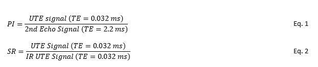
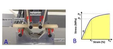
A total of 135 cortical bone specimens were harvested from the human tibial and femoral midshafts of 37 donors (61±24 years old). Bone strips were randomly distributed into eight groups and placed in 30-mL syringes filled with perfluoropolyether (Fomblin, Ausimont, Thorofare, NJ) during the MRI scans to minimize dehydration and susceptibility artifacts. MRI scans were performed on a 3T clinical scanner (GE Healthcare, Waukesha, WI) using a homemade 1-inch diameter transmit/receive birdcage coil. The UTE-MRI scans involved a) the dual-echo 3D Cones UTE sequence (repetition time (TR)=100ms, TE=0.032 and 2.2ms, flip angle (FA)=10°) for PI measurement (Eq.1 in Fig.1) (3) and, b) the 3D Cones IR-UTE sequence (TR=100ms, TI=45ms, and TE=0.032ms, FA=20°) for SR calculation ((Eq.2 in Fig.1) (4). Other imaging parameters included: field of view (FOV)=40 mm, matrix size=160×160, in-plane pixel size=0.25 mm, slice-thickness=2mm, receiver bandwidth=125 kHz, and total scan time ≈ 10 mins. Specimens were later scanned on a μCT scanner (Skyscan 1076, Belgium) at 9 μm isometric voxel size. Average bone porosity, pore size, and BMD were measured from μCT images. Next, the tensile mechanical properties of each bone strip were measured using a four-point bending failure test (Fig.2) (5). Spearman’s rank correlations were calculated between the UTE-MRI indices, microstructural parameters, and mechanical properties (Young’s modulus, yield stress, ultimate stress, and failure energy). Correlations with P-values below 0.05 were considered significant. All measurements and models were performed using MATLAB software (The Mathworks Inc., Natick, MA, USA).Results
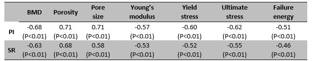
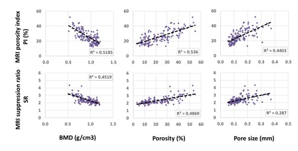
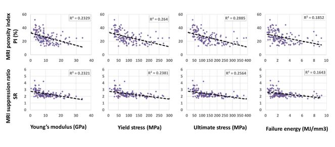
Spearman’s correlations of MRI-based measures (PI and SR) with microstructural and mechanical properties are presented in Figure3 (Table1). The µCT measures showed significant correlations with PI (moderate to strong, R=0.68-0.71) and SR (moderate, R=0.58-0.63). Young’s modulus, yield stress, and ultimate stress demonstrated significant moderate correlations with PI and SR (R=0.52-0.62). PI correlates higher than SR with microstructural and mechanical properties. Figures 3 demonstrates the scatter plots and linear regressions of PI and SR on the µCT-based BMD, porosity, and pore size. Figures 4 illustrates the scatter plots and linear regressions of PI and SR on Young’s modulus, yield stress, ultimate stress, and failure energy.DISCUSSION
PI and SR, two recently developed rapid UTE-MRI-based indices, showed significant correlations with microstructural and mechanical properties of cortical bone specimens. PI correlated better than SR with the microstructural and mechanical properties of bone strips. However, there might be an optimal TR/TI combination that could further improve the correlations. This should be investigated in future studies. This study highlighted PI and SR, as potential rapid techniques to assess cortical bone mechanical properties and intracortical bone microstructure. PI and SR from UTE-MRI are noninvasive, ionizing-radiation-free, and clinically translatable due to their simplicity and time efficiencyConclusion
PI and SR can potentially serve as fast and noninvasive (non-ionizing radiation) biomarkers for evaluating cortical bone in various bone diseases.Acknowledgements
The authors acknowledge grant support from the National Institutes of Health (R01AR068987, R01AR062581, R01AR075825, K01AR080257, R01AR079484, and 5P30AR073761), Veterans Affairs Clinical Science and Rehabilitation R&D (I01CX001388, I01RX002604, and I01CX000625), and GE Healthcare.References
1. Ma Y-J, Jerban S, Jang H, Chang D, Chang EY, Du J. Quantitative Ultrashort Echo Time (UTE) Magnetic Resonance Imaging of Bone: An Update. Front Endocrinol (Lausanne) 2020;11:667–676 doi: 10.3389/fendo.2020.567417.
2. Jerban S, Chang DG, Ma Y, Jang H, Chang EY, Du J. An Update in Qualitative Imaging of Bone Using Ultrashort Echo Time Magnetic Resonance. Front Endocrinol (Lausanne) 2020;11:677–689 doi: 10.3389/fendo.2020.555756.
3. Rajapakse CS, Bashoor-Zadeh M, Li C, Sun W, Wright AC, Wehrli FW. Volumetric Cortical Bone Porosity Assessment with MR Imaging: Validation and Clinical Feasibility. Radiology 2015;276:526–35 doi: 10.1148/radiol.15141850.
4. Li C, Seifert AC, Rad HS, et al. Cortical Bone Water Concentration: Dependence of MR Imaging Measures on Age and Pore Volume Fraction. Radiology 2014;272:796–806 doi: 10.1148/radiol.14132585.
5. ASTM. Standard Test Methods for Flexural Properties of Unreinforced and Reinforced Plastics and Electrical Insulating Materials 1. Annual Book of ASTM Standards 2011:1–11 doi: 10.1520/D0790-10.
Figures