4536
Application of Artificial Intelligence (AI)-assisted compressed sensing technology in ankle joint
Nan Wang1, Qingwei Song1, Ailian Liu1, Guobin Li2, Shuheng Zhang2, Yunfei Zhang3, and Yongming Dai3
1the First Affiliated Hospital of Dalian Medical University, Dalian, China, 2Shanghai United Imaging Healthcare Co., Ltd, Shanghai, China, 3MR Collaboration, Central Research Institute, United Imaging Healthcare, Shanghai, China
1the First Affiliated Hospital of Dalian Medical University, Dalian, China, 2Shanghai United Imaging Healthcare Co., Ltd, Shanghai, China, 3MR Collaboration, Central Research Institute, United Imaging Healthcare, Shanghai, China
Synopsis
Keywords: Bone, Joints
ACS combines CS, half Fourier HF and parallel imaging (PI), and introduces deep learning neural network as AI module into the reconstruction process. Millions of fully sampled data are used to train AI models, so as to suppress various reconstruction artifacts introduced by traditional acceleration methods under high acceleration factors without affecting anatomy and pathological structures. ACS is used for noise suppression, artifact reduction and information recovery. ACS can effectively correct any major errors of single acceleration methods, thus providing a higher acceleration level for MRI imaging.Summary of Main Findings
Compared with conventional parallel acquisition, the application of ACS technology can save 30%-50% of the scanning time in routine sequence scanning of ankle joint.Introduction
MRI is indispensable for the diagnosis of osteoarthrosis. The limitation of clinical MRI application lies in the scanning time, especially for trauma patients, who can't cooperate with the examination process for a long time because of pain. The objective of our study was to explore the effect of different acceleration factors of Artificial Intelligence (AI)-assisted compressed sensing (ACS) technology in ankle joint MRI.Materials and methods
Ten healthy volunteers were enrolled in this study. All patients underwent the ankle joint MRI examinations (coronal PDWI, sagittal T2WI and axial T1WI) with a recently-developed ACS technology while various acceleration factors were configured (2.0 – 4.0 times, ACS2.0 to ACS4.0). Besides, the conventional acceleration method: parallel imaging (PI) with 2.0 times acceleration (PI2.0) was used as the reference. All MR examinations were conducted with a 3.0 T scanner (uMR Omega, United Imaging Healthcare). Two experienced radiologists were invited to evaluate the image quality. The signal-to-noise ratio (SNR) and contrast-to-noise ratio (CNR) of different anatomic locations containing the talus, achilles tendon, anterior tibial ligament, posterior talofibular ligament and peroneal brevis muscle were measured and statistically compared.Results
The variation in acceleration factors didn’t result in statistical differences in SNR and CNR for PDWI, T2WI and T1WI (p > 0.05). There are statistical differences in subjective scores among images with different acceleration factors for PDWI, T2WI and T1WI (P < 0.05). Post-hoc multiple comparisons suggested that the subjective image quality scores of PDWI for PI2.0, ACS2.0, ACS2.5 and ACS3.0 are all significantly higher than those of ACS3.5 and ACS4.0 (P < 0.05). The subjective image quality scores of T2WI and T1WI for PI2.0, ACS2.0, ACS2.5, ACS3.0 and ACS3.5 are all higher than those of ACS4.0 (P < 0.05).Discussion and Conclusions
ACS holds great clinical potential which can greatly improve patient comfort due to the integration of parallel imaging, half Fourier imaging, compressed sensing, and deep learning. During clinical practice, in consideration of the scanning time and image quality, the ankle PDWI was recommended to be with 3.0 times acceleration factor for ACS, while T2WI and T1WI sequences were recommended to be with 3.5 times acceleration factor for ACS , which saved the scanning time by 34%, 42% and 51% respectively compared with the conventional parallel acquisition.Acknowledgements
No acknowledgement found.References
No reference found.Figures
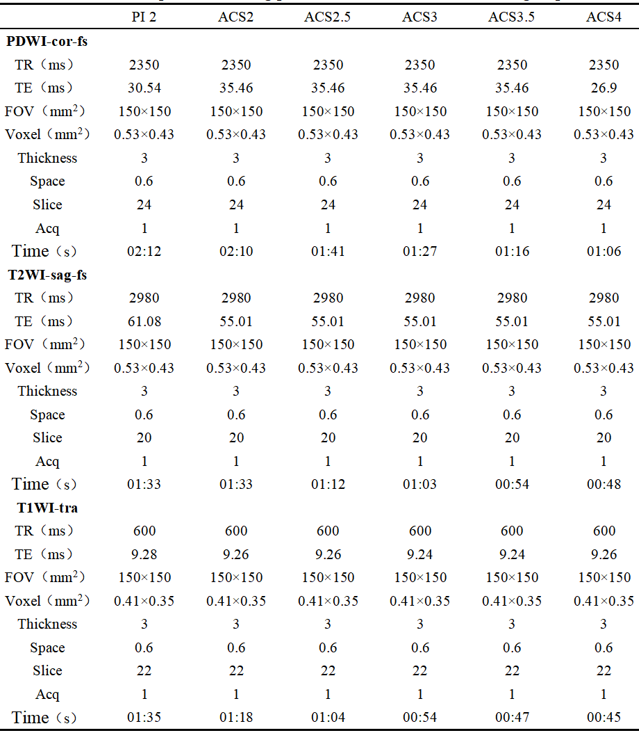
Table1: Sequence scanning parameter table between different groups
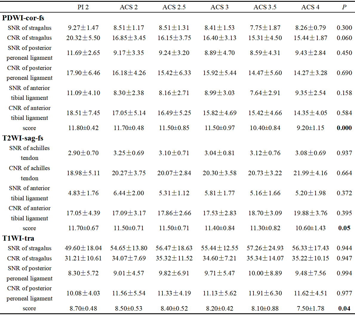
Table2: Statistical results of SNR, CNR and subjective score between different groups
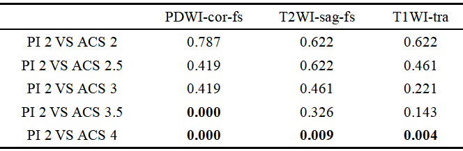
Table3: Subsequent pairwise comparison results
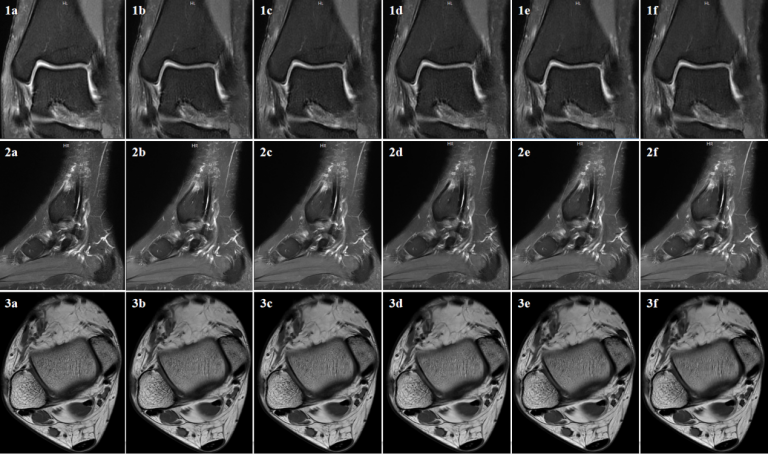
Fig1. 1a-1f show that the acceleration factors of coronal PDWI images of ankle joint (PI2, ACS2/2.5/3/3.5/4).
Fig2. 2a-2f show that the acceleration factors of sagittal T2WI images of ankle joint (PI2, ACS2/2.5/3/3.5/4)..
Fig3. 3a-3f show that the acceleration factors of transverse T1WI images of ankle joint (PI2, ACS2/2.5/3/3.5/4).
DOI: https://doi.org/10.58530/2023/4536