4532
3D T2 and T1ρ Mapping of Ischemic Injury to the Femoral Head: An In Vivo Piglet Model Study1Veterinary Clinical Sciences, University of Minnesota, Saint Paul, MN, United States, 2Center for Magnetic Resonance Research, University of Minnesota, Minneapolis, MN, United States
Synopsis
Keywords: Bone, Ischemia
Legg-Calvé-Perthes disease (LCPD) is a pediatric hip disorder caused by femoral head ischemia and osteonecrosis. In this work, we investigated whether 3D T2 and T1ρ relaxation time mapping are sensitive to ischemic injury to the bone and marrow of the femoral epiphysis and metaphysis following surgical induction of femoral head ischemia in a piglet model of LCPD. We found that T2 and T1ρ increased in the ischemic femoral epiphysis and decreased in the perfused metaphyseal spongiosa. Our findings support the potential clinical use of T2 and T1ρ mapping to assess the severity of femoral head injury in patients with LCPD.Introduction
Legg-Calvé-Perthes disease (LCPD) is a childhood hip disorder caused by disruption of blood supply to the femoral head, which can result in joint deformation and osteoarthritis.1 T2 and T1ρ relaxation time mapping have been shown to be sensitive in detecting early-stage ischemic injury to the femoral head in ex vivo and in vivo studies of a piglet model of LCPD.2-5 T2 and T1ρ increase in the secondary ossification center (SOC, i.e., the bone and bone marrow of the femoral epiphysis, proximal to the growth plate) as early as 48 hours following surgical induction of complete femoral head ischemia.2-5 While it has been previously assumed that the metaphysis (i.e., the bone and bone marrow distal to the growth plate) remains unaltered by the surgery,2-5 a recent study in the same model found T2 and T1ρ decreased within the primary spongiosa (i.e., the metaphyseal bone adjacent to the growth plate).6 Previous studies have relied on analysis of single 2D slices that might not provide an accurate one-to-one comparison between ischemic and contralateral-control femoral heads. Furthermore, only primary metaphyseal spongiosa and global changes within the SOC have been studied. Therefore, the purpose of this study was two-fold: (i) to determine whether 3D T2 and T1ρ mapping are sensitive to changes in the SOC and metaphyseal spongiosa following ischemic injury to the femoral epiphysis in the piglet model one week post-operatively; and (ii) to investigate potential regional differences in the response of the relaxation times to ischemic injury within the SOC and metaphyseal spongiosa.Methods
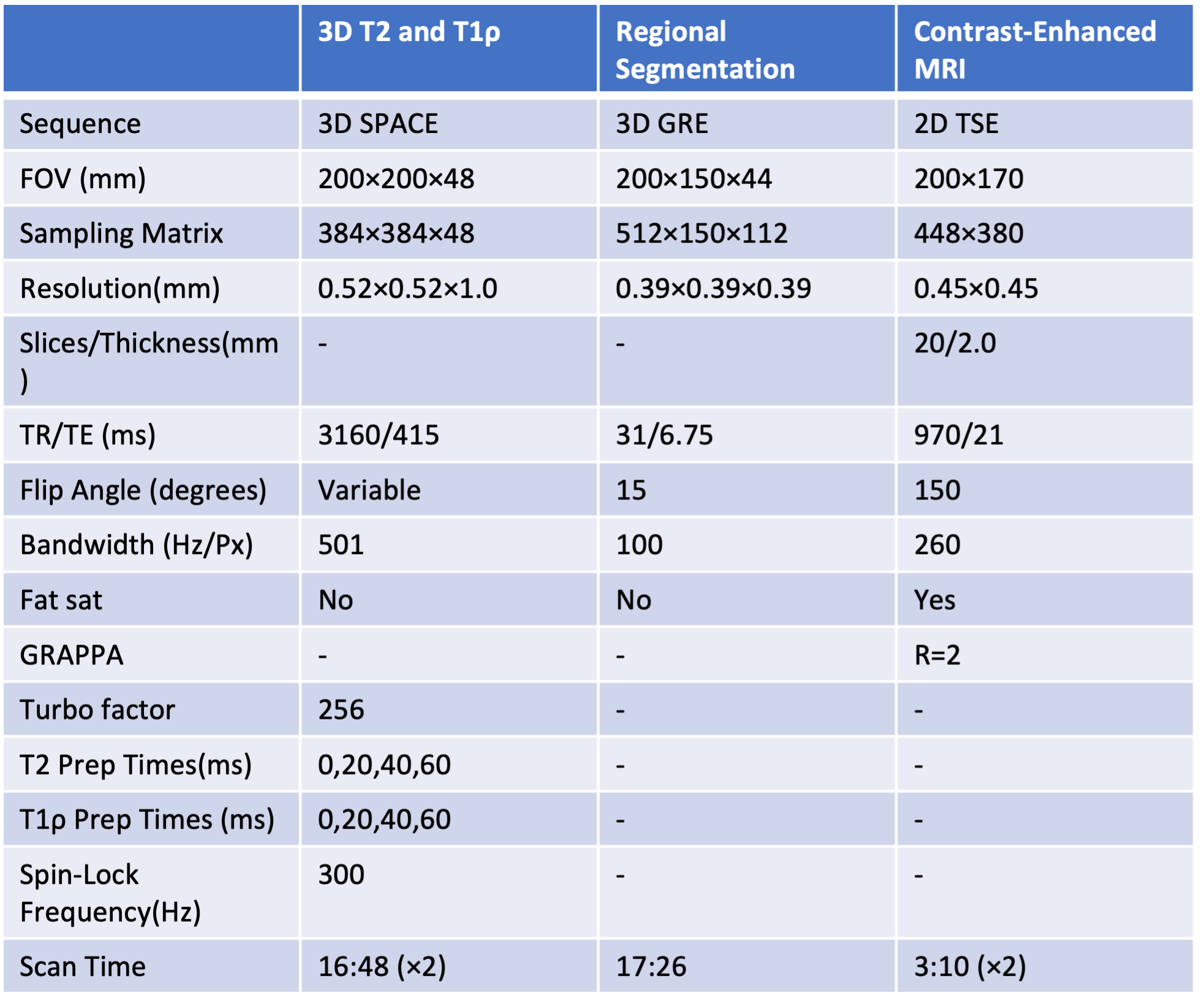
Animal Model: This study was approved by our institution’s IACUC. Seven piglets underwent surgery at the age of six weeks to induce complete ischemia in one of the femoral heads by interrupting its blood supply. This was achieved by placing a ligature around the femoral neck and transecting the ligamentum teres.7 The unoperated contralateral femoral head served as a control.In Vivo, 3T MRI: One week after surgery, the bilateral hips of each piglet were imaged in vivo at 3T MRI using: (i) T2 and T1ρ mapping using a magnetization-prepared 3D SPACE sequence (T2: MLEV-4 preparation; T1ρ: 300 Hz continuous-wave spin-lock preparation); and (ii) subtraction contrast-enhanced MRI (CE-MRI) following intravenous injection of 0.2 mmol/kg gadoteridol to confirm complete ischemia induction in the operated femoral heads. MRI acquisition parameters are summarized in Table 1.
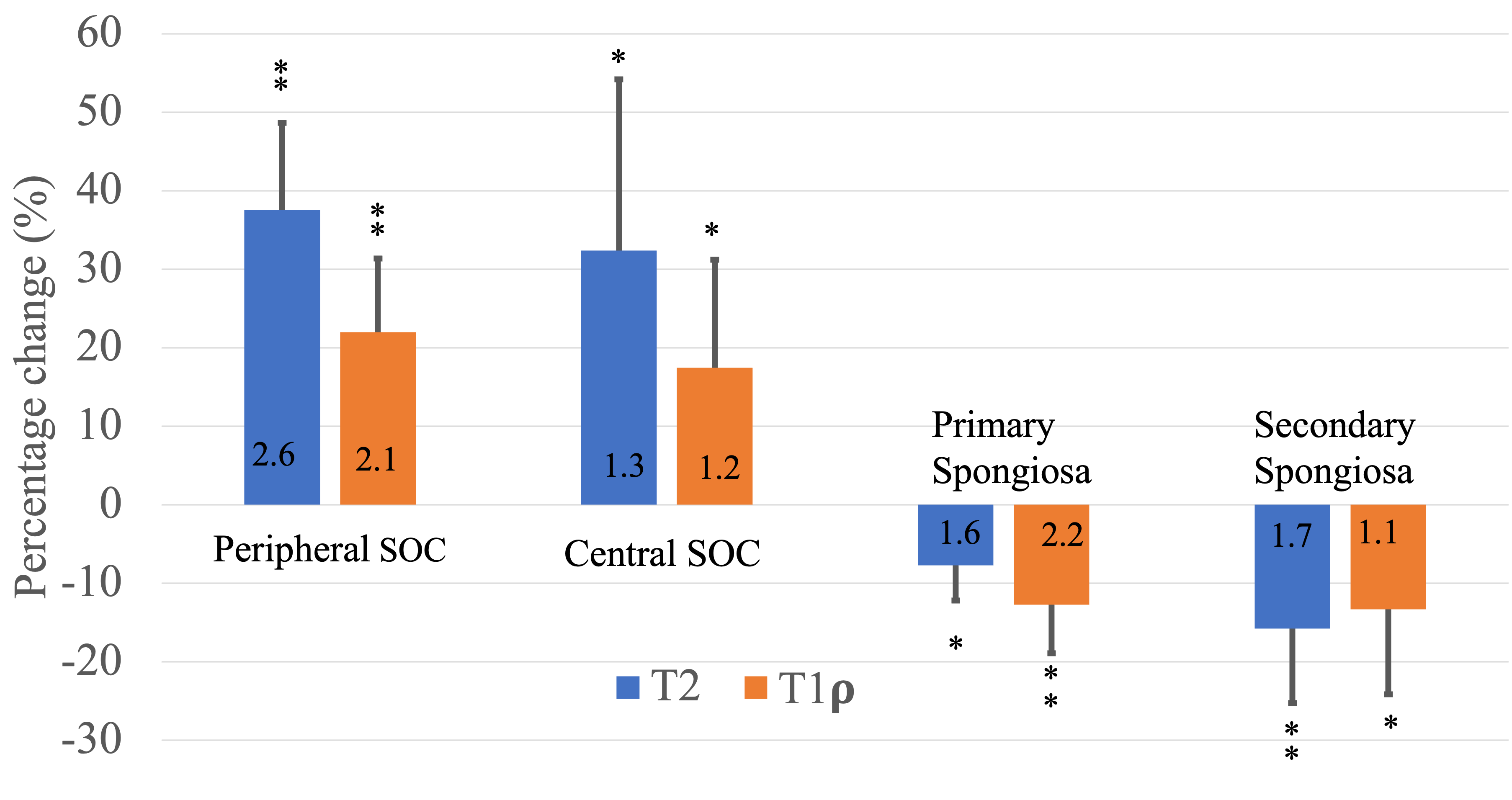
Data Analysis: Four regions of interest (ROIs) were manually defined using the high-resolution 3D GRE images: (i) Central SOC (inner 2/3 of SOC), (ii) Peripheral SOC (Outer 1/3 of SOC), (iii) Primary spongiosa (metaphysis immediately adjacent to and twice the thickness of the growth plate), and (iv) Secondary spongiosa (adjacent to primary spongiosa and two times thicker than the primary spongiosa). Median T2 and T1ρ relaxation times were measured in each ROI, and their percent changes in the ischemic vs. control femoral heads were calculated and statistically analyzed using paired t-tests with an uncorrected significance threshold of p<0.05. Effect sizes were also calculated using Cohen’s d.
Results
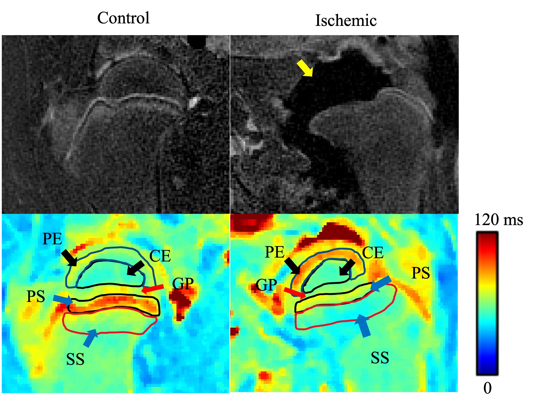
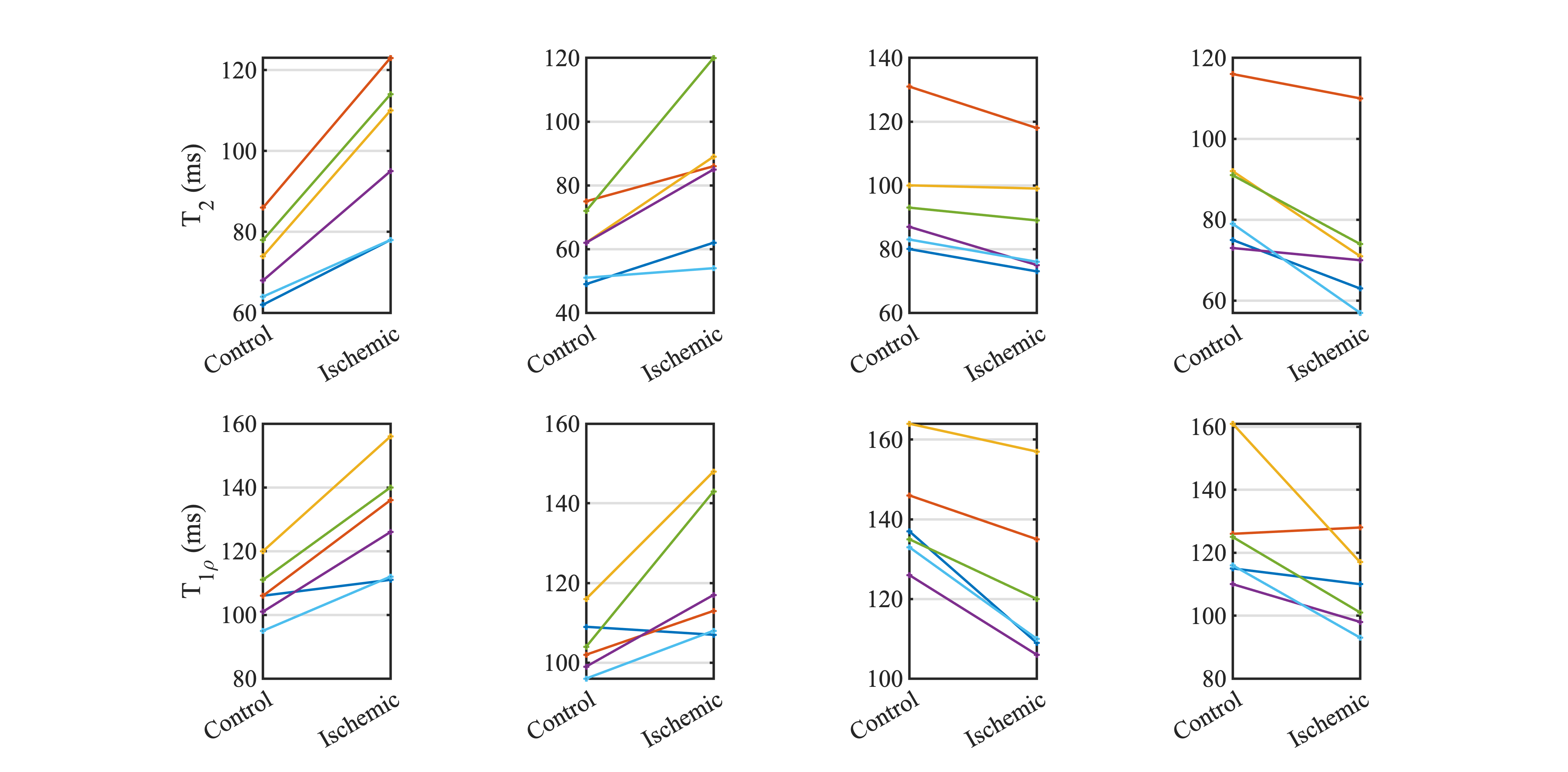
CE-MRI confirmed the global induction of ischemia in the operated limb in 6/7 piglets (Figure 1); one piglet had only partially induced ischemia in the femoral head and thus was excluded from the analysis. T2 and T1ρ relaxation times were increased in the SOC of the ischemic vs. control femoral heads (Figures 1-3), with a greater increase in the peripheral vs. central SOC. On average, T2 and T1ρ increased in peripheral SOC by 37.5±11.1% (p=0.0013) and 22.0±9.4% (p=0.0034) and in the central SOC by 32.4±21.8% (p=0.0210) and 17.5±13.8% (p=0.0301), respectively. Conversely, T2 and T1ρ were decreased in the primary and secondary spongiosa of the ischemic vs. control femoral heads, with the secondary spongiosa showing a relatively greater decrease (Figures 2 and 3). On average, T2 and T1ρ decreased in primary spongiosa by 7.7±4.5% (p=0.0113) and 12.8±6.2% (p=0.0028) and in secondary spongiosa by 15.8±9.5% (p=0.0085) and 13.3±10.8% (p = 0.0459), respectively.Discussion
Our findings demonstrate that 3D T2 and T1ρ mapping are sensitive to changes to both SOC and metaphyseal spongiosa following ischemic injury to the femoral head. The results indicate that the peripheral SOC is more vulnerable to ischemic damage than the central SOC. We speculate that this is due to the peripheral SOC being more metabolically active, vascularized, and cellular than the central SOC. We found T1ρ to be more sensitive to changes in the primary spongiosa than T2, which is in agreement with the previous speculation that T1ρ might be particularly useful in assessing changes in this region due to its reduced sensitivity to mineralization and blood products compared to T2.3Conclusion
T2 and T1ρ mapping are sensitive to changes in the SOC and metaphyseal spongiosa due to the ischemic injury to the femoral head in a piglet model. These methods may be clinically valuable to help assess the extent of injury and growth disturbance to the developing femoral head in children with LCPD.Acknowledgements
This project was supported by NIH grants R56AR078315, K01AR070894, UL1TR002494, and P41EB027061. We thank Kathleen Stuebner, Amber Winter, Kelly Bergsrud, Andrea Chehadeh, Sara Pracht, Katalin Kovacs, Paula Overn, and Dee Koski for their assistance.References
1. Kim HK. Pathophysiology and new strategies for the treatment of Legg-Calve-Perthes disease. J Bone Joint Surg Am 2012; 94:659-669.
2. Johnson CP, Wang L, Tóth F, Aruwajoye O, Carlson CS, Kim HK, Ellermann JM. Quantitative MRI helps to detect hip ischemia: a preclinical model of Legg- Calvé-Perthes disease. Radiology 2018; 289(2):386-395.
3. Johnson CP, Tóth F, Carlson CS, Armstrong AR, Zbýň Š, Wu B, Ellermann JM, Kim HK. T1ρ and T2 mapping detect acute ischemic injury in a piglet model of Legg–Calvé–Perthes disease. J Orthop Res 2022; 40(2):484-494.
4. Johnson CP, Toth F, Armstrong AR, Kim HK, Ellermann JM. Quantitative T2, T1ρ, and diffusion mapping of early-stage ischemic osteonecrosis of the femoral head: an in vivo piglet model study at 3T MRI. Proc 28th ISMRM Annual Meeting 2020; No. 1145.
5. Johnson CP, Bhave S, Armstrong AR, Tóth F. Utility of adiabatic T1ρ and T2ρ mapping to detect ischemic injury to the femoral head: an in vivo piglet model study at 3T MRI. Proc 29th ISMRM Annual Meeting 2021; No. 0154.
6. Buko E, Armstrong AR, Johnson CP. MRI Relaxation Times Change in the Metaphysis Following Ischemic Injury to the Femoral Head: An In Vivo Piglet Model Study. Proc 31st ISMRM Annual Meeting 2022; No. 1602.
7. Kim HK, Su PH. Development of flattening and apparent fragmentation following ischemic necrosis of the capital femoral epiphysis in a piglet model. J Bone Joint Surg Am 2002; 84-A(8):1329-34.
Figures