4527
A clinical application of ZTE-MRI in diagnosis of knee using joint space and angles1Hubei University of Medicine, Department of Radiology, Taihe Hospital,, Hubei, China, 2GE Healthcare, Beijing, China, 3Shiyan Taihe Hospital, Hubei, China, 4Biomedical Engneering College, Hubei University of Medicine, Hubei, China
Synopsis
Keywords: Bone, Bone
X-ray showed no significant abnormalities at early stage of OA, but obvious manifestation appeared in the middle and late stages. Our study demonstrated that joint space and joint angle can be measured on ZTE-MRI in evaluation of knee mechanical function and further support anatomical evaluation using Kellgren-Lawrence grading system that is the most widely used method for osteosurgeons in diagnosis of knee osteoarthritis. We also found both JS and CD were correlated with KLZTE score and both showed significant difference between mild/moderate and advanced groups. Therefore, ZTE-MRI has great potential in displaying structure abnormality.Introduction
Osteoarthritis (OA) is one of the most common arthritis and mainly manifested by hyperosteogeny, sclerosis and joint space stenosis[1]. X-ray showed no significant abnormalities at early stage of OA, but obvious manifestation appeared in the middle and late stages. Application of zero echo time magnetic resonance imaging (ZTE-MRI) in display of narrowed joint space, osseous surface defects, sclerosis growth have shown equivalent performance to computed tomography and better than T1-weighted imaging[2,3]. With the advantages of better soft-tissue contrast and radiation-free feature, to explore applications in musculoskeletal MRI is essential and benefits to patients for time-saving, convenience and one-stop check-up. Conventional radiograph is used to measure joint space stenosis in reflection of meniscus injury[4]. However, joint space is the sum of articular cartilage, meniscus, a small amount of synovial fluid and potential anatomic relationship between the two bony articular surfaces[5]. Among various grading systems for knee osteoarthritis, Kellgren-Lawrence grading system is the most widely used method for osteosurgeons in diagnosis of knee osteoarthritis[6]. Therefore, we attempted to investigate the clinical application of ZTE-MRI in diagnosis of knee using joint space and angles and compare inter-group difference using KL grading as a classifier.Materials and methods
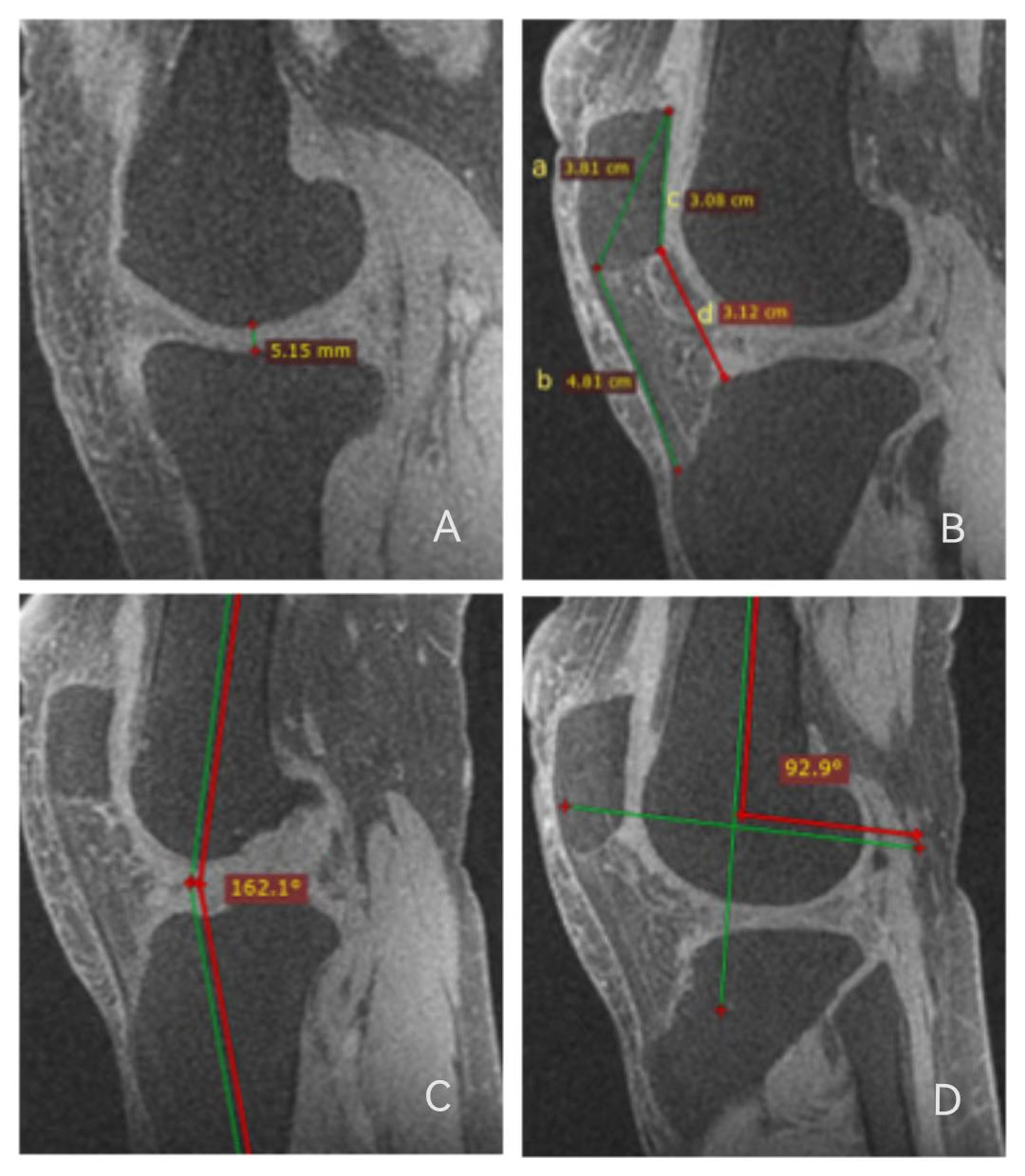
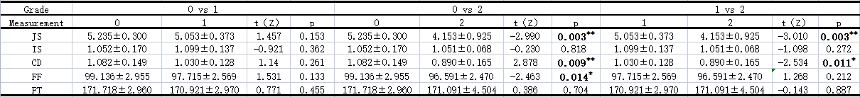
This prospective study recruited 87 patients with suspected OA from May 2022 to October 2022. All participants underwent conventional MR scan and ZTE sequence (repetition time = 4000, flip angle = 3°, NEX = 2, spokes per segment = 288, voxel resolution = 1×1 ×1 mm2) on 1.5T (Signa Voyager, GE Healthcare). The joint space, ratio of the length of patellar tendon to the length of patella (IS), distance from the lowest point of patella surface to the anterior upper angle of tibial plateau (CD), the angle between the long axis of femoral shaft and that of femoral condyle (FF), and the angle between the long axis of femoral shaft and the long axis of tibial shaft (FT) were measured on ZTE-MRI (Figure 1). Inter-modality consistency of KL grading on radiograph and ZTE-MRI was analyzed using Intraclass correlation coefficient (ICC). Between-group comparisons of the abovementioned values were analyzed using independent sample T test and Mann-Whitney U T test depending on data normality and equality of variance. P <0.05 was considered statistically difference.Results
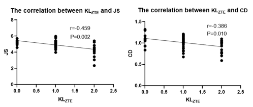
Inter-modality agreement of KL grading based on radiograph and ZTE was good ( k =0.872, P<0.05). There were significantly correlated between IS and CD (r=0.300, p =0.048), between JS and CD (r=0.403, p =0.007), and between JS and FF (r=0.298, p =0.049). In addition, the association of KL grading on ZTE (KLZTE) was correlated with JS and CD (r=0.-459, p=0.002; r=-0.386, p=0.010, respectively)(Figure 2). According to KLZTE, there were statistically significant differences of JS and CD between mild/moderate and advanced groups as well as FF between mild and advanced groups (Table 1).Discussion and conclusions
Our study demonstrated that joint space and joint angle can be measured on ZTE-MRI in evaluation of knee mechanical function and further support anatomical evaluation using Kellgren-Lawrence grading system. Quantitative information is beneficial for long-term tracking and patient comprehension about KOA progression. We found both JS and CD were correlated with each other and also with KLZTE score, and both also showed significant difference between mild/moderate and advanced groups. High patella is reckoned as the primary cause of non-traumatic anterior knee pain, patellofemoral joint instability and patella cartilage injury, while low patella is mainly a common complication after knee injury, surgery or artificial knee replacement, which can cause joint structure change, movement restriction, pain and other symptoms, which seriously affect patients' life and work. The commonly used clinical methods for measuring patella height include Insall-Salvati and Caton-Deschamps methods (normal value is 0.8-1.2, >1.2 is high patella, < 0.8 is low patella). When IS or CD does not fall in the normal range, KOA patients would have the manifest of knee pain and possibly reflects osseous degradation. Radiation-free ZTE-MRI not only provides CT-like images in great delineation of joint space and bone structure but also can simultaneously be acquired with routine MRI for regular knee MRI exam[7]. It would be convenience for patients to have one-stop knee check-up using ZTE-MRI and routine MR imaging.Acknowledgements
I would like to express my gratitude to all those who have helped me during the writing of this abstract. I gratefully acknowledge the help of my supervisor Professor Wen Chen. I do appreciate his patience, encouragement,and professional instructions during my abstract writing.Also,I would like to thank Weiyin Vivian Liu, for her instructive advice and useful suggestions on my abstract.I am deeply grateful of her help in the completionof this abstract.I am also deeply indebted to all the other tutors and teachers in this study for their direcct or indirect help to me.Last but not the least,my gratitude also extends to my parents for their continuous support and encouragement all of my life.References
[1]Turmezei TD, B Low S, Rupret S, Treece GM, Gee AH, MacKay JW, Lynch JA, Poole KES, Segal NA. Quantitative Three-dimensional Assessment of Knee Joint Space Width from Weight-bearing CT. Radiology. 2021 Jun;299(3):649-659. doi: 10.1148/radiol.2021203928. Epub 2021 Apr 13. PMID: 33847516; PMCID: PMC9490554.
[2]Jansen MP, Mastbergen SC, Eckstein F, van Heerwaarden RJ, Spruijt S, Lafeber FPJG. Comparison between 2D radiographic weight-bearing joint space width and 3D MRI non-weight-bearing cartilage thickness measures in the knee using non-weight-bearing 2D and 3D CT as an intermediary. Ther Adv Chronic Dis. 2021 Aug 21;12:20406223211037868. doi: 10.1177/20406223211037868. PMID: 34434539; PMCID: PMC8381425.
[3]Li Y, Xiong Y, Hou B, Liu C, Wang J, Liu WV, Li X. Comparison of zero echo time MRI with T1-weighted fast spin echo for the recognition of sacroiliac joint structural lesions using CT as the reference standard. Eur Radiol. 2022 Jun;32(6):3963-3973. doi: 10.1007/s00330-021-08513-5. Epub 2022 Jan 21. PMID: 35059805.
[4]Hou B, Liu C, Li Y, Xiong Y, Wang J, Zhang P, Liu J, Liu WV, Li X. Evaluation of the degenerative lumbar osseous morphology using zero echo time magnetic resonance imaging (ZTE-MRI). Eur Spine J. 2022 Mar;31(3):792-800. doi: 10.1007/s00586-021-07099-2. Epub 2022 Jan 11. PMID: 35015138.
[5]Mortensen JF, Mongelard KBG, Radev DI, Kappel A, Rasmussen LE, Østgaard SE, Odgaard A. MRi of the knee compared to specialized radiography for measurements of articular cartilage height in knees with osteoarthritis. J Orthop. 2021 May 12;25:191-198. doi: 10.1016/j.jor.2021.05.014. PMID: 34045822; PMCID: PMC8141415.
[6]Madan-Sharma R, Kloppenburg M, Kornaat PR, Botha-Scheepers SA, Le Graverand MP, Bloem JL, Watt I. Do MRI features at baseline predict radiographic joint space narrowing in the medial compartment of the osteoarthritic knee 2 years later? Skeletal Radiol. 2008 Sep;37(9):805-11. doi: 10.1007/s00256-008-0508-6. Epub 2008 Jun 20. PMID: 18566813; PMCID: PMC2491711.
[7]Bharadwaj UU, Coy A, Motamedi D, Sun D, Joseph GB, Krug R, Link TM. CT-like MRI: a qualitative assessment of ZTE sequences for knee osseous abnormalities. Skeletal Radiol. 2022 Aug;51(8):1585-1594. doi: 10.1007/s00256-021-03987-2. Epub 2022 Jan 28. PMID: 35088162; PMCID: PMC9198000.
Figures