4526
Application of Multiecho Dixon Technique and IVIM-DWI in Patients with Primary Osteoporosis : A Preliminary Study with 3.0 T MRI1Affiliated Hospital of Shaanxi University of Chinese Medicine,, xianyang, China, 2MR senior Scientific Marketing Specialist,Siemens Healthineers, Shang hai, China
Synopsis
Keywords: Bone, Quantitative Imaging, Multiecho Dixon Technique , IVIM-DWI
Bone mineral density (BMD) is a common standard for evaluating osteoporosis, but it can only reflect the change of bone mass, not the microscopic nodules of tissueChange of structure.Multi-echo Dixon and IVIM-DWI were used to evaluate the changes of spinal bone marrow microstructure in osteoporosis, osteopenia and normal subjects.The results showed that there were significant differences in FF, D and D* between osteoporosis, osteopenia and normal subjects.These results indicate that IVIM-DWI can quantitatively reflect the changes of lumbar microcirculation and fat content, and can be used as a biomarker for the progression of osteoporosis.Introduction:
Osteoporosis (OP) has become a global public health problem and cutting-edge research problem. Microvessel reduction and microcirculatory disturbance are important factors of the genesis and progression of OP. Quantitative MRI is of critical importance in identifying individuals at risk for OP and also in monitoring response to treatment. Purpose: The purpose of our study was to investigate the role of multi-echo Dixon technique and IVIM-DWI in assessing vertebral marrow changes among subjects with osteoporosis, osteopenia and normals.Purpose:
The purpose of our study was to investigate the role of multi-echo Dixon technique and IVIM-DWI in assessing vertebral marrow changes among subjects with osteoporosis, osteopenia and normals.Methods:
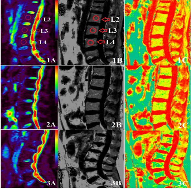
Totally 56 subjects, who underwent QCT of the spine, were divided into three groups (normal, osteopenia, and osteoporosis) based on T-score. All data were collected on a MAGNETOM Skyra 3T MR scanner (Siemens AG, Erlangen, Germany) including IVIM and 3D gradient multiecho Dixon chemical shift sequence using an 18-channel table-mounted spine matrix coil with the following parameters. IVIM-DWI: TE/TR of 72.4/1600ms, and 8 b values of 0, 50, 100, 150, 200,250, 300, 400, 600 and 800 sec/mm2 on 3 gradient directions. Multiecho Dixon: TR: 9.0 msec; TE: 1.23 ms, 2.46 ms, 3.69 ms 4.92 ms, 6.15 ms and 7.38 ms; flip angle: 4.0°, readout echo bandwidth 1080 Hz/pixel; slice thickness: 2.5 mm; FOV: 400 mm; matrix 256 × 256. The regions of interest (ROIs) were applied in lumbar 2-4 (areas 1.0 cm2 ) in sagittal IVIM images and fat_fraction mappings. One-way ANOVA were performed to evaluate the significance of the inte-group difference in FF and IVIM parameters (f value, D value and D* value) between different groups.Results:
Totally 56 subjects, including a osteoporosis group (n=16, 7 males, age= 61.0 ± 9.5 years, T-score = -3.32 ± 0.68 ), a osteopenia group (n=20,9 males, age= 54.2 ± 10.3 years, T-score = -1.84 ± 0.29) and a normal group (n=20, 6 males, age= 45.9 ± 10.7 years, T-score = 0.15 ± 0.83) were enrolled. The FF, D and D* of normals were 43.63±7.88, 0.393±0.105, 78.19±16.06, respectively; and osteopenia were 49.58±5.02, 0.356±0.097, 87.36±21.39; and osteoporosis were 57.88±10.01, 0.303±0.069, 97.27±29.65. Furthermore, the FF, D and D* among osteoporosis, osteopenia and normals were significantly different (p < 0.05). A statistically significant positive correlation between D value and T-score (r=0.854, P <0.001). The D* value (r=–0.785, P < 0.001) and FF (r= -0.882, P < 0.001) were negatively correlated to the T-score.Conclusion:
The multiecho Dixon technique IVIM-DWI can quantitatively reflect the change of lumbar microcirculatory and fat contant, which can be used as biomarkers for disease progression in osteoporosis.Acknowledgements
Thanks to the Affiliated Hospital of Shaanxi University of Traditional Chinese MedicineReferences
1.Qiu X, Fu Y, Chen J, Ye Y, Wang Z, Ming X. The Correlation between Osteoporosis and Blood Circulation Function Based on Magnetic Resonance Imaging. J Med Syst. 2019 Mar 2;43(4):91
2.Zhu J, Xiong Z, Zhang J, Qiu Y, Hua T, Tang G. Comparison of semi-quantitative and quantitative dynamic contrast-enhanced MRI evaluations of vertebral marrow perfusion in a rat osteoporosis model. BMC Musculoskelet Disord. 2017 Nov 14;18(1):446.
3.Lasbleiz J, Le Ster C, Guillin R, Saint-Jalmes H, Gambarota G. Measurements of Diffusion and Perfusion in Vertebral Bone Marrow Using Intravoxel Incoherent Motion (IVIM) With Multishot, Readout-Segmented (RESOLVE) Echo-Planar Imaging. J Magn Reson Imaging. 2019 Mar;49(3):768-776
Figures