4525
Assessment of vertebral and intervertebral disc microenvironment changes in postmenopausal women using IVIM and fat-water MRI
Shuo Zhang1, Qingwei Song1, Hongbo Feng1, Qianrui Guo2, and Yang Yang3
1The First Affiliated Hospital of Dalian Medical University, Dalian, China, 2Dalian Medical University, Dalian, China, 3Beijing United Imaging Research Institute of Intelligent Imaging, Beijing, China
1The First Affiliated Hospital of Dalian Medical University, Dalian, China, 2Dalian Medical University, Dalian, China, 3Beijing United Imaging Research Institute of Intelligent Imaging, Beijing, China
Synopsis
Keywords: Bone, Data Acquisition
Intravoxel incoherent motion (IVIM) and fat-water magnetic resonance imaging (FWMRI) are rarely used for vertebral and intervertebral disc. To search for predictive value of them in assessing the lumbar spine microenvironment changes, we performed a study with postmenopausal women combined the parameters with bone mineral density (BMD) and major fracture risk assessment (FRAX) score. We report that fat fraction (FF) has significant correlation with FRAX, FF was moderately negatively correlated with BMD; ADCslow of intervertebral discs showed moderately negatively correlation with FRAX score and FF. IVIM and FWMRI have potential to become new biomarkers in predicting lumbar spine microenvironment changes.Purpose
To investigate the predictive value of intravoxel incoherent motion (IVIM) and fat-water magnetic resonance imaging (FWMRI) parameters in evaluating the vertebral and intervertebral disc microenvironment changes in postmenopausal women.Methods
In this study, all enrolled women underwent MRI and dual-energy X-ray absorptiometry (DXA) imaging. Major fracture risk was also evaluated for all participants using Fracture Risk Assessment Tool (FRAX) by a senior radiologist with International Society for Clinical Densitometry (ISCD) qualification. Participants were divided into premenopausal (n=10) and postmenopausal (n=10) group. The exclusion criteria were as follows: (1) spine surgery; (2) bone diseases; (3) malignant tumors; (4) long-term use of drugs that affect bone metabolism. MRI examinations were performed through a commercially available 3.0 T MRI system. Routine MR lumbar spine protocol was performed using a 32-channel spine matrix coil for pretreatment planning. IVIM images were acquired on the basis of ss-EPI method with 16 b values (0, 10, 20, 40, 80, 110, 140, 170, 200, 300, 400, 500, 600, 800, 1000, 1200s/mm2). As traditional monoexponentially ADC values are unable to distinguish the true diffusion and perfusion effects in tissues, a biexponential model was used to calculate the true apparent diffusion coefficient (ADCslow), pseudo–apparent diffusion coefficient (ADCfast) and perfusion fraction (f) maps. FWMRI images were obtained using a fat analysis and calculation technique(FACT) sequence. To overcome the limitations of traditional region-growing methods and multiresolution methods, variable projection was implemented for robust fat-water separation in this study (Hao et al. 2019; Cheng et al. 2017). FWMRI images were acquired with the following parameters: FOV = 400 x 300 mm, matrix = 256 x 192 x 22, pixel size = 1.56 x 1.56 x 8 mm3, contrast = 6, flip angle = 3°, Bandwidth = 900 Hz/pixel, TE = 1.73/3.26/4.79/6.32/7.85/9.38 ms, TR = 10.94 ms. IVIM images were measured with a region of interest-based approach on corresponding maps of the vertebral and intervertebral discs for ADCslow, ADCfast and f respectively. Bone marrow fat fraction (FF) and R2* values of vertebral were measured on FWMRI images as shown in Figure 1. MRI parameters were tested for significant differences between two groups using T test for normal distributions or Mann-Whitney test if the data was not normally distributed as shown in Table 1. Spearman’s rank correlation was performed to test correlation between MRI parameters and BMD, FRAX score respectively as shown in Table 2.Results
There were significant differences in FF of vertebral (P < 0.001) and ADCslow of intervertebral discs (P = 0.003) between premenopausal and postmenopausal group. Moreover, there was significant correlation between bone marrow FF and FRAX (r = 0.824, P < 0.0001), and FF was moderately negatively correlated with BMD (r = -0.409, P = 0.073). ADCslow of intervertebral discs showed moderately negatively correlation with FRAX score and FF.Conclusions
IVIM and fat-water MRI parameters have potential to become new biomarkers in effectively predicting vertebral and intervertebral disc microenvironment changes in postmenopausal women.Acknowledgements
NoneReferences
[1] Hao Peng, Chao Zou, Chuanli Cheng, et al. Fat-water separation based on Transition Region Extraction (TREE). Magnetic Resonance in Medicine, 2019, 82(1): 436-448. 3.
[2] Chuanli Cheng, Chao Zou, Changhong Liang, et al. Fat-water separation using a region-growing algorithm with self- feeding phasor estimation. Magnetic Resonance in Medicine 2017: 77: 2390-2401.
Figures
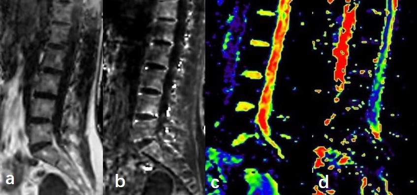
Figure 1. Different parameter maps of vertebral and intervertebral disc. (a) FF; (b) R2*; (c) ADCslow; (d) ADCfast.
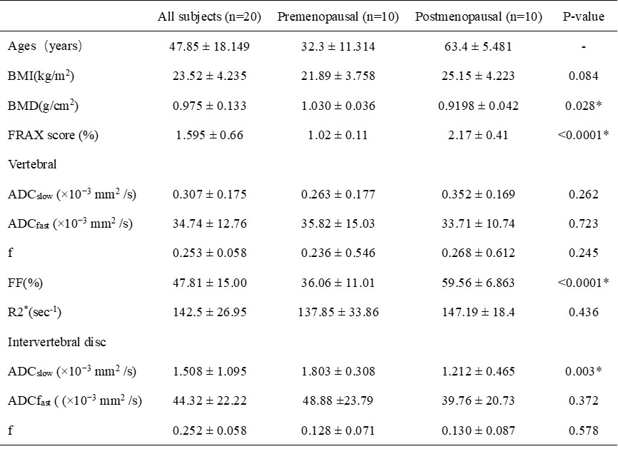
Table 1. Characteristics of patients in premenopausal and postmenopausal group. Data are presented as mean ± SD. BMD, FRAX score, bone marrow FF and ADCslow of intervertebral disc were significant different between premenopausal and postmenopausal group (P < 0.01). BMD, bone mineral density; FRAX, Fracture Risk Assessment Tool; FF, fat fraction; ADCslow, true apparent diffusion coefficient; ADCfast, pseudo–apparent diffusion coefficient; f, perfusion fraction.
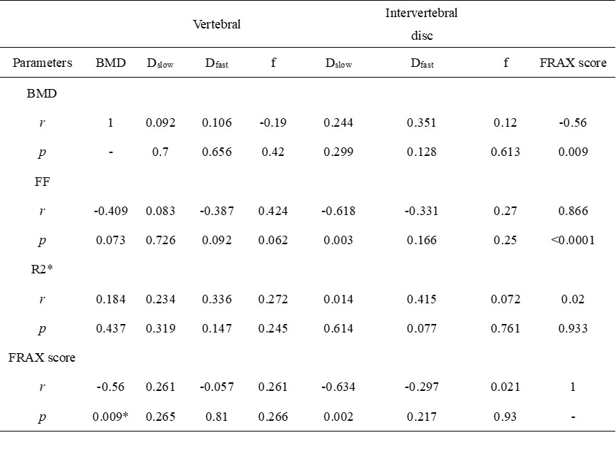
Table 2. The correlations among different parameters.
DOI: https://doi.org/10.58530/2023/4525