4508
Chemical Shift Inversion Recovery (CSIR) spectroscopy with hyperpolarized xenon-129 MRI: A comparison with CSSR spectroscopy
Kai Ruppert1, Luis Loza1, Faraz Amzajerdian1, Hooman Hamedani1, Mostafa K Ismail1, Ryan J Baron1, Ian F Duncan1, Harrilla Profka1, Stephen Kadlecek1, and Rahim R Rizi1
1Radiology, University of Pennsylvania, Philadelphia, PA, United States
1Radiology, University of Pennsylvania, Philadelphia, PA, United States
Synopsis
Keywords: Hyperpolarized MR (Gas), Pulse Sequence Design
Chemical shift saturation recovery (CSSR) MR spectroscopy using hyperpolarized xenon-129 provides metrics of pulmonary physiology by saturating the xenon dissolved-phase magnetization in the lung with a 90° RF pulse and measuring the subsequent signal recovery via gas exchange. Our measurements in a rat demonstrate that chemical shift inversion recovery (CSIR) spectroscopy, which replaces the saturation with an inversion pulse, produces equivalent results to CSSR but with greater robustness with respect to both low signal amplitudes at short delay times and incomplete saturation.Purpose
Chemical shift saturation recovery (CSSR) MR spectroscopy using hyperpolarized xenon-129 (HXe) has been shown to yield important metrics of pulmonary function in humans as well as various animal species1-9. This is accomplished by selectively saturating the magnetization of xenon dissolved in the lung parenchyma at a chemical shift of approximately 200 ppm with a narrow-bandwidth 90° RF pulse while leaving the gas phase (GP) magnetization at 0 ppm largely unaffected. Regrowth of the signal of the membrane (Mem) and red blood cell resonances is measured by collecting spectra at a variable delay time after the saturation pulse and then fitting to a theoretical gas exchange model. Especially at short delay times, however, the dissolved-phase signals are small and potentially contaminated by residual magnetization due to imperfect saturation, leading to significant relative errors for these data points. In this work, we investigated whether the use of chemical shift inversion recovery (CSIR) spectroscopy, which substitutes the saturation with an inversion RF pulse, can increase measurement accuracy.Methods
A healthy male Sprague-Dawley rat (340g) was anesthetized, intubated, and mechanically ventilated using a home-built ventilator with either air (during non-imaging periods) or a HXe gas mixture (during imaging; 21% O2, 79% N2/HXe) at a tidal volume of 10 ml/kg and breathing rate of 43 breaths per minute. Imaging was performed using a 3T horizontal-bore animal imaging system (Bruker Biospec); 1L of enriched xenon gas was used for each experiment, polarized using a prototype commercial optical pumping system (XeBox-E10, Xemed LLC, NH). Proton T2-weighted fast spin-echo images were acquired for localization prior to spectroscopy experiments. All studies were approved by the Institutional Animal Care and Use Committee of the University of Pennsylvania.Data was acquired using a customized MR spectroscopy pulse sequence. During a 5-s end-inspiratory breath hold, the dissolved xenon magnetization was either saturated or inverted with a 90° or 180° Gaussian RF pulse (0.913 ms pulse duration), respectively. Following a variable delay time Δτ ranging from 1.15 to ~100 ms, a 90° Gaussian RF excitation pulse (0.913 ms pulse duration) was applied, and a free induction decay was acquired (50 kHz receiver bandwidth, 31.56 ms sampling duration). The next saturation/inversion pulse was applied after a fixed gas exchange time of 80 ms. This sequence was repeated 20 times during the same breath hold for each Δτ. Following Fourier transform, spectra in steady state were averaged and the areas underneath the gas phase (GP, 0 ppm), membrane (Mem, 197 ppm), and red blood cell (RBC, 211 ppm) peaks were numerically integrated. Septal wall thickness and capillary transit time were calculated using the analytical uptake model of Patz et al.10, based on the recovery of the averaged membrane signal following each saturation/inversion pulse.
Results and Discussion
Figure 1 depicts the Mem-GP ratios following either saturation or inversion as a function of Δτ and the fitted Patz model function (solid lines). The apparent alveolar septal wall thickness was 6.8 μm for both measurement types, with respective capillary transit times of 0.34 s (CSSR) and 0.17 s (CSIR). However, given that no data was acquired for delay times longer than 100 ms, these transit times are not reliable. Although CSSR and CSIR produced identical values for the septal wall thickness, the inversion measurements appear to be much smoother—and not just at short delay times, as anticipated, but over the entire Δτ interval, confirming the potential for improved measurement accuracy when using CSIR rather than CSSR spectroscopy. Additional studies in a larger animal cohort and human subjects will be required to validate our observations, but our measurements already indicate the equivalence of the two acquisition techniques. A potential drawback of CSIR spectroscopy is the challenge of implementing it on a human MRI scanner due to the high power requirements of RF inversion pulses with sufficiently broad bandwidth to invert all dissolved-phase resonances.Conclusion
Our preliminary study indicates that the proposed CSIR spectroscopy technique yields pulmonary function metrics comparable to those obtained with the established CSSR spectroscopy technique while also being less sensitive to incomplete saturation of the dissolved-phase resonances and the low signal amplitudes at short delay times.Acknowledgements
Supported by NIH grant R01 HL142258.References
[1] Ruppert K et al. NMR of hyperpolarized 129Xe in the canine chest: spectral dynamics during a breath-hold. NMR Biomed 2000;13:220-228. [2] Butler JP et al. Measuring surface-area-to-volume ratios in soft porous materials using laser-polarized xenon interphase exchange nuclear magnetic resonance. J Phys Condens Matter 2002;14:L297-L304. [3] Qing et al. Assessment of lung function in asthma and COPD using hyperpolarized 129Xe chemical shift saturation recovery spectroscopy and dissolved-phase MRI. NMR in Biomed 2014;27(12):1490-1501. [4] Zhong et al. Simultaneous assessment of both lung morphometry and gas exchange function within a single breath‐hold by hyperpolarized 129Xe MRI. NMR in Biomed 2017; 30(8). [5] Doganay et al. Quantification of regional early stage gas exchange changes using hyperpolarized 129Xe MRI in a rat model of radiation-induced lung injury. Med Phys 2016;43(5):2410-2420. [6] Kern et al. Regional investigation of lung function and microstructure 129Xe chemical shift saturation recovery parameters by localized and dissolved‐phase imaging: A reproducibility study. MRM 2019; 81(1):13-24. [7] Ruppert et al. Using Hyperpolarized Xenon-129 MRI to Quantify Early-Stage Lung Disease in Smokers. Acad Radiol 2019; 26(3):355-366. [8] Kern et al. Mapping of regional lung microstructural parameters using 129 Xe dissolved‐phase MRI in healthy volunteers hyperpolarized and patients with chronic obstructive pulmonary disease. MRM 2018; 81(4):2360-2373. [9] Zanette et al. Physiological gas exchange mapping of hyperpolarized 129Xe using spiral-IDEAL and MOXE in a model of regional radiation-induced lung injury. Med Phys 2018;45(2):803-816. [10] Patz et al. Diffusion of hyperpolarized 129Xe in the lung: a simplified model of 129Xe septal uptake and experimental results. New J Physics 2011;13:015009.Figures
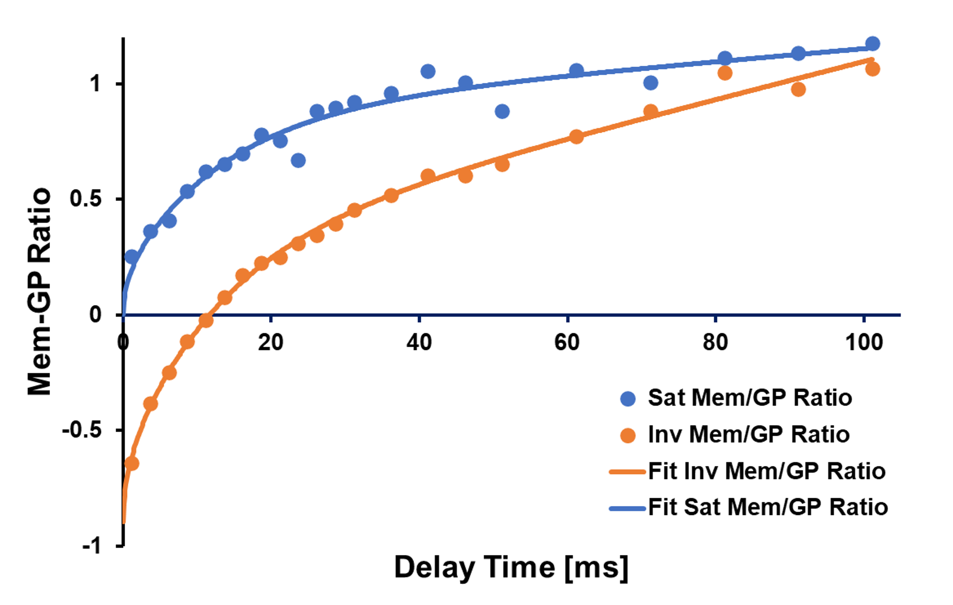
Figure 1. Mem-GP ratios as a function of the delay time
following either saturation (blue) or inversion (orange) of the dissolved-phase
magnetization in a rat. The apparent alveolar septal wall thickness derived
from fitting a theoretical gas uptake model to the measurement data (solid
lines) was found to be 6.8 μm for both acquisition types although
the capillary transit times differed, presumably because no data points were
collected for very long delay times.
DOI: https://doi.org/10.58530/2023/4508