4506
Batch-mode production of hyperpolarised xenon gas with a continuous-flow polariser for preclinical and clinical human lung ventilation images1MR Research Centre, Department of Clinical Medicine, Aarhus University, Aarhus N, Denmark, 2GE Healthcare, Brøndby, Denmark, 3POLARIS group, University of Sheffield, Sheffield, United Kingdom, 4GE Healthcare, Munich, Germany
Synopsis
Keywords: Hyperpolarized MR (Gas), Hyperpolarized MR (Gas), Lung ventilation imaging
Hyperpolarised 129Xe gas was accumulated using spin-exchange optical pumping (SEOP) by continuous-flow of xenon gas directly from the cell without the use of cryogenic separation. 3D ventilation images were acquired in human and porcine models to determine clinical diagnostic image quality. Xenon ventilation image quality indicates that on demand batch production of xenon is feasible for both preclinical and clinical lung ventilation imaging examinations.
Introduction
Hyperpolarised (HP) xenon-129 (129Xe) lung MRI has developed to the point of providing clinical value in evaluation of lung diseases [1]. HP 129Xe gas polarisation with spin-exchange optical pumping (SEOP) is conventionally performed using one of two approaches: “batch-mode” and “continuous-flow” [2], [3]. Batch-mode operation involves extracting one batch of xenon gas from the SEOP cell after sealed-cell polarisation build-up; it typically uses high xenon concentrations (>10%), is cryogen-free, can yield high polarisations (up to 90%), yet usually requires long production times (up to 60min) [4]. Continuous-flow production typically uses a lower concentration of xenon gas (between 1-3%) that is flowed through and cryogenically separated from buffer gases (He, N2) with reported production times ranging between 10 minutes to an hour for doses needed for ventilation imaging [2]. Faster production rate of a rapid batch-mode polariser would provide an attractive prospect for both preclinical and clinical applications of 129Xe lung MRI in terms of throughput and imaging workflow.The aim of this study was to evaluate the use of a high-performance continuous-flow polariser [2] in batch-mode operation to produce HP 129Xe lung ventilation images with low-concentration xenon gas collected without the use of time-consuming cryogenic separation.
Methods
For all experiments 129Xe gas was polarised using SEOP with a custom-built polariser (POLARIS, University of Sheffield). The gas mixture (3% enriched xenon, 10% N2, 87% He) was dispensed into a 1000ml Tedlar bag and administered following a 1-2min walk to the MRI scanner.Pig experiments: Xenon images of porcine lungs were acquired on a 3T MRI scanner (MR750, GE Healthcare, Waukesha, WI, USA) using a 129Xe transmit-receive quadrature vest coil (Clinical MR Solutions, Brookfield, WI, USA) tuned to 35.3MHz. A 3D coronal steady-state free precession (SSFP) sequence was used for ventilation imaging [5]; voxel size of 5x5x10 mm, matrix size of 80x80x30, repetition time (TR) = 3.2ms, echo time (TE) = minimum (~1ms), flip angle (FA) = 10 degrees, bandwidth = 31.25 kHz, total acquisition time of 6 sec. Ventilation images were acquired from 8 anaesthetised pigs (6 healthy and 2 with lung emboli) weighing 40 and 60kg, respectively.
Human experiments: 129Xe images of human lungs were acquired on a 1.5T MRI scanner (MR450W, GE Healthcare, Waukesha, WI, USA) using a 129Xe transmit-receive quadrature vest coil (Clinical MR Solutions, Brookfield, WI, USA) tuned to 17.7 MHz. Sequence parameters: acquired voxel size = 4.75x4.75x15 mm, matrix size = 80x80x20, TR = 3.6ms, TE = 1.3ms, FA = 13 degrees, bandwidth = 31.25 kHz, total acquisition time = 5 sec. Ventilation images were acquired in one healthy volunteer (male, 36 years old, total lung capacity ~6L). Images were post-processed with a FASTA (Fast Adaptive Shrinkage/Thresholding Algorithm) total variation denoising algorithm (λ=0.3) [6].
Results and Discussion
Results show ventilation images from 8 pigs (Fig. 1) and their corresponding SNR measurements from healthy pigs vs. pigs with lung emboli, with inhaled volumes of 800ml and 1000ml of the SEOP cell gas mixture (just 3% of which is enriched xenon). SNR measurements indicate diagnostic image quality in all ventilation images except in healthy pig no 5 where SNR is borderline but sufficient to identify significant defects with the batch dose. Fig. 2 demonstrates feasibility in a healthy human volunteer with a 1000ml dose. In contrast to conventional batch-mode polarisers, where the long 129Xe polarisation build-up times prevent true “back-to-back” dose production, our results demonstrate that low xenon concentration batch-mode doses could be produced on-demand with a minimal waiting time between.Fig. 3 shows plots of modelled 129Xe polarisations achieved after 5 min of sealed-cell polarisation build up, as well as dose equivalence (DE) volumes over a range of xenon concentrations and oven temperatures. Fig. 3b shows that it is possible to achieve ~2-fold MR signal enhancement going from 3% (33mL, DE = 11mL) to 25% (250mL, DE = 20mL) xenon concentration assuming a 1L bag of gas. Given that it is possible to generate 250mL of polarised 129Xe within 5 min with a DE volume of 75mL during continuous-flow production of polarised 129Xe, it may be more practical and cost-efficient to use lower (3-10%) concentrations of xenon for back-to-back (<5 min) batch-mode dispenses of polarised 129Xe for gas-phase lung imaging.
Conclusion
Ventilation images acquired using hyperpolarised 129Xe gas in low concentration (3% Xe) batches from a continuous-flow polariser show promise for on-demand production in a preclinical setting. Further human data is required to assess the feasibility of the method for application in a clinical setting, and to determine its potential beyond ventilation imaging where increased 129Xe SNR is necessary for accurate quantification.Acknowledgements
No acknowledgement found.References
[1] J. P. Mugler and T. A. Altes, “Hyperpolarized 129Xe MRI of the human lung,” J. Magn. Reson. Imaging, vol. 37, no. 2, pp. 313–331, 2013, doi: 10.1002/jmri.23844.
[2] G. Norquay, G. J. Collier, M. Rao, N. J. Stewart, and J. M. Wild 129Xe-Rb Spin-Exchange Optical Pumping with High Photon Efficiency Phys. Rev. Lett vol. 121, no. 153201, 2018, doi: 10.1103/PhysRevLett.121.153201.
[3] H. Marshall, N. J. Stewart, H. F. Chan, M. Rao, G. Norquay, and J. M. Wild, “In vivo methods and applications of xenon-129 magnetic resonance,” Prog. Nucl. Magn. Reson. Spectrosc., vol. 122, p. 42, Feb. 2021, doi: 10.1016/J.PNMRS.2020.11.002.
[4] J. R. Birchall et al., “XeUS: A second-generation automated open-source batch-mode clinical-scale hyperpolarizer,” J. Magn. Reson., vol. 319, p. 106813, Oct. 2020, doi: 10.1016/J.JMR.2020.106813.
[5] N. J. Stewart, G. Norquay, P. D. Griffiths, and J. M. Wild, “Feasibility of human lung ventilation imaging using highly polarized naturally abundant xenon and optimized three-dimensional steady-state free precession,” Magn. Reson. Med., vol. 74, no. 2, pp. 346–352, 2015, doi: 10.1002/mrm.25732.
[6] T. Goldstein, C. Studer, and R. Baraniuk, “FASTA: A Generalized Implementation of Forward-Backward Splitting,” Jan. 2015, doi: 10.48550/arxiv.1501.04979.
Figures
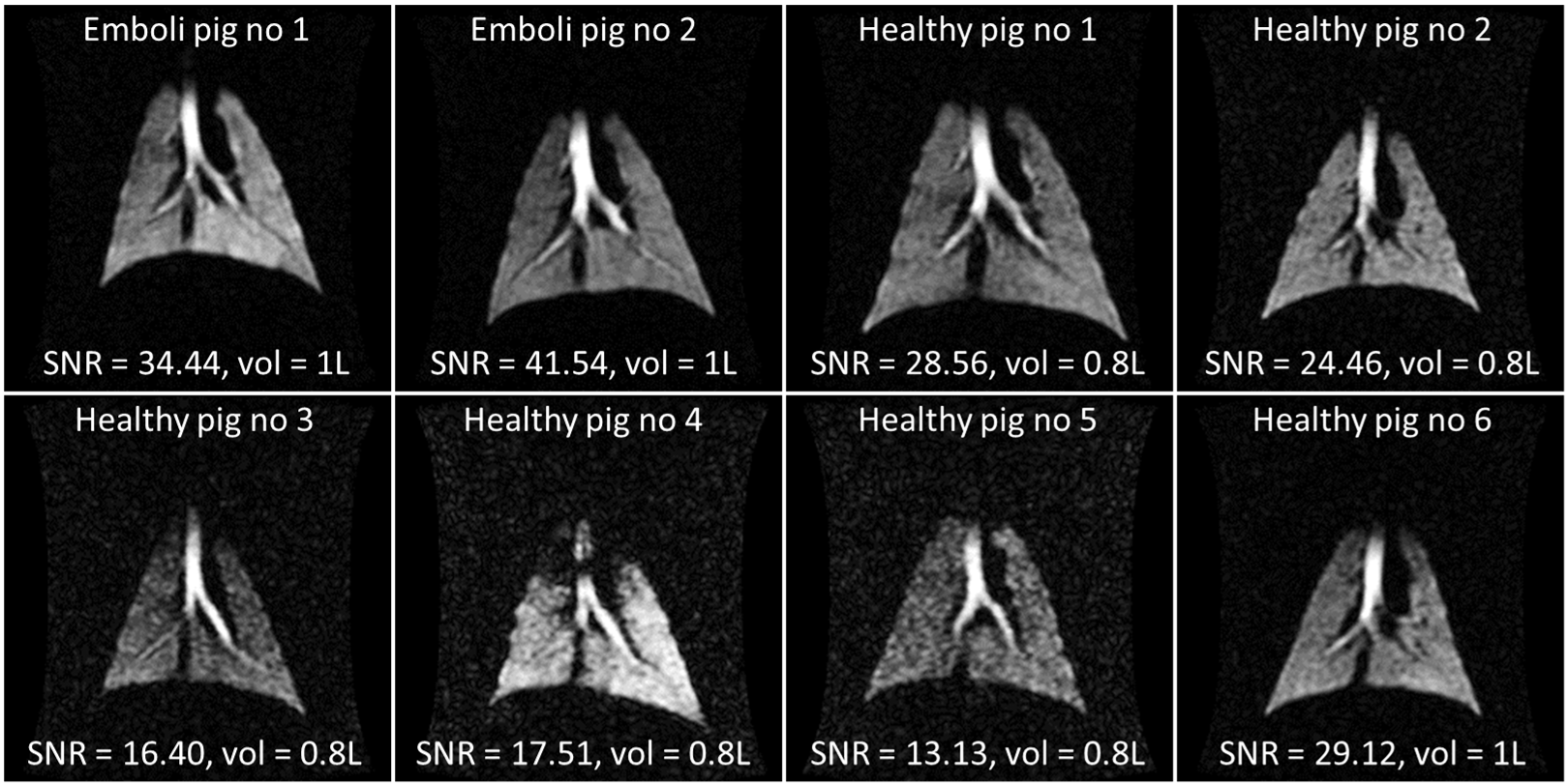
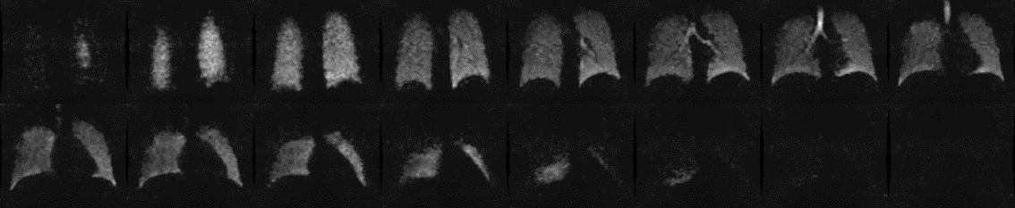
Figure 2 - 129Xe images acquired in a healthy human volunteer.
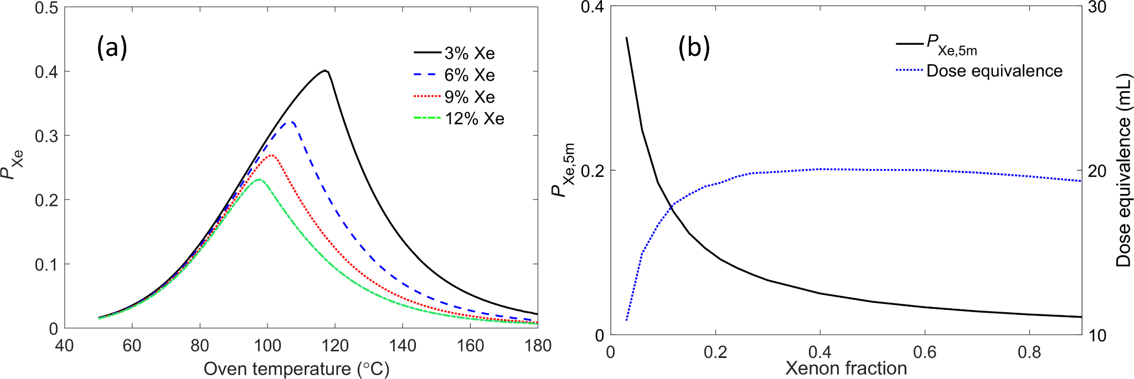
Figure 3 - Batch-mode 129Xe polarisation modelling. (a) 129Xe polarisation as a function of oven temperature for different xenon concentrations. (b) PXe,5m represents the 129Xe polarisation after 5 min of polarisation build-up with a spin-up time constant specific to the optimal temperature for each xenon concentration. The dose equivalence is a product of the 129Xe polarisation and the volume of xenon dispensed (i.e. the equivalent volume of 100% polarised 129Xe), representing the relative magnetisation/signal intensity available in a dispensed batch of polarised 129Xe.