4501
Improving Xenon-129 Lung Ventilation Image Quality with a Commercial Deep-Learning Based Image Reconstruction1The University of Sheffield, Sheffield, United Kingdom, 2GE Healthcare, Cambridge, United Kingdom, 3GE Healthcare, San Francisco, CA, United States, 4GE Healthcare, Calgary, AB, Canada
Synopsis
Keywords: Hyperpolarized MR (Gas), Hyperpolarized MR (Gas)
The utility of a deep learning based reconstruction tool for improving the quality of hyperpolarized 129Xe lung ventilation images was assessed. DL-reconstructed 129Xe ventilation image quality and SNR was improved compared with conventionally reconstructed images. In a cohort of patients with asthma and/or COPD, a small bias towards increased ventilation defect percentage, and a bias towards decreased coefficient of variation, in DL-reconstructed vs. conventionally-reconstructed images, was observed. Initial feasibility of utilising this tool for reduced-cost 129Xe ventilation imaging using natural-abundance xenon, and improved spatial resolution imaging with 129-enriched xenon, is demonstrated.Introduction
Hyperpolarized 129Xe images can be signal-to-noise limited, especially in patients with severe lung disease who struggle to fully inhale the gas dose. For this reason, most human 129Xe imaging studies are currently performed with 129-enriched Xe (>85% 129Xe), as it provides ~3-fold SNR benefits over natural-abundance Xe (26% 129Xe) (1); yet, the enrichment process is expensive (~5–10-fold cost increase per litre). As such, methods to improve image quality and SNR are economically desirable.A deep convolutional neural network (CNN)-based image reconstruction tool that acts on raw MR data and produces images with increased sharpness and reduced noise (2) has been recently commercialised, and applied to imaging of various organs (3, 4). The CNN was trained with supervised learning using pairs of low-noise, high resolution images and typical noisy lower resolution counterparts.
The purpose of this work was to assess the utility of this DL reconstruction to improve the quality of hyperpolarized 129Xe lung ventilation images, and whether the fidelity of quantitative metrics were preserved.
Methods
Retrospective analysis: N=34 129Xe ventilation MRI datasets that were acquired from patients with asthma, COPD and combined asthma-COPD were randomly selected from a database of >100 clinical 129Xe MRI examinations performed between 2020–2022. In all examinations, the xenon dose was a 50:50 mix of 129-enriched Xe (polarisation ~25% (5)) and N2 of total volume 1L (or less depending on patient height (6)). Images were reconstructed using the DL-recon tool without re-training (2), with a denoising “level” (scalar factor: 0–1) of 0.25, 0.5, 0.75 and 1. For comparison, images were also reconstructed using the conventional manufacturer recon pipeline (i.e. Fermi filtering, FFT etc.). For DL-reconstructed images at a nominal denoising level of 0.75, quantitative metrics of lung ventilation, namely; ventilation defect percentage (VDP) (7) and coefficient of variation (CV) (8), were calculated for comparison with conventional images. The original mask of the lungs derived from the conventional 129Xe images with same-session 1H MR anatomical scans was used to process the DL-reconstructed images. Bland-Altman plots were produced to identify differences in each metric. Image quality was compared qualitatively between reconstructions, and the structural similarity index measure (SSIM) was evaluated for each pair of images at all denoising levels to quantify their similarity.Prospective acquisition: to explore the effect of DL-recon on low SNR images, and the feasibility for low-dose natural-abundance Xe MRI, N=3 healthy volunteers were scanned with a 50:50 mix of natural-abundance Xe and N2 in a 1L Tedlar bag. N=1 of these healthy volunteers was also scanned with the usual dose of 129-enriched Xe described above, but with increased spatial resolution.
For both retrospective, and prospective natural-abundance Xe studies, acquisition parameters were: 1.5 T GE Healthcare scanner (HDx/450w), T-R vest coil (CMRS), 3D SSFP acquisition with in-plane FOV between 36–48cm, matrix 100x100 (in-plane resolution 3.6–4.8cm2), slice thickness 10mm, flip angle ~10°, bandwidth 31.25kHz. For the prospective enriched Xe acquisition, the slice thickness was halved (5mm) and flip angle decreased (~7°).
Results & Discussion
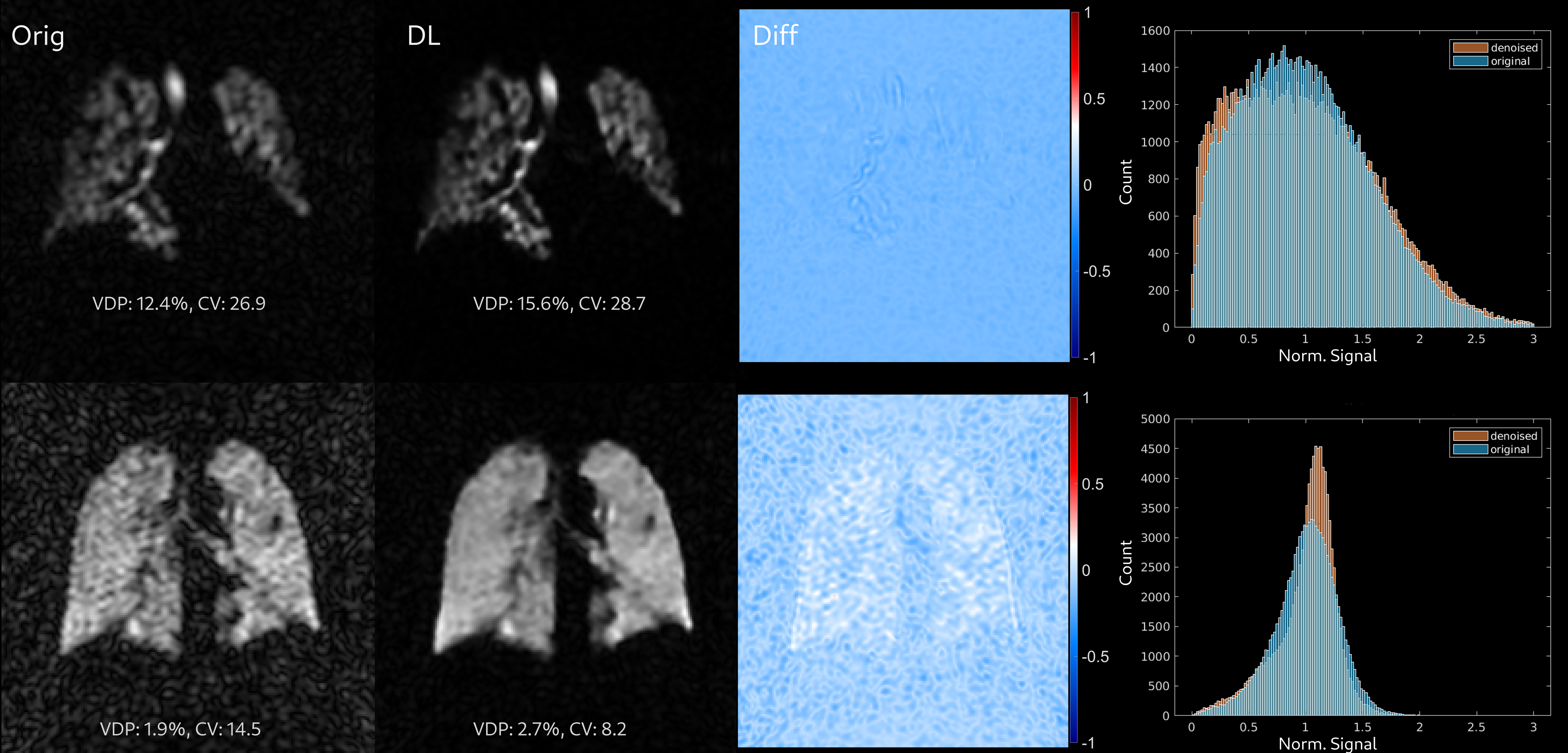
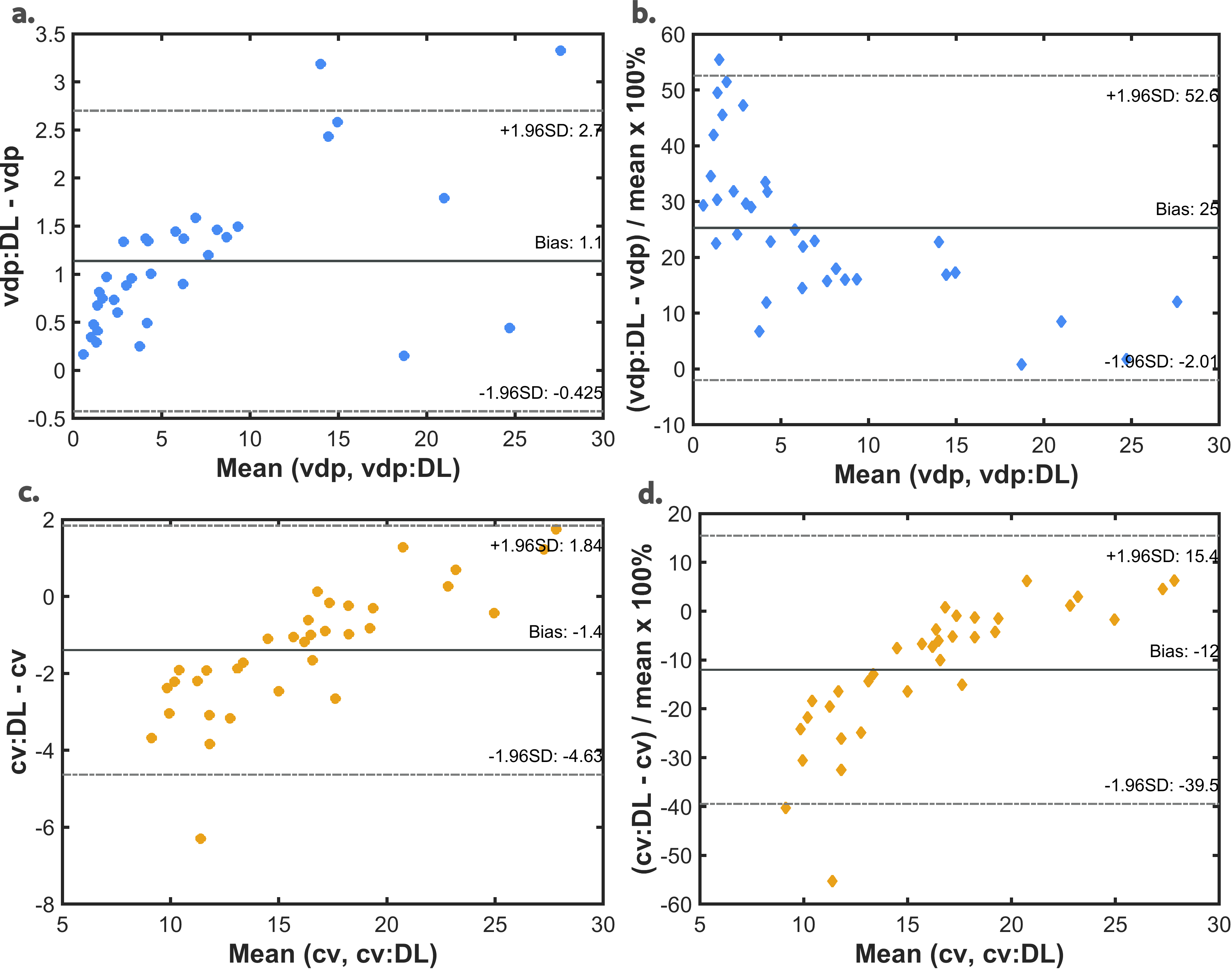
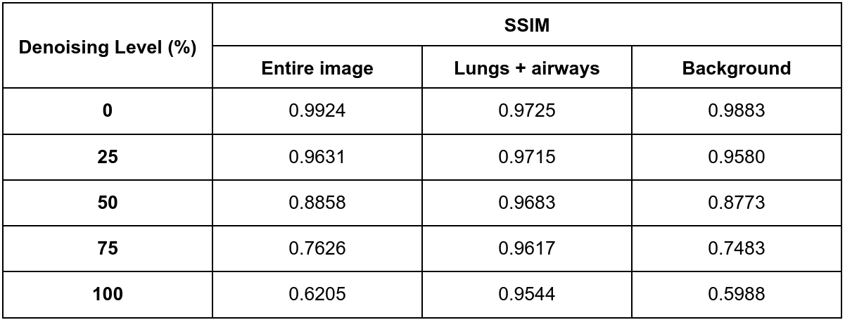
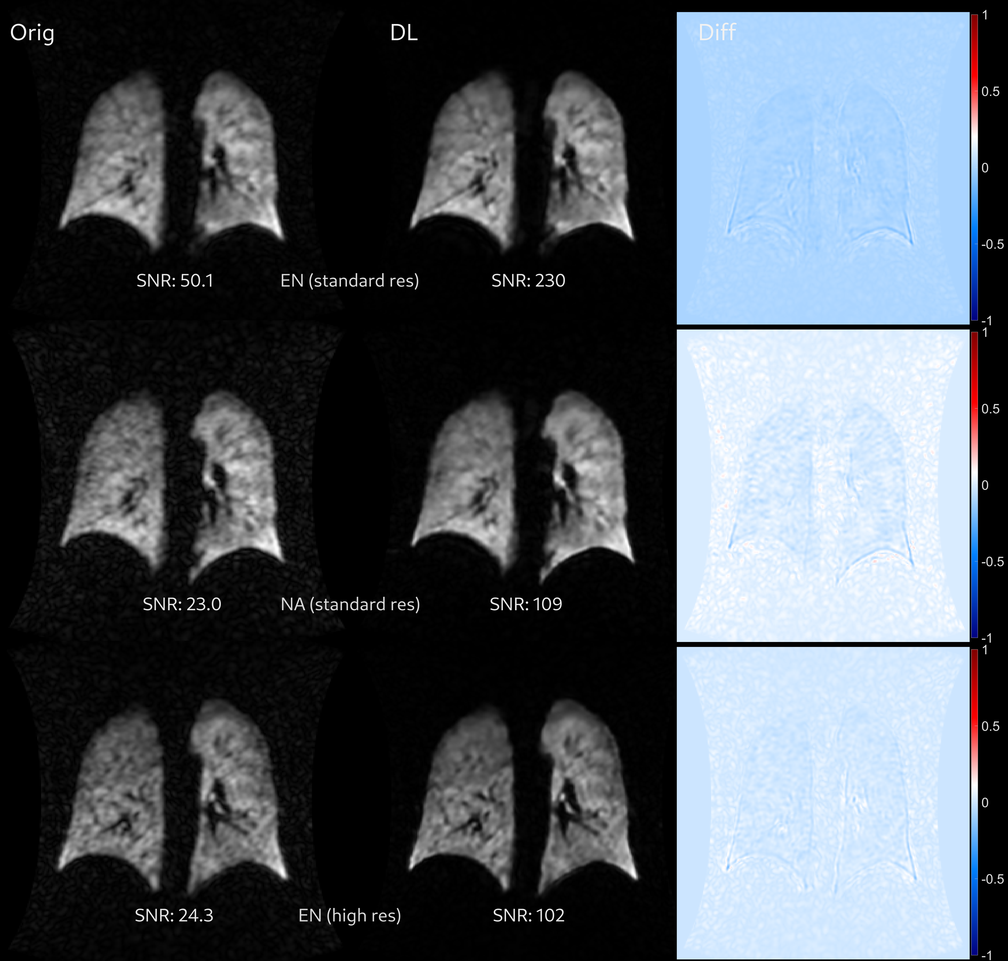
Example images from the retrospective DL reconstructions of images from subjects with asthma and asthma+COPD are shown in Figure 1 alongside the conventional reconstructions, and their difference image. In all cases, an increase in image SNR and sharpness of the lung boundaries were found, and qualitatively, the physiological ventilation distribution remained visually unchanged. VDP was increased for DL-reconstructed images when compared with conventional images; Bland-Altman analysis revealed a positive bias of 1.1% (Figure 2). Part of this bias appears to arise from the sharper edges of the lungs on DL-reconstructed 129Xe images; i.e. the reduced VDP may be partially artefactual due to using the same lung cavity mask for both images, rather than adapting the mask for DL-reconstructed images.An opposing trend was observed in CV; a negative bias (-12%) towards reduced CV for DL-reconstructed images. This implies a reduced local heterogeneity, or smoothing, corresponding visually to reduced noise granularity of the images, rather than a smoothing out of physiological variations in ventilation (see e.g. difference images and pixel signal histograms in Figure 1). Median SSIM values are reported in Table 1. As expected, SSIM decreased with increasing denoising level. When separately assessing the SSIM for the lungs+airway region and for the background only, it was found that the SSIM was relatively unchanged in the lungs+airway, but decreased sharply in the background. This is a promising indication that the main shapes / structural features of the image are unchanged by the DL reconstruction.
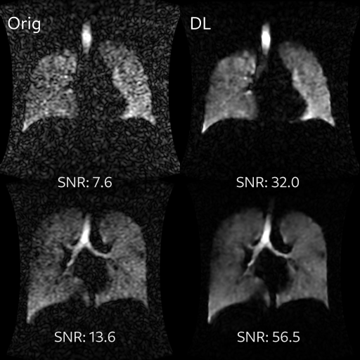
In Figures 3&4, the results of applying the DL reconstruction to natural-abundance Xe and high-resolution enriched Xe images are shown. Whilst the conventional reconstructions exhibit borderline SNR for clinical use, the DL-reconstructed images exhibit sufficient SNR and contrast for clinical evaluation.
Conclusion
DL-based image reconstruction was found to improve 129Xe ventilation image quality and SNR. Further application of this tool on images acquired from patients with a range of lung pathologies is required to fully evaluate the physiological interpretation of the resulting images. This approach could enable routine low-cost 129Xe ventilation imaging using natural-abundance xenon, and / or improved spatial resolution imaging with 129-enriched xenon.Acknowledgements
No acknowledgement found.References
1. Stewart NJ, Norquay G, Griffiths PD, Wild JM: Feasibility of human lung ventilation imaging using highly polarized naturally abundant xenon and optimized three-dimensional steady-state free precession. Magnetic Resonance in Medicine 2015; 74:346–352.
2. Lebel RM: Performance characterization of a novel deep learning-based MR image reconstruction pipeline. arXiv 2020.
3. Park JC, Park KJ, Park MY, Kim M, Kim JK: Fast T2-Weighted Imaging With Deep Learning-Based Reconstruction: Evaluation of Image Quality and Diagnostic Performance in Patients Undergoing Radical Prostatectomy. Journal of Magnetic Resonance Imaging 2022; 55:1735–1744.
4. Sun S, Tan ET, Mintz DN, et al.: Evaluation of deep learning reconstructed high-resolution 3D lumbar spine MRI. Eur Radiol 2022; 32:6167–6177.
5. Norquay G, Collier GJ, Rao M, Stewart NJ, Wild JM: Xe 129 -Rb Spin-Exchange Optical Pumping with High Photon Efficiency. Physical Review Letters 2018; 121:153201.
6. Chan H-F, Smith LJ, Biancardi AM, et al.: Image Phenotyping of Preterm-born Children using Hyperpolarised 129Xe Lung MRI and Multiple-breath Washout. Am J Respir Crit Care Med 2022.
7. Eddy RL, Svenningsen S, McCormack DG, Parraga G: What is the minimal clinically important difference for helium-3 magnetic resonance imaging ventilation defects? European Respiratory Journal 2018; 51.
8. Tzeng Y-S, Lutchen K, Albert M: The difference in ventilation heterogeneity between asthmatic and healthy subjects quantified using hyperpolarized 3He MRI. Journal of Applied Physiology 2009; 106:813–822.
Figures