4469
Comparison of cardiac DWI with and without ZOOM by using second-order motion compensated spin echo for healthy control and HCM patients1Xijing Hospital, Fourth Military Medical University, Xi 'an, China, 2Philips Healthcare,, Beijing, China
Synopsis
Keywords: Myocardium, Diffusion/other diffusion imaging techniques, Hypertrophic Cardiomyopathy(HCM)
Hypertrophic cardiomyopathy (HCM) is a hereditary cardiomyopathy with a relatively high rate of sudden death. Diffusion-weighted imaging (DWI) is widely used for evaluating motion of water protons as one of the functional magnetic resonance imaging methods. Second-order motion-compensated spin echo (M2SE) has been applied for cardiac DWI, but there are few reports of M2SE combined with ZOOM for HCM. The results showed that both signal-to-noise ratio (SNR) and contrast-to-noise ratio (CNR) of ZOOM-M2SE were significantly higher than those of M2SE only, and the ADC value of hypertrophic cardiomyopathy was also significantly higher than that of healthy controls. Cardiac DWI using ZOOM-M2SE would be a more appropriate method for investigating the myocardial microenvironment in healthy and HCM subjects.
Introduction
Hypertrophic cardiomyopathy(HCM) is the most common inherited cardiomyopathy,which is associated with abnormal hypertrophy of the myocardium and disordered arrangement of cardio myocytes[1]. Diffusion-weighted imaging (DWI) is a vital technology to assess the changes of the microenvironment. But cardiac beat and respiratory movement have always been the challenges of cardiac DWI, leading to severe artifacts that cannot be used for clinical diagnosis. Second-order motion-compensated spin echo (M2SE) is an efficient technology that can be used to reduce motion-induced signal loss [2]. M2SE was used on myocardial fibrosis in HCM to investigate the correlation between ADC and late gadolinium enhancement (LGE) [3]. However, studies on cardiac DWI using M2SE combined with ZOOM (ZOOM-M2SE) are still limited. The aim of this study is to investigate the feasibility of ZOOM-M2SE for clinical applications, especially for the diagnosis of HCM.Methods
Who underwent cardiac magnetic resonance examination from May to October 2022 were collected and analyzed. Detailed information of patients was shown in Table 2.All images were acquired on a 3.0T Philips scanner (Ingenia CX, Best, the Netherlands) with a 16-channel abdominal coil. Conventional sequences included the acquisition of cine images by steady-state free precession (SSFP) sequence in 2-, 3-, and 4- chamber and short-axis views, and an inversion-recovery gradient-echo sequence for late gadolinium enhancement (LGE). DWI with ZOOM-M2SE and M2SE only were scanned. Detailed parameters were listed in Table 1. For image quality assessment, signal-to-noise ratio (SNR) and contrast-to-noise ratio (CNR) of the two groups of images corresponding to high b value images were evaluated. The mean and variance of the six regions of interest (ROI) of the left ventricular anterior wall, anterior lateral wall, posterior lateral wall, posterior wall, posterior septum, and anterior septum were respectively taken, and SNR and CNR were calculated as follows[4]:$$SNR=\frac{MEAN(SI Myocardial Wall)}{MEAN(SD Myocardial Wall)}$$,$$CNR Wall-Cavity=\frac{MEAN(SI Myocardial Wall)-MEAN(SI Ventricular Cavity)}{√(〖(SD(SI Myocardial Wall))〗^2+〖(SD (SI Ventricular Cavity))〗^2 )}$$.Apparent diffusion coefficient (ADC) values of hypertrophic cardiomyopathy and normal control group were measured. Two independent sample T tests were used for the mean values obtained. The diagnostic efficiency was statistically analyzed by Youden’s analysis. SPSS 21.0 was used for statistical analysis, p<0.05 was considered as significant difference.
Results
12 patients with HCM (49.30±10.22 years;75% men) and 10 normal controls(31.80±10.65;80% men) were included in this study. The SNR and CNR of ZOOM-M2SE were significantly higher than M2SE in both the control group (t=-2.915, p=0.013 and t=-3.568, p=0.002) and in HCM group (t=-2.251, p=0.035 and t=-2.849, p=0.009),which were shown in Table 3. M2SE and ZOOM-M2SE and the corresponding ADC values of the HCM group were shown in Figure 1. The ADC map generated by DWI-ZOOM has a significant difference (1.51±0.18 VS =1.74±0.26X10⁻³mm2/sec, p=0.03). ROC results showed that the AUC of HCM compared with the control group were 0.800(P=0.018 as shown in Figure 2), with the sensitivity and specificity were 0.917and 0.60, respectively.Discussion
Cardiac DWI with and without ZOOM by using M2SE method for healthy control and hypertrophic cardiomyopathy were used in this study This study demonstrated that DWI-ZOOM sequence showed better SNR and CNR for both the control group and the HCM group when compared with the DWI-no-ZOOM with M2SE. Moreover, the ADC map generated by DWI-ZOOM can also significantly distinguish the control group from the HCM group, which were consistent with the researches that DWI could offer noncontract alternative for the evaluation of microstructure change[5]. Heartbeat and respiratory movements have limited the application of conventional diffusion sequences in cardiac magnetic resonance. M2SE could provide efficiently way for sable imaging acquisition, especially could achieve better image quality when combined with ZOOM method. This may have potential value in the diagnosis and treatment of HCM, and even further investigate cardiac diffusion tensor imaging (DTI).Conclusion
DWI-ZOOM with M2SE can be used for cardiac magnetic resonance imaging, especially for the diagnosis of HCM, which may improve the diagnostic efficiency of cardiomyopathy.Acknowledgements
No acknowledgement found.References
1. Antunes, MO, Scudeler, TL. Hypertrophic cardiomyopathy. Int J Cardiol Heart Vasc. 2020; 27 100503. doi: 10.1016/j.ijcha.2020.100503
2. Teh, I, Romero R, WA, Boyle, J, et al. Validation of cardiac diffusion tensor imaging sequences: A multicentre test-retest phantom study. NMR BIOMED. 2022; 35 (6): e4685. doi: 10.1002/nbm.4685
3. Sharifian, M, Rezaeian, N, Asadian, S, et al. Efficacy of Novel Noncontrast Cardiac Magnetic Resonance Methods in Indicating Fibrosis in Hypertrophic Cardiomyopathy. CARDIOL RES PRACT. 2021; 2021 9931136. doi: 10.1155/2021/9931136
4. Ratering, D, Baltes, C, Dörries, C, et al. Accelerated cardiovascular magnetic resonance of the mouse heart using self-gated parallel imaging strategies does not compromise accuracy of structural and functional measures. J CARDIOVASC MAGN R. 2010; 12 43. doi: 10.1186/1532-429X-12-43
5. Sharifian, M, Rezaeian, N, Asadian, S, et al. Efficacy of Novel Noncontrast Cardiac Magnetic Resonance Methods in Indicating Fibrosis in Hypertrophic Cardiomyopathy. CARDIOL RES PRACT. 2021; 2021 9931136. doi: 10.1155/2021/9931136
Figures
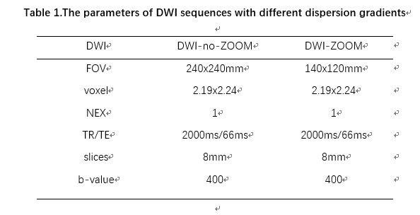
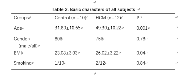
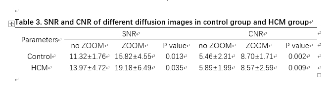
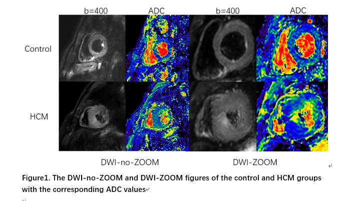
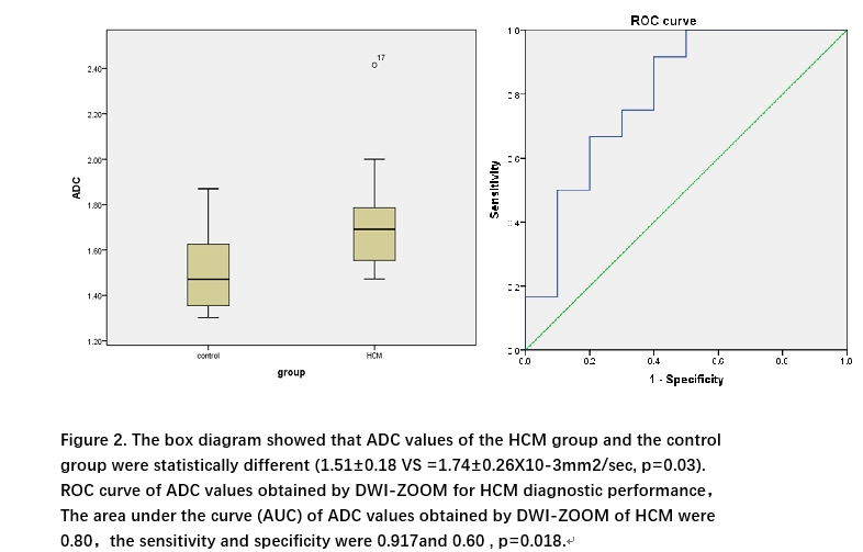
Figure 2. The box diagram showed that ADC values of the HCM group and the control group were statistically different (1.51±0.18 VS =1.74±0.26X10⁻³mm2/sec, p=0.03). ROC curve of ADC values obtained by DWI-ZOOM for HCM diagnostic performance,The area under the curve (AUC) of ADC values obtained by DWI-ZOOM of HCM were 0.80,the sensitivity and specificity were 0.917and 0.60 , p=0.018.