4465
Deep Learning Algorithm Based on LTSM for Automated Bi-Ventricle Segmentation of CMR Images1Department of Radiology, Peking Union Medical College Hospital, Chinese Academy of Medical Sciences and Peking Union Medical College, Beijing, China, 2Shukun (Beijing) Technology Co., Ltd, Beijing, China
Synopsis
Keywords: Heart, Heart
Cardiac Magnetic Resonance(CMR) imaging is an advanced cardiovascular imaging modality to evaluate cardiac structure and function. Therefore, the accuracy of segmentation directly affects the clinical evaluation and diagnosis. In this study, we used Long Short-Term Memory(LSTM) network, proposing a novel deep learning-based model for accurate automated bi-ventricle segmentation of CMR images.Introduction
Cardiovascular diseases are a leading cause of morbidity and mortality worldwide1. Cardiac magnetic resonance imaging(CMR) is an advanced cardiovascular imaging modality. It has a significant role in evaluating cardiac structure and function, such as left ventricular (LV) volume, right ventricular (RV) volume, wall thickness, and ejection fraction (EF) analysis2,3. In recent years, the deep-learning method has been developed and applied in clinical practice, assisting radiologists with the time-consuming process of imaging reconstruction4. In CMR, deep learning approaches are mainly used for right and left ventricle segmentation5,6. However, because of low contrast, motion artifacts, and blurry boundaries, the segmentation of cardiac structures using CMR has still been challenging7. Most of the existing research segments the CMR data of each phase separately and outputs the myocardial segmentation mask of each phase8, which is simple and direct. Therefore, our study aimed to propose a deep learning algorithm based on LTSM for accurate CMR imaging segmentation.Methods
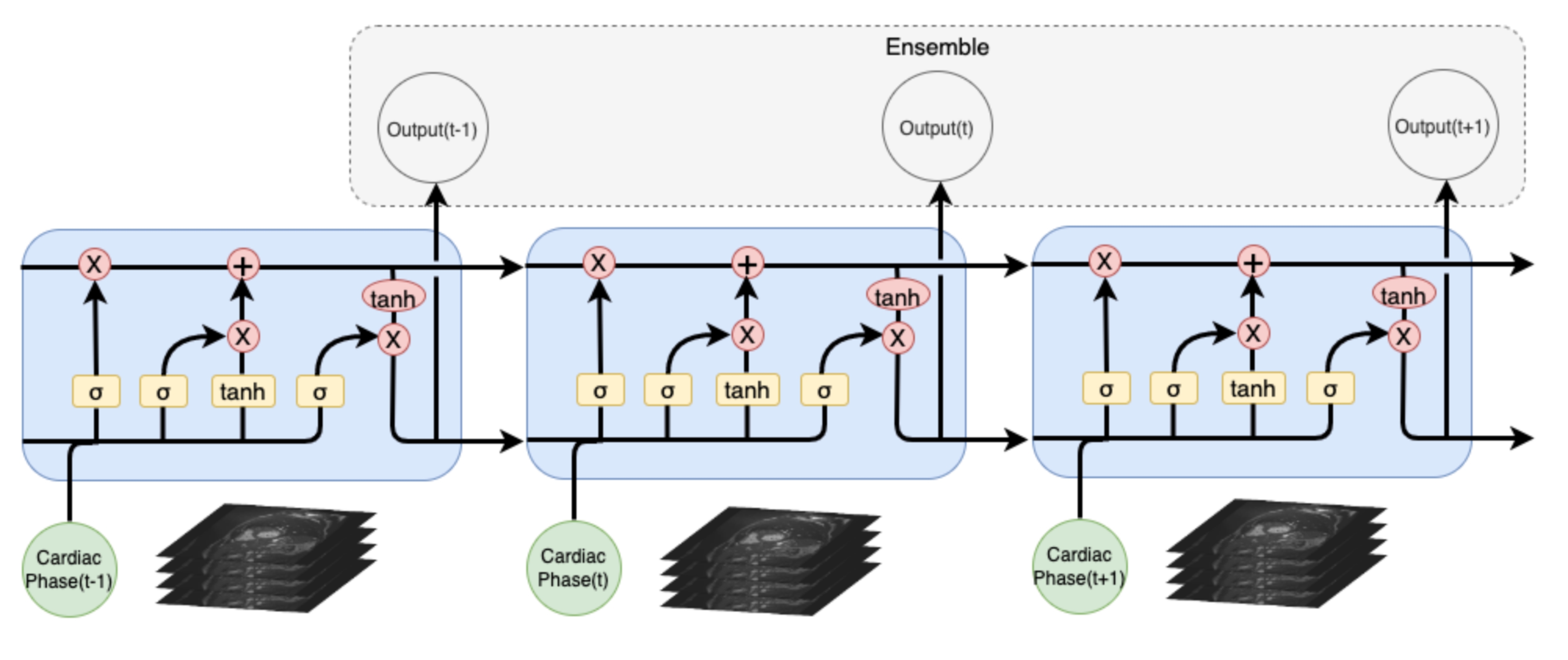
Our study retrospectively included 100 consecutive patients who underwent 1.5T CMR examinations from multi-centers. 85 patients were randomly selected as the training set, while 25 patients as the validation set. Scans with poor image quality were excluded by manual inspection. Considering that the accuracy of 3D segmentation in medical images is generally higher than that of 2D segmentation, our work used the Long Short-Term Memory(LSTM) network to segment the images by integrating CMR segmentation prediction of adjacent cardiac phases. The overall framework of our study is shown in Figure.1. Because of the high requirements for boundary segmentation accuracy, compared with the commonly used Dice Loss, we used dynamically adjusted Dice Loss and Hausdorff distance loss to optimize the model for better performance. The segmentation accuracy of ventricles and myocardium was evaluated by dice similarity coefficient (DSC).Results
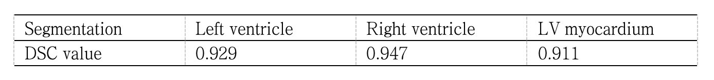
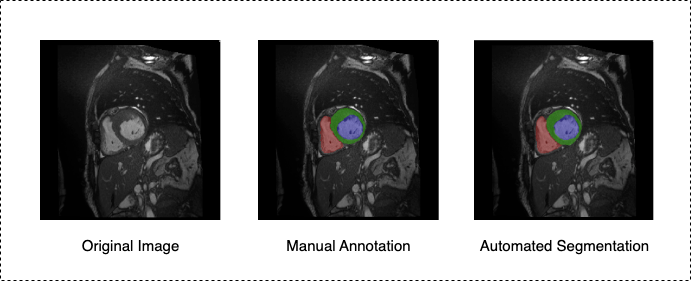
The dice similarity coefficient (DSC) of the model is shown in Table 1. The DSC value of the left ventricle, right ventricle, and LV myocardium are 0.929, 0.947, and 0.911, respectively. A representative case with the original image, manual annotation, and automated segmentation is shown in Figure 2.Discussion
Our study proposed a novel DL-based automatic segmentation method for CMR. The results demonstrate that it can automatically obtain accurate segmentation of the left ventricle, right ventricle, and LV myocardium. Most of the existing research segments the CMR data of each phase separately. The disadvantage is that it does not use the relative relationship between the myocardial positions of adjacent cardiac phases. Therefore, the Long Short-Term Memory(LSTM) network we used could solve this problem, obtaining better segmentation results. Future research may be required to evaluate the model's performance on different scanner manufacturers with larger amounts of data.Conclusion
Our study presents a novel deep learning model based on LTSM for accurate bi-ventricle segmentation on CMR images. It has the potential to optimize the post-processing of CMR images in clinical workstations.Acknowledgements
No acknowledgement found.References
1. Mortality, G. B. D. & Causes of Death, C. Global, regional, and national life expectancy, all-cause mortality, and cause-specifc mortality for 249 causes of death, 1980–2015: a systematic analysis for the Global Burden of Disease Study. Lancet 388(1459–1544), 2016.
2. Stokes MB, Nerlekar N, Moir S, et al. The evolving role of cardiac magnetic resonance imaging in the assessment of cardiovascular disease. Aust Fam Physician. 2016 Oct;45(10):761-764.
3. Seraphim A, Knott KD, Augusto J, et al. Quantitative cardiac MRI. J Magn Reson Imaging. 2020 Mar;51(3):693-711.
4. Chen C, Qin C, Qiu H, et al. Deep Learning for Cardiac Image Segmentation: A Review. Front Cardiovasc Med. 2020 Mar 5;7:25.
5. Avendi MR, Kheradvar A, Jafarkhani H. A combined deep-learning and deformable-model approach to fully automatic segmentation of the left ventricle in cardiac MRI. Med Image Anal. 2016 May;30:108-119.
6. Shaaf ZF, Jamil MMA, Ambar R, et al. Automatic Left Ventricle Segmentation from Short-Axis Cardiac MRI Images Based on Fully Convolutional Neural Network. Diagnostics (Basel). 2022 Feb 5;12(2):414.
7. Lara Hernandez KA, Rienmüller T, Baumgartner D, et al. Deep learning in spatiotemporal cardiac imaging: A review of methodologies and clinical usability. Comput Biol Med. 2021 Mar;130:104200.
8. Bernard O, Lalande A, Zotti C, et al,.Deep Learning Techniques for Automatic MRI Cardiac Multi-Structures Segmentation and Diagnosis: Is the Problem Solved? IEEE Trans Med Imaging. 2018 Nov;37(11):2514-2525.
Figures