4462
Cardiac MRI Quantitative Analysis of Native T1 and Enhanced T1 Maps in Patients with Acute Heart Failure
Yanhui Hao1, Jianxin Guo2, Yi Zhu3, Rui Zhang1, Lihong Chen2, and Jiantao Liu4
1THE FIRST AFFILIATED OF XI'AN JIAOTONG UNIVERSITY, Xi'an, China, 2THE FIRST AFFILIATED OF XI'AN JIAOTONG UNIVERSITY, XI'an, China, 3Philips Healthcare, Beijing, China, 4Philips Healthcare, XI'an, China
1THE FIRST AFFILIATED OF XI'AN JIAOTONG UNIVERSITY, Xi'an, China, 2THE FIRST AFFILIATED OF XI'AN JIAOTONG UNIVERSITY, XI'an, China, 3Philips Healthcare, Beijing, China, 4Philips Healthcare, XI'an, China
Synopsis
Keywords: Heart, Body
The purpose of this study was to investigate the predictive value of native T1 and enhanced T1 in the diagnosis of acute heart failure. Using echocardiographic ejection fraction and clinical symptoms as reference standards, The statistical results of our data showed that the native T1 value was found to have high sensitivity and specificity in predicting acute heart failure.Introduction
Heart failure (HF) constitutes an imposing health care problem, which impaires the ability of the heart to supply physiological perfusion to organs[1]. The diagnosis of heart failure by ultrasound is misdiagnosed in up to 50% of cases[1]. Myocardial T1 mapping is emerging as the noninvasive method of choice in assessment of myocardial disease allowing quantification of altered magnetization properties, which relate to the pathophysiological changes in the myocardium. [2]. We hypothesized that acute heart failure could be predicted based on native T1 and enhanced T1, and explored the application value of T1mapping value in acute heart failure. The aim of this study is to assess the diagnostic potential applied to Native T1 and Enhanced T1 obtained with cardiac MRI for the diagnosis of acute heart failure.Methods
This retrospective study from February 2022 to October 2022 included 42 participants (overall mean age 6 standard deviation, 34.7 years±12.2 [range, 18–63 years]; mean age of women, 46.1 years 6 10.8 [range, 24–63 years]; mean age of men, 29.8 years±9.2 [range, 18–56 years]) from the Magnetic Resonance Imaging in clinical suspicion of acute heart failure. Participants underwent cardiac ultrasonography, cardiac MRI at 3.0T, in which native T1 and enhanced T1 mapping as well as were assessed. The ejection fraction of echocardiography was used as the reference standard.Results
Echocardiography confirmed the diagnosis of acute heart failure in 29 patients, whereas 13 participants had no signs of acute heart failure. Mean native T1 and enhanced T1 values showed a different diagnostic performance, with the area under the curve in receiver operating curve analyses as follows: 0.95 (95% confidence interval [CI]: 0.87, 1.00) for native T1, 0.26 (95% CI: 0.00, 0.56) for enhanced T1, native T1 show a higher diagnostic performance and a sensitivity and specificity of 83% [95% CI: 81%, 99%].Discussion
Early diagnosis of acute heart failure is very important for the prognosis of patients. The active treatment of early acute heart failure is closely related to the readmission rate and survival rate [3]. Non-invasive methods of diagnosis are what we pursue. With the more and more extensive application of cardiac magnetic resonance in clinical practice and the development of magnetic resonance technology, the determination of quantitative T1 value has been widely used in a variety of diseases [4]. The pathophysiology of AHF is highly heterogeneous. Left ventricular diastolic or systolic dysfunction leads to increased preload and afterload, which in turn leads to pulmonary congestion. Fluid retention and redistribution lead to systemic congestion and eventually organ dysfunction due to hypoperfusion. Heart failure can cause myocardial injury due to any cause, such as myocardial infarction, cardiomyopathy, hemodynamic overload, inflammation, and so on, resulting in changes in myocardial T1 values. In this study, native T1 has an excellent predictive effect on acute heart failure, which confirms the clinical value of T1mapping in acute heart failure. The low sensitivity and specificity of enhanced T1 may be related to the contrast agent.Conclusion
Native T1 shows high sensitivity and specificity for the diagnosis of acute heart failure.Acknowledgements
No acknowledgement found.References
1. Heidenreich P A, Albert N M, Allen L A, et al. Forecasting the impact of heart failure in the United States: a policy statement from the American Heart Association[J]. Circulation: Heart Failure, 2013, 6(3): 606-619. 2 Puntmann V O, Carr-White G, Jabbour A, et al. T1-mapping and outcome in nonischemic cardiomyopathy: all-cause mortality and heart failure[J]. JACC: Cardiovascular Imaging, 2016, 9(1): 40-50. 3.Arrigo M,et al. Acute heart failure. Nat Rev Dis Primers. 2020 Mar 5;6(1):16. 4.Muscogiuri G, et al. Cardiac Magnetic Resonance T1-Mapping of the Myocardium: Technical Background and Clinical Relevance. J Thorac Imaging. 2018 Mar;33(2):71-80.Figures
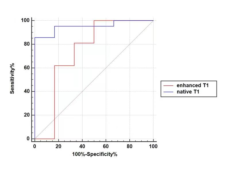
Figure1:Mean native T1 and enhanced T1 values showed a
different diagnostic performance, with the area under the curve in receiver
operating curve analyses as follows: 0.95 (95% confidence interval [CI]: 0.87,
1.00) for native T1, 0.26
(95% CI: 0.00, 0.56) for enhanced T1, native T1 show a higher diagnostic
performance and a sensitivity and specificity of 83%
DOI: https://doi.org/10.58530/2023/4462