4453
MR-based body composition analysis, resting state fMRI and autonomic function evaluation for phenotyping biomarkers1NUCLEAR MAGNETIC RESONANCE, ALL INDIA INSTITUTE OF MEDICAL SCIENCES, NEW DELHI, NEW DELHI, IN, India, 2PHYSIOLOGY, ALL INDIA INSTITUTE OF MEDICAL SCIENCES, NEW DELHI, NEW DELHI, India, 3PHYSIOLOGY, ALL INDIA INSTITUTE OF MEDICAL SCIENCES, NEW DELHI, NEW DELHI, IN, India, 4NUCLEAR MAGNETIC RESONANCE, ALL INDIA INSTITUTE OF MEDICAL SCIENCES, NEW DELHI, NEW DELHI, India
Synopsis
Keywords: Screening, Quantitative Imaging, MR body composition analysis, phenotyping, subcutaneous fat, resting state fMRI
Phenotyping individuals are gaining importance in personalized medicine. This MR based body composition analysis and resting state (R-fMRI) along with autonomic functions (AFT) evaluation have determined parameters for physical, functional and physiological phenotyping in healthy volunteers (n=30). MR studies (at 3T) were R-fMRI using EPI sequence, subcutaneous fat evaluation by mDIXON sequence and AFT included parasympathetic (deep breathing, HUT) and sympathetic (HUT, hand grip test, cold pressor test). Increased functional activation, higher abdominal fat and fall in Baroreflex independent sympathetic reactivity were observed in volunteers having BMI ≥25 Kg/m2 compared to those with BMI <25Kg/m2.Introduction
As the importance of precision medicine increases, phenotyping is becoming as important as genotyping (1). Phenotyping refers to physical, functional and physiological characteristics of an individual and it plays an important role in preventive and personalized medicine, and also precision nutrition (2). This study has used MR-based body composition analysis and resting state fMRI (R-fMRI) for physical and functional phenotyping, respectively. In addition, physiological phenotyping has been evaluated using Autonomic Function tests (AFT), namely parasympathetic and sympathetic reactivity tests (3).Material and Methods
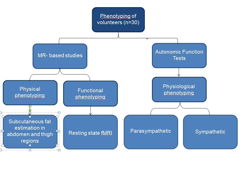
The workflow of the study is shown in Figure 1.Volunteers: Healthy volunteers (15 females, 15 males, 20-40 years age) were recruited for the study after obtaining Institute Ethical Clearance. Exclusion criteria were pregnant women, lactating mothers and history of systemic illness. The volunteers were categorized into two groups on the basis of their BMI values - Group I (< 25 kg/m2; n=15) and Group II (≥ 25kg/m2; n=15) (4).
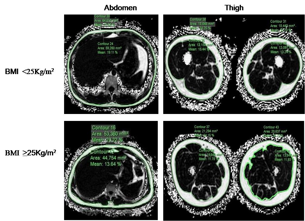
MR studies: The studies were carried out in a 3T MR scanner (Ingenia, Philips). mDIXON- Quant sequence (5) was used for evaluating the subcutaneous fat in abdomen (T7-L3 vertebra) and thigh (superior surface of hip joint to inner surface of medial) regions. The following acquisition parameters were used: FOV of 400x329x188mm3, voxel of 2x2.5x5mm3 and matrix of 200x312x74. EPI sequence was used for acquiring resting-state (R-fMRI) data with the following parameters: FOV of 230x230x153mm3, voxel of 3x3x3mm3 and 240x240x51 matrix.
Autonomic Function Tests: The following autonomic reactivity tests were carried out, Parasympathetic evaluation: deep breathing test (change in heart rate, ratio of exhalation and inhalation) and Head-up tilt (HUT); Sympathetic evaluation: HUT, hand grip test and cold pressor test (CPT).
Data analysis: Percentage subcutaneous fat in abdomen and thigh regions were evaluated by drawing contours on fat fraction maps obtained from m-DIXON quant sequence. R-fMRI data was evaluated using MRIcron and CONN (R2018b). AFT data was scored using Post-hoc analysis (Labchart analyzer). Independent T-test was used for statistical analysis of physical and physiological phenotyping markers.
Results
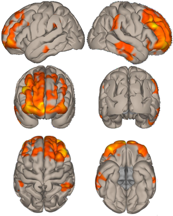
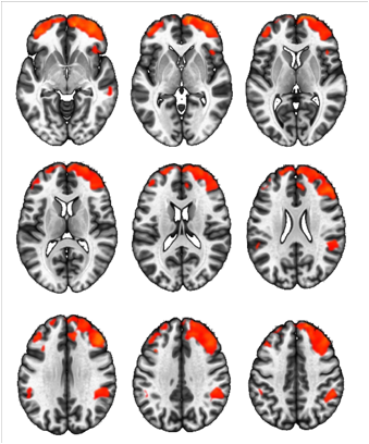
Figure 2 shows representative contoured fat fraction MR images for Group I and Group II volunteers. Increased R-fMRI activation areas in Group II volunteers are shown in the sagittal, coronal and axial planes (Figure 3). These are the difference images of Group I and Group II volunteers showing the increased activation areas in the volunteers with BMI ≥ 25kg/m2. The differences in the resting state activation between the two groups can be seen more clearly in the axial sections (Figure 4). Statistically significant differences (p < 0.02) were observed between Group I and Group II volunteers in the abdominal % subcutaneous fat and the Cold- Pressor test. Percentage subcutaneous fat in abdomen - Group I (8.63±35%) and Group II (15.66±7.8%).In the autonomic functions, the cardiovascular evaluation using Cold-Pressor test showed a showed significant (p < 0.05) fall in Baroreflex independent sympathetic reactivity in Group II (8.78±6.4 mmHg) volunteers as compared to Group I (15.5±8.5 mmHg).
Conclusion
With increasing interest in personalized medicine and nutrition, the role of phenotypig an individual is beginning to be taken cognisance of. Although there are other techniques like Dual Engery X-ray absorptiometry (DEXA) used for phenotyping (6), MR offers a non-invasive methodology for the same. This study has demonstrated the potential of MR-based body composition analysis and resting state fMRI for physical and functional phenotyping, respectively. An additional novelty of the study is evaluation of autonomic functions for physiological phenotyping. Further in-depth studies and correlation analysis of the dataset are underway.Acknowledgements
The work was supported by Science and Engineering Research Board (SERB) and Indian Council of Medical Research (ICMR), Goverment of IndiaReferences
(1) Dudley JT, et al. Personalized medicine: from genotypes, molecular phenotypes and the quantified self, towards improved medicine. Pac Symp Biocomput, 1, 342-346, 2015.
(2) König IR, et al. What is precision medicine? Eur Respir J, 50, 1700391, 2017.
(3) Ziemssen T, et al. The Investigation of the Cardiovascular and Sudomotor Autonomic Nervous System- A Review. Frontiers Neurol, 10, 1-13, 2019.
(4) Gallagher D, et al. Healthy percentage body fat ranges: an approach for developing guidelines based on body mass index. Am J Clin Nutr, 3, 694-701, 2000.
(5) Zhang Y, et al. Reliability of measuring the fat content of the lumbar vertebral marrow and paraspinal muscles using MRI mDIXON-Quant sequence. Diagn Interv Radiol, 5, 302-307, 2018.
(6) Chad K, et al. Regional Body Composition Analysis Using DEXA, Med & Sci in Sports & Exercise, 37, 707-715, 2005.
Figures