4450
ADC histogram features of rectal cancer before and after neoadjuvant radiation to predict heterochronic hematogenous metastasis
Xiaoxian Zhang1, Chunmiao Xu1, Zhiwei Shen2, Ke Jiang2, and Xuejun Chen1
1Henan tumor hospital, Zhengzhou, China, 2Philips healthcare,Beijing,China, Beijing, China
1Henan tumor hospital, Zhengzhou, China, 2Philips healthcare,Beijing,China, Beijing, China
Synopsis
Keywords: Cancer, Diffusion/other diffusion imaging techniques
Bloodstream metastasis to the liver, lung, and bone is the most prevalent mechanism of rectal cancer metastasis. However, there are currently no good indications for diagnosing heterochronic hematogenous metastases at the early stage. Therefore, this study aimed to explore whether ADC histograms might predict new heterochronic hematogenous metastases before and after neoadjuvant radiation for rectal cancer. Results showed that the first-order histogram feature ADC5% derived from post-treatment ADC maps might be utilized as an imaging biomarker and an independent predictor of new heterochronic hematogenous metastases after surgery following neoadjuvant chemotherapy for rectal cancer.Introduction
Rectal cancer is one of the most frequent tumors in the world, accounting for five of the top five incidence rates in both men and women. Approximately 70% of patients will develop metastatic colorectal cancer, and tumor metastasis is a primary cause of mortality in colorectal cancer patients. Bloodstream metastasis to the liver, lung, and bone is the most prevalent mechanism of rectal cancer metastasis. As a result, early detection of metastases in rectal cancer is important and is linked to patient prognosis and survival. However, there are currently no good indications for diagnosing heterochronic hematogenous metastases at the early stage. Magnetic resonance diffusion weighted imaging (DWI) has been demonstrated to be useful in measuring rectal cancer grading, staging, therapy effectiveness, and gene mutation.Therefore, the purpose of this research was to determine whether pre-treatment and post-treatment ADC histograms might predict new heterochronic hematogenous metastases after neoadjuvant radiation for rectal cancer.Methods
From February 2018 to May 2021, clinical, pathological, imaging, treatment choices, and follow-up data for patients diagnosed with rectal cancer in our hospital were collected retrospectively. Our hospital's ethics committee authorized the trial. This study comprised 87 patients who had been followed for more than three years.All the patients were divided into two groups: (1) Metastasis group (34 patients): patients without metastasis at the time of diagnosis and new hematogenous metastases discovered during surgical follow-up. (2) Metastases-free group (53 cases): patients with no metastasis at any point throughout the three-year follow-up period. The study was conducted on 3T MR system from multiple manufacturers (Philips, Semiens, GE) with abdominal coils. Conventional T2WI, DWI, and enhanced T1WI images were acquired. EnhancedT1WI: matrix 320;T2WI: slice thickness 5mm,matrix 384;DWI: slice thickness 5mm,matrix 160,b=50,800 s /mm2.All raw data were analyzed with the ITK-Snap, and the lesions' regions of interest (ROI) were outlined by two pelvic radiologists with at least 5 years of experience respectively. The ITK-Snap post-processed maps were subsequently analyzed in MATLAB to derive the following parameters: percentile ADC values (ADC5th, ADC25th, ADC50th, ADC75th, ADC95th), minimum ADC (ADCmin), maximum ADC (ADCmax), mean ADC (ADCmean), variance, standard deviation, interquartile distance, full distance, kurtosis, skewness, mean absolute deviation. The data were reported as mean±standard deviation if they conformed to a normal distribution, and as median (Superior and inferior quartiles) if not. The data were statistically analyzed using t-test, corrected t-test, or rank sum test.Results
The differences between the two groups in ADC histogram parameters were not statistically significant before treatment,while the post-treatment ADC histogram metrics ADC5% and interquartile distance differed significantly between the two groups, with the metastatic group having a greater ADC5% and a lower interquartile distance.Discussion
The advantages and innovations of this study, compared to previous studies, are as follows: 1) a total of 87 patients were enrolled, with complete pre- and post-treatment MRI images, as well as clinicopathological parameters; 2) the treatment protocols of the enrolled patients were uniform, with all of them undergoing neoadjuvant radiotherapy plus total mesorectal excision (TME) radical surgical resection after diagnosis and postoperative radiotherapy; 3) In order to portray the whole information of the tumor inside, the ROI in this study includes the cystic necrosis inside the lesion. There was no statistically significant difference in the distribution of ADC5% between the two groups before treatment, but there was a statistically significant difference in the distribution of ADC5% between the two groups after treatment, and the ADC in the group with metastasis was higher than that in the group without metastasis. It is essentially compatible with Bianca Boca's findings (3). Jin Li (4) investigated the difference between the groups with and without metastasis in the lymph nodes of stage T3 rectal cancer and discovered that the distribution of ADC5% and ADC10% with metastasis was relatively higher than that of the group without metastasis, which is consistent with the findings of this study.Conclusion
This study demonstrated that the first-order histogram feature ADC5% and a lower interquartile distance derived from post-treatment ADC maps might be utilized as an imaging biomarker and an independent predictor of new heterochronic hematogenous metastases after surgery following neoadjuvant chemotherapy for rectal cancer.Acknowledgements
No acknowledgement found.References
1. R. L. Siegel, K. D. Miller, H. E. Fuchs, A. Jemal, Cancer Statistics, 2021. Ca-cancer j clin 71, 7-33 (2021). 2. J. Wang et al., Metastatic patterns and survival outcomes in patients with stage IV colon cancer: A population-based analysis. Cancer Med 9, 361-373 (2020).
3. B. Boca Petresc et al., The Utility of ADC First-Order Histogram Features for the Prediction of Metachronous Metastases in Rectal Cancer: A Preliminary Study. Biology (Basel) 11, (2022)
4. J. Li et al., Histogram Analysis of Diffusion-Weighted Magnetic Resonance Imaging as a Biomarker to Predict Lymph Node Metastasis in T3 Stage Rectal Carcinoma. Cancer Manag Res 13, 2983-2993 (2021)
Figures
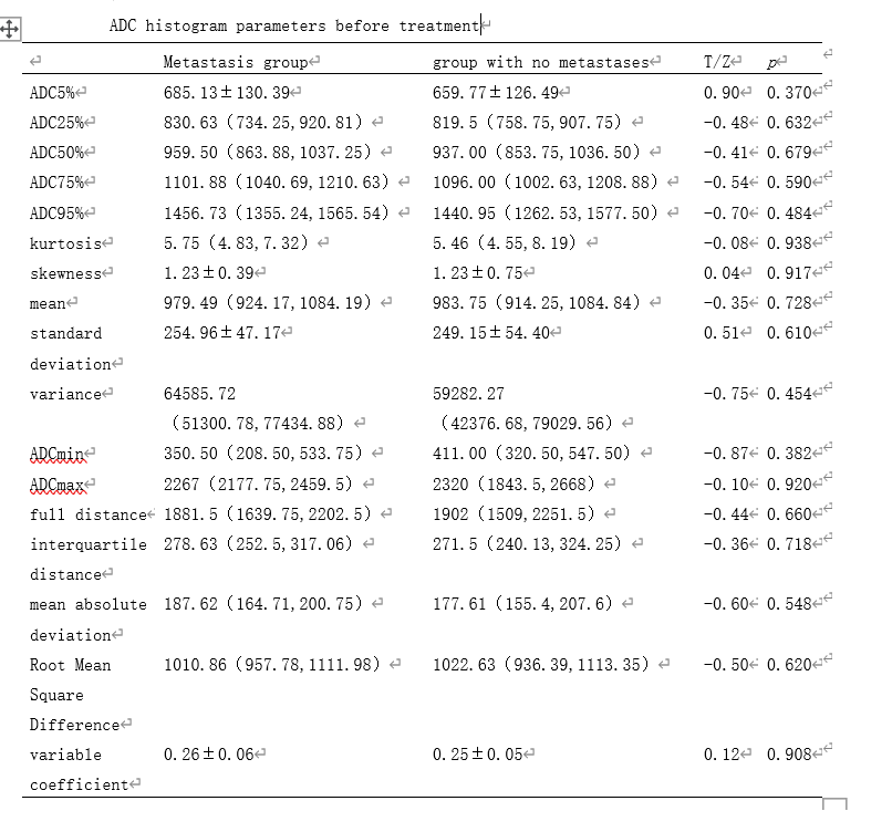
ADC histogram parameters before
treatment
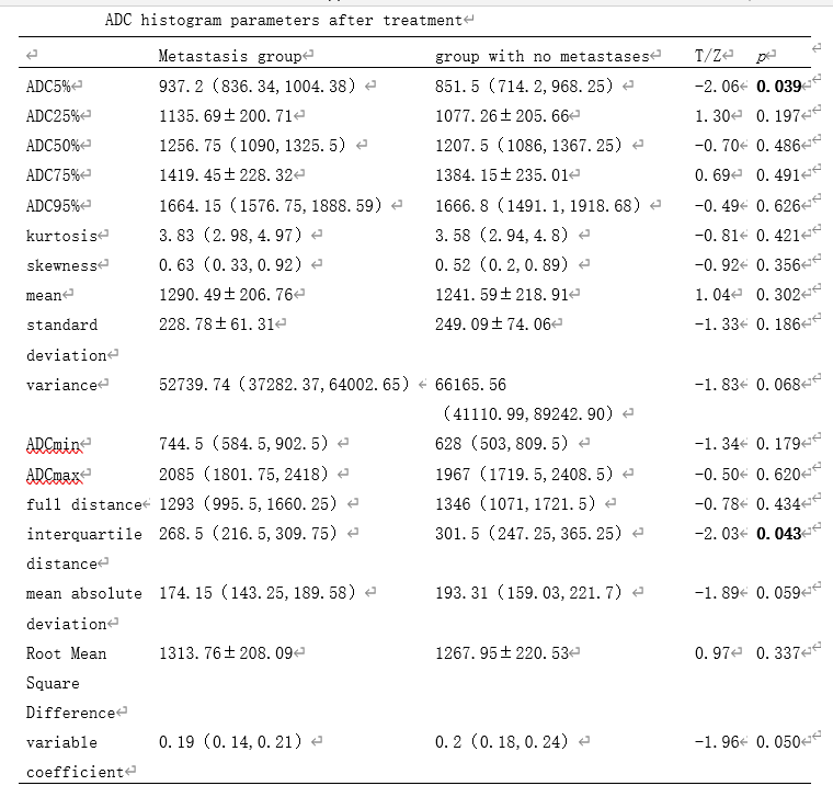
ADC histogram parameters after
treatment
DOI: https://doi.org/10.58530/2023/4450