4447
Oxygen-enhanced high-resolution 3D ultrashort echo time MRI for patients with connective tissue diseases: a preliminary study1Renji Hospital, School of Medicine, Shanghai Jiao Tong University, Shanghai, China, 2United Imaging Research Institute of Intelligent Imaging, Beijing, China, 3Ningbo Hangzhou Bay Hospital, Ningbo, China, 4Central Research Institute, United Imaging Healthcare, Shanghai, China
Synopsis
Keywords: Lung, Lung
This preliminary study investigates the utility of high-resolution 3D-UTE to assess patients with CTD-ILD and their pulmonary function, including both pulmonary parenchyma and lesions, using OE-UTE-MRI. Our results suggest that high-resolution 3D UTE and OE-UTE MRI could potentially be as useful as thin-section CT for lesion detection of CTD patients with ILD and provide additional pulmonary functional loss assessment.
Introduction
Interstitial lung disease (ILD) encompasses a heterogeneous group of lung diseases characterized by diffuse involvement of the pulmonary parenchyma by varying degrees of inflammation and/or fibrosis. Among many causes, connective tissue disease (CTD), the systemic autoimmune disorder caused by excessive immune-activated inflammation that targets the connective tissues of the body, is one of the most common systemic diseases related causes of ILD.Currently, computed tomography (CT) has been the gold standard imaging method for ILDs evaluation. However, CT is associated with ionizing radiation exposure, which should be a concern, especially for patients with CTD-ILD, as multiple CT scans might be necessary through the chronic course of these pathologies.
More recently, three-dimensional radial ultrashort echo time (3D-UTE) MRI has shown promise for noninvasively assessment of lung diseases 1,2. The 3D-UTE-based oxygen-enhanced (OE) MRI has also been used to assess regional ventilation abnormalities of the lung 3. This preliminary study aims at investigating the utility of high-resolution 3D-UTE to evaluate patients with CTD-ILD and their pulmonary function, including both pulmonary parenchyma and lesions, using OE-UTE-MRI.
Methods
This study included 14 CTD-ILD patients. All patients underwent thin-section CT. Pulmonary MR imaging was performed at 1.43 T (uMR 586, United Imaging Healthcare, Shanghai, China). Respiratory-gated 3D radial UTE pulse sequences (TR: 2.5 msec, TE: 0.08 msec, flip angle: 8°, slice thickness: 2 mm, FOV: 350 × 350 mm2, matrix: 512 × 512) were acquired twice for each patient. The first was acquired during free-breathing with 21% oxygen (normoxic), while the second was acquired with 100% oxygen (hyperoxic). Percent signal enhancement (PSE) was used to quantify pulmonary ventilation.Lesion detectability agreement between 3D-UTE and CT was used to identify the representative image findings of CTD-ILD. The difference in the region of interest (ROI) mean PSE value between normal pulmonary parenchyma and lesions was derived for regional pulmonary function assessment.
Results
The high-resolution 3D-UTE MRI achieved very good agreement in lesion detectability compared to thin-section CT (k = 1.00), as shown in Figure 1. The mean PSE of CTD-ILD lesions (3.7 ± 0.9%) was significantly lower than that of the pulmonary parenchyma (5.9 ± 1.2%) (Figure 2).Discussion
It is known that the pulmonary application of MRI has been severely limited by respiratory motion artifact, low proton density, and fast signal decay caused by short tissue T2*. Our results suggest that the capability of high-resolution 3D-UTE pulmonary MR imaging for assessing CTD-ILD is potentially as good as that of standard thin-section CT. The measurements of regional ventilation defects from PSE maps depict local areas of structural abnormality. The results are in agreement with the previous study 4 but with higher resolution and shorter scan time, which further demonstrates that the OE MRI has superior potential for the assessment of lung diseases.Conclusions
High-resolution 3D UTE was found to be as useful as thin-section CT for lesion detection for CTD patients with ILD. OE-UTE MRI was able to evaluate regional pulmonary function loss for CTD-ILD patients.Acknowledgements
No acknowledgement found.References
1. Ohno Y, Koyama H, Yoshikawa T, et al. Pulmonary high-resolution ultrashort TE MR imaging: Comparison with thin-section standard- and low-dose computed tomography for the assessment of pulmonary parenchyma diseases. J Magn Reson Imaging. Feb 2016;43(2):512-32. doi:10.1002/jmri.25008
2. Yang S, Zhang Y, Shen J, et al. Clinical Potential of UTE-MRI for Assessing COVID-19: Patient- and Lesion-Based Comparative Analysis. J Magn Reson Imaging. Aug 2020;52(2):397-406. doi:10.1002/jmri.27208
3. Zhao F, Zheng L, Shan F, et al. Evaluation of pulmonary ventilation in COVID-19 patients using oxygen-enhanced three-dimensional ultrashort echo time MRI: a preliminary study. Clinical Radiology. 2021;76(5):391. e33-391. e41.
4. Ohno Y, Nishio M, Koyama H, et al. Oxygen-enhanced MRI for patients with connective tissue diseases: comparison with thin-section CT of capability for pulmonary functional and disease severity assessment. Eur J Radiol. Feb 2014;83(2):391-7. doi:10.1016/j.ejrad.2013.11.001
Figures
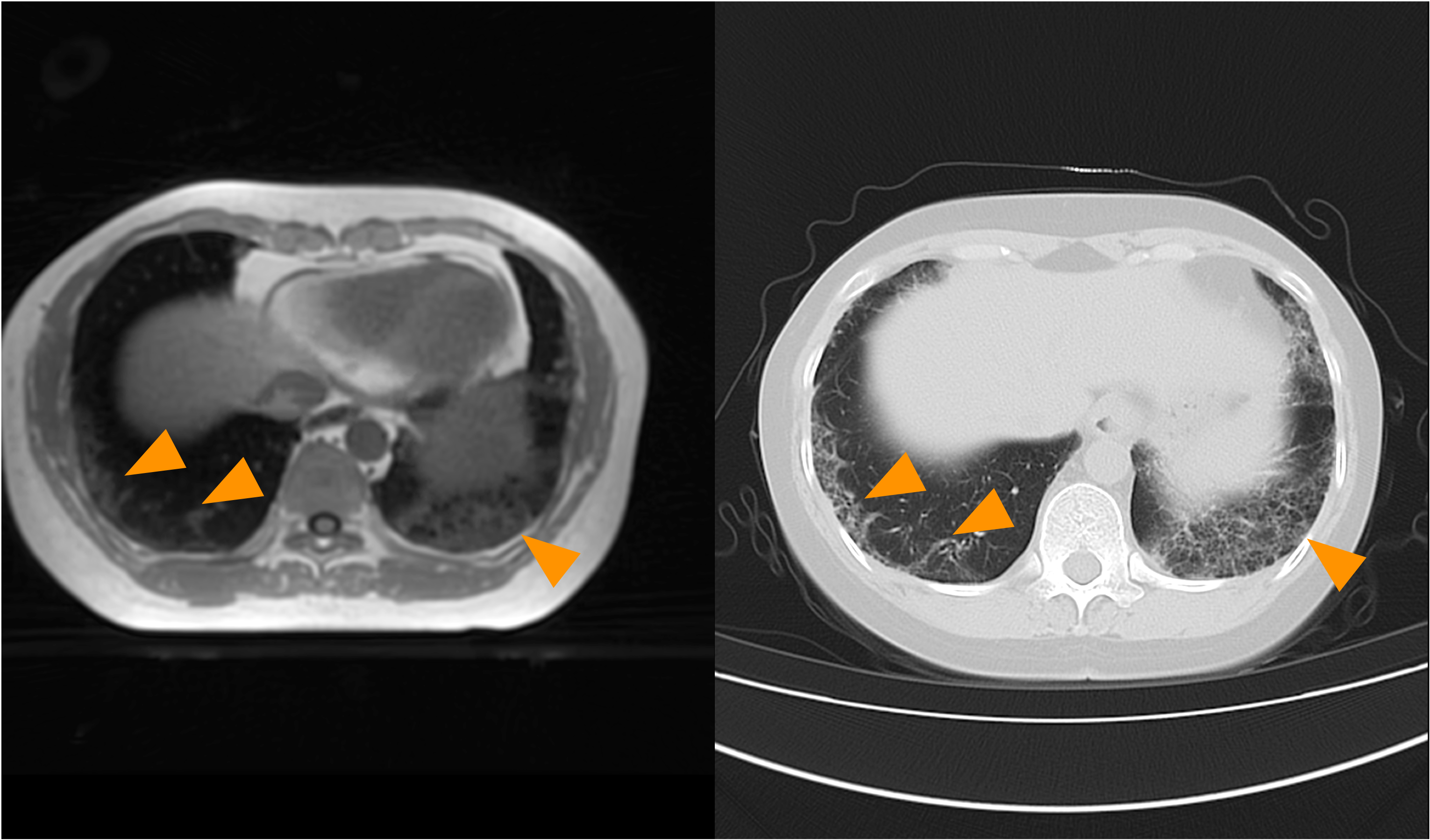
Figure 1. Representative CT and UTE-MRI images of a patient with lesions caused by CTD-ILD (arrows).
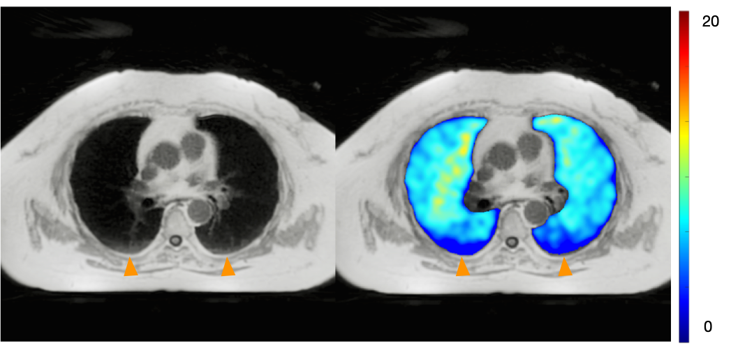
Figure 2. Representative UTE-MRI and PSE map of a patient with lesions caused by CTD-ILD. The mean PSE of lesions was lower than that of the pulmonary parenchyma (arrows).