4444
MRI Radiomics Model Preoperatively Predicts Key Gene Mutation in Pancreatic Ductal Adenocarcinoma Patients1Radiology, West China Hospital, Sichuan University, Chengdu, China, 2Affiliated Hospital of North Sichuan Medical College, Nanchong, China
Synopsis
Keywords: Pancreas, Cancer
Individualized management of pancreatic ductal adenocarcinoma (PDAC) is difficult and understanding it underlying biology is important. Radiomics model based on MRI was developed to predict status of KRAS and TP53 in PDAC, univariate analysis, MRMR and LASSO were applied for feature selection and multivariable logistic regression used for modeling. The classification metrics were applied to assess these models discrimination. Results suggested radiomics model based on MRI outperformed clinical model for predicting gene mutations of PDAC. This study confirmed that radiomics model based on MRI had a potentiality to predict the KRAS and TP53 mutation in PDAC.
Introduction
Pancreatic ductal adenocarcinoma (PDAC) is one of the most aggressive cancers with a 5-year survival rate inferior 10% and is predicted to become the second leading cause of death in Untied Stated by 2030 1. Due to the heterogeneity of tumors, the therapy and prognosis evaluation of PDAC is challenging. Thus, understanding the underlying biology of PDAC is important to optimize the treatment strategies and evaluate prognosis.Large scale studies in PDAC have demonstrated high-frequency alterations in KRAS, TP53, CDKN2A and SMAD4 2, the most common is KRAS activation followed by TP53 inactivation. These mutated genes are related to patient survival and have been used to develop new therapies to overcome PADC resistance 3-5.
Magnetic resonance imaging (MRI) can noninvasively detect pancreatic malignancy but it is difficult to detect tumor microenvironment. Radiomics 6 can obtain large number of potential characteristic data in a noninvasive way, and quantitatively analyze the obtained feature data to provide a basis for treatment decision-making. Radiogenomics is gradually emerging to study the relationship between radiological features and genetic characteristics for better achieving patient individualized management 7. The aim of this study is to evaluate the capability of radiomics features based on MRI to predict status of KRAS and TP53 in PDAC.
Methods
Consecutive patients with pathologically confirmed PDAC and preoperative multi-sequence MRI available were recruited. The paraffin sections of each patient were collected for whole exon genomic sequencing, mutated and non-mutated group were classified according to KRAS and TP53 alteration status. Demographic, clinical and pathological data were obtained from medical records, the over survival (OS) was acquired. OS was defined the operation date to death. Take the follow-up for one year as the boundary. We compared the clinical data and OS between the mutated and non-mutated group.An experienced radiologist manually delineated the entire PDAC on the arterial phase on MRI blind to the clinical and pathological data. The process was implemented using an open source software IBEX (β1.0, http://bit.ly// IBEX MD Anderson) on MATLAB 2016b (The MathWorks Inc). 350 radiomics features including intensity histogram, texture (gray-level co-occurrence matrix GLCM; the gray-level run-length matrix, GLRLM) and shape features were contained.
All radiomics features tested by independent samples t-test or Mann-Whiney U test to selected useful features between mutated and non-mutated group. Then, minimum redundancy maximum relevance (MRMR) algorithm was used to select the top 15 features with nonredundant and highly informative. The Least absolute shrinkage and selection operator (LASSO) was applied for dimensionality reduction and feature selection. LASSO regression adjusts the model complexity by controlling the regularization parameter (λ) to obtain fewer optimal features for radiomics modeling. Clinical characteristics were measured based on the variable type. Continuous variables were compared using independent t tests or Wilcoxon based on the distribution. Categorical variables were compared using chi-square tests or Fisher’s exact tests. The selected radiomics and clinical features modeled by multivariable logistic regression. A joint model was combined radiomics with clinical features modeled by multivariable logistic regression. A ten-fold cross-validation method was used to verify the performance of the joint model. Delong test was used to compare the AUC of the joint, radiomics and clinical models.
Results
36 patients (15 patients with non-mutated and 21 patients with mutated) were included. Genetic analysis revealed alteration in KRAS is 19 (52%) and TP53 is 13 (36%). The baseline characteristics were recorded in table1. Mutated group lesion had a higher tumor size and had a shorter 1-year survival rate compared with non-mutated group. 7 radiomics features were included for radiomics modeling, one clinical feature tumor size was modeled for clinical model, the joint model included radiomics and clinical data. The classification metrics of different models were showed in table2. Among these models, the joint and radiomics models achieved satisfactory performance, no significant difference between the two but both outperformed the clinical model. These results were shown in Fig.1A. The results of the joint model after ten-fold cross-validation was shown in Fig.1B.Discussion
Our study developed a non-invasive, quantitative radiomics model to preoperatively predict the mutation status of KRAS and TP53 in PDAC. The prediction performance of radiomics and joint models were outstanding the clinical model, this may be associated with radiomics features can extract more quantitative information from images compared with naked eye. Another reason of the radiomics model for good performance was the optimization of feature selection and modeling. Univariate analysis, MRMR and LASSO were applied for feature selection to ensure the importance of each feature in the final model. The joint model had excellent performance for combining the radiomics and clinical feature. The limitations of our study were the small sample and retrospective study leads to no external validation, however, a 10-fold cross validation which is the most frequently used internal validation approach 8 was applied to validate our results. A prospective, multi-center and large-scale research will need to be performed to support our finding.Conclusion
Our study confirmed that radiomics model based on arterial sequence MRI had a potentiality to predict the KRAS and TP53 mutation in PDAC. Our results laid the foundation for developing a non-invasive diagnosis method and better management of patients with PDAC.Acknowledgements
We would like to acknowledge all those who helped us during our thesis writing.
References
1. Rahib L, Smith BD, Aizenberg R et al. Projecting cancer incidence and deaths to 2030: the unexpected burden of thyroid, liver, and pancreas cancers in the United States. Cancer Res. 2014;74(11):2913-21.
2. Bailey P, Chang DK, Nones K et al. Genomic analyses identify molecular subtypes of pancreatic cancer. Nature. 2016;531(7592):47-52.
3. Chand S, O'Hayer K, Blanco FF et al. The Landscape of Pancreatic Cancer Therapeutic Resistance Mechanisms. Int J Biol Sci. 2016;12(3):273-82.
4. Chini CC, Guerrico AM, Nin V et al. Targeting of NAD metabolism in pancreatic cancer cells: potential novel therapy for pancreatic tumors. Clin Cancer Res. 2014;20(1):120-30.
5. Penny HL, Sieow JL, Adriani G et al. Warburg metabolism in tumor-conditioned macrophages promotes metastasis in human pancreatic ductal adenocarcinoma. Oncoimmunology. 2016;5(8):e1191731.
6. Gillies RJ, Kinahan PE, Hricak H. Radiomics: Images are more than pictures, they are data. RADIOLOGY 2016;278:563-577.
7. Bai HX, Lee AM, Yang L, Zhang P, Davatzikos C, Maris JM et al. Imaging genomics in cancer research: Limitations and promises. The British journal of radiology 2016;89:20151030
8. Kocak B, Durmaz ES, Erdim C, Ates E, Kaya OK, Kilickesmez O. Radiomics of renal masses: Systematic review of reproducibility and validation strategies. AJR. American journal of roentgenology 2020;214:129-136.
Figures
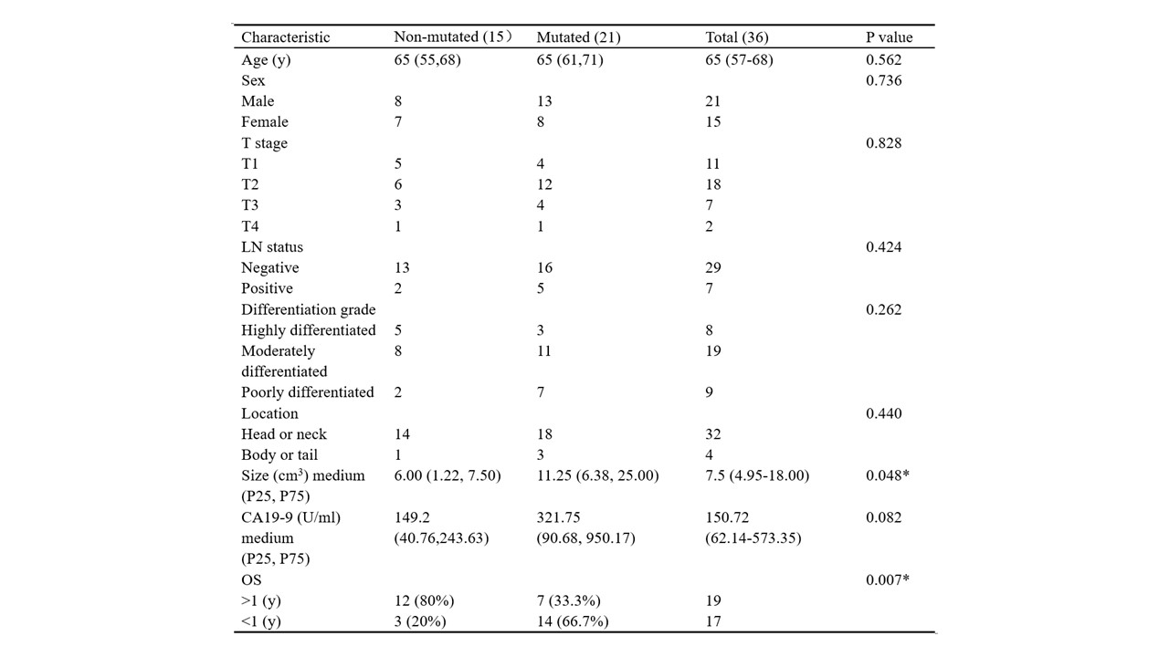
Table 1: Baseline Characteristics of Patients with Pancreatic Ductal Adenocarcinoma.
LN Lymph node, OS Overall survival
* indicates a statistically significant difference.
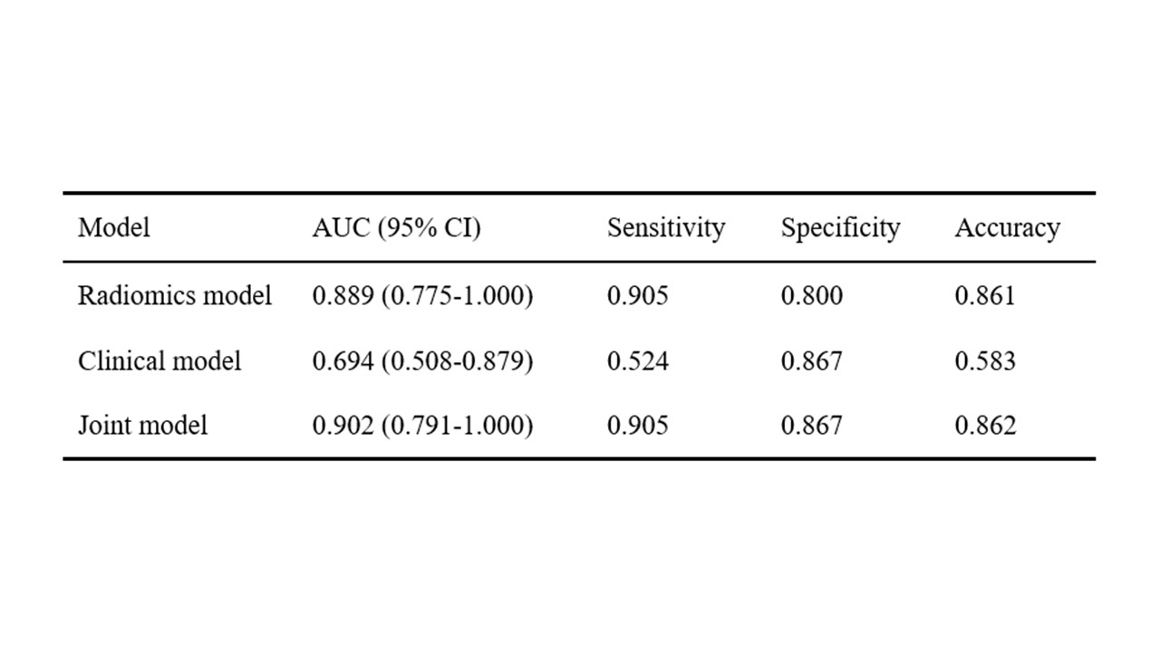
Table 2: The performance of different models in distinguish absence or presence mutation of KRAS and TP53.
AUC area under the receiver operating characteristic curve, CI confidence interval
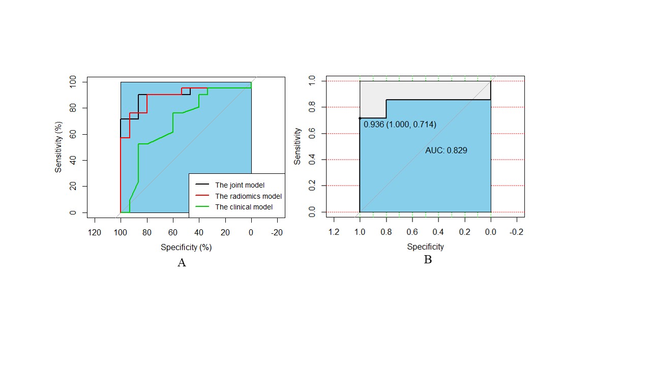
Figure 1: The ROC curve of AUC comparison between the joint model, radiomics model and clinical model for predicting the mutations of KRAS and TP53 (A). The ROC curve of the joint model after ten-fold cross-validation (B).