4442
The feasibility of 4D Flow in evaluating hemodynamic changes of transplant renal artery stenosis1Third Affiliated Hospital of Soochow University, Department of Radiology, Changzhou, China, 2Philips Healthcare, Shanghai, China, Shanghai, China
Synopsis
Keywords: Kidney, Transplantation
Although 4D-Flow MRI is capable of detecting hemodynamic abnormalities, it is rarely employed in transplanted kidneys. Using 4D-Flow MRI, we assessed total volume, net flux, maximum flow, and mean flow rate and performed a statistical analysis of the correlation in this study. Total volume, net flux, maximum flow, and mean flow rate were shown to be inversely linked to the degree of stenosis in the transplanted renal artery. As a result, 4D-Flow MRI may be applied to study the hemodynamic changes of the transplanted renal artery noninvasively.Introduction
Renal transplant artery stenosis (TRAS) is a rather common complication after transplantation. Previous research has discovered hemodynamic changes in the transplanted kidney1. 4D-Flow MRI is a useful technique to detect hemodynamic changes2,3,4. For TRAS hemodynamic research, 4D-Flow MRI is rarely employed. The current study's objective is to explore whether 4D-Flow MRI can evaluate hemodynamic changes of transplant renal artery stenosis (TRAS).Material and Methods
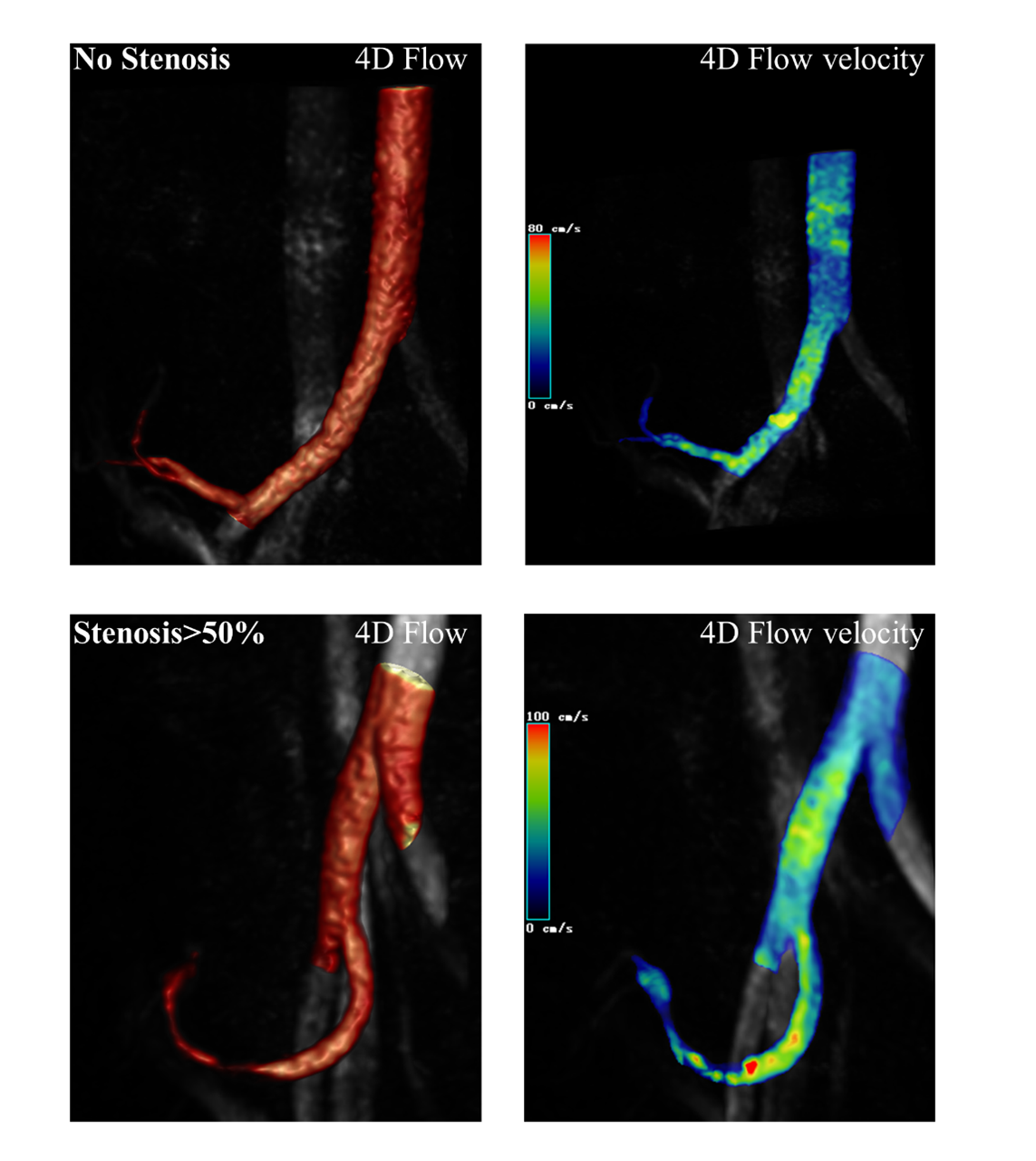
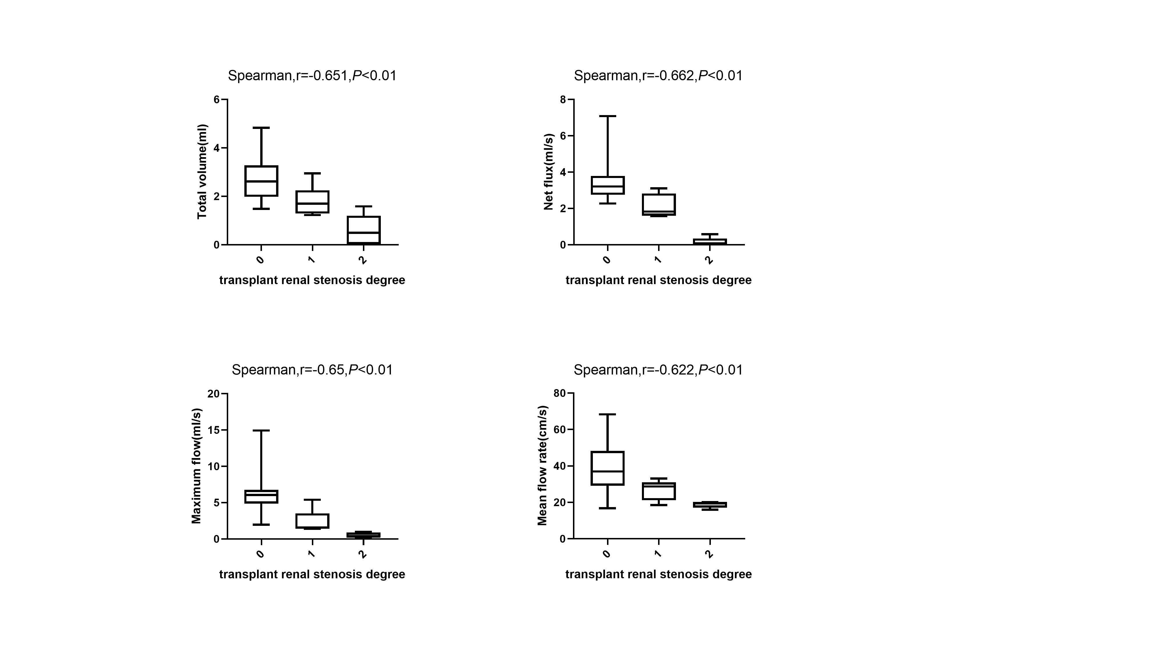
This study encompassed 44 patients (30 males and 14 females; age range, 22-62 years; median age, 42.5 years) who underwent allogeneic kidney transplantation between June 1999 and March 2022. Conventional MRI, non-contrast enhanced magnetic resonance angiography (NC-MRA), and 4D-Flow MRI were all performed on all patients5,6,7. 4D-Flow coronal oblique acquisitions were performed in transplant renal artery8,9. The degree of TRAS (0, no stenosis; 1, 0<stenosis≤50%; 2, stenosis>50%) was evaluated in the NC-MRA images. The total volume, net flux, maximum flow, and mean flow rate of the transplanted renal artery were calculated using 4D-Flow software (cvi42, Circle CVI, Canada). Hemodynamic parameters (total volume, net flux, maximum flow) were compared between renal transplant patients using the non-parametric Kruskal-Wallis H test. The one-way ANOVA was used to compare mean flow rate between kidney transplant patients. Spearman correlation coefficients were utilized to assess the transplanted kidney's total volume, net flux, maximum flow, mean flow rate in relation to the degree of stenosis of the transplanted renal artery.Results
For the no stenosis, the total volume of the transplanted kidney was 2.86±1.09 ml, the net flux was 3.30±1.37 ml/s, the maximum flow was 6.32±2.71 ml/s, and the mean flow rate was 39.41±14.18 cm/s. The total volume of the transplanted kidney artery stenosis was 1.80±1.31 ml, with a net flux of 1.87±2.32 ml/s and a maximum flow of 3.71±3.35 ml/s. The mean flow rate measured 24.73±9.32 cm/s. These results are shown in Figure 1. The degree of stenosis in the transplant renal artery was negatively correlated with total volume, net flux, maximum flow, mean flow rate (r=0.651, P<0.01; r=0.662, P<0.01; r=0.65, P<0.01; r=0.622, P<0.01).Discussion
When comparing the stenotic transplanted renal artery stenosis group to the non-stenotic transplanted renal artery group, 4D-Flow MRI reveals changes in the total volume, the net flux, the maximum flow and the mean flow rate. Total volume and maximum flow had a negative correlation with the stenosis of the transplanted renal artery.Conclusion
Non-invasive examination of hemodynamic changes in the transplanted renal artery is possible with 4D-Flow MRI.Acknowledgements
No acknowledgement found.References
1. Wystrychowski G, Kolonko A, Chudek J, et al. Systemic vascular hemodynamics and transplanted kidney survival. Transplant Proc. 2011;43(8):2922-2925.
2. Soulat G, Scott MB, Pathrose A, et al. 4D flow MRI derived aortic hemodynamics multi-year follow-up in repaired coarctation with bicuspid aortic valve. Diagn Interv Imaging. 2022;103(9):418-426.
3. Bane O, Said D, Weiss A, et al. 4D flow MRI for the assessment of renal transplant dysfunction: initial results. Eur Radiol. 2021;31(2):909-919.
4. Soulat G, McCarthy P, Markl M. 4D Flow with MRI. Annu Rev Biomed Eng. 2020;22:103-126.
5. Zhang LJ, Peng J, Wen J, et al. Non-contrast-enhanced magnetic resonance angiography: a reliable clinical tool for evaluating transplant renal artery stenosis. Eur Radiol. 2018;28(10):4195-4204.
6. Fan M, Ni X, Li Y, et al. Assessment of transplant renal artery stenosis with diffusion-weighted imaging: A preliminary study. Magn Reson Imaging. 2019 Jul;60:157-163.
7. Li X, Wang W, Cheng D, Yu Y, et al. Perfusion and oxygenation in allografts with transplant renal artery stenosis: Evaluation with functional magnetic resonance imaging. Clin Transplant. 2022 Aug 27:e14806.
8. Baird DP, Williams J, Petrie MC, et al. Transplant renal artery stenosis. Kidney Int Rep. 2020;5(12):2399-2402.
9. Fan M, Xing Z, Du Y, et al. Quantitative assessment of renal allograft pathologic changes: comparisons of mono-exponential and bi-exponential models using diffusion-weighted imaging. Quant Imaging Med Surg. 2020 Jun;10(6):1286-1297.
Figures