4428
Assessment and mitigation of EMI from in-room equipment in the setting of interventional 0.55T MRI
Bilal Tasdelen1, Ecrin Yagiz1, Rajiv Ramasawmy2, Ahsan Javed2, Dogangun Uzun2, Dursun Korel Yildirim2, Adrienne Campbell-Washburn2, and Krishna S Nayak1
1Ming Hsieh Department of Electrical and Computer Engineering, University of Southern California, Los Angeles, CA, United States, 2Cardiovascular Branch, Division of Intramural Research, National Heart, Lung, and Blood Institute, National Institutes of Health, Bethesda, MD, United States
1Ming Hsieh Department of Electrical and Computer Engineering, University of Southern California, Los Angeles, CA, United States, 2Cardiovascular Branch, Division of Intramural Research, National Heart, Lung, and Blood Institute, National Institutes of Health, Bethesda, MD, United States
Synopsis
Keywords: System Imperfections: Measurement & Correction, System Imperfections: Measurement & Correction, EMI, interference, pilot tone
Five devices that are relevant in interventional setting and a Pilot-Tone generator are assessed in terms of their electromagnetic interference with 0.55T MRI. Two of the devices (ergometer and flat-panel television) generated significant interference, however acceptable image quality was achieved when using the EDITER interference cancellation technique. EDITER also enabled the use of higher Pilot-Tone amplitudes and use of cheap, general-purpose signal generator for Pilot-Tone generation.Introduction
MRI scanner rooms are fitted with Faraday cages to dramatically attenuate (~90dB)[1] electromagnetic interference (EMI) from external sources. Sophisticated MRI applications may require auxiliary devices inside the cage (e.g., patient monitoring during interventional procedures, or pilot tone hardware for physiological gating). These devices are designed to be MRI-safe, but making them EMI compliant increases their cost. Additionally, in the case of pilot tone (PT) hardware[4], low-cost signal generators cause imperfections in pulse generation (e.g., jitter) that result in artifacts. Therefore, PT can also benefit from interference cancellation.In this work, we characterize EMI from several in-room devices and test the effectiveness of an EMI cancellation algorithm, External Dynamic InTerference Estimation and Removal (EDITER)[2]. The development of interference removal algorithms has been largely motivated by cage-free and point-of-care MRI[2-3], however, we demonstrate they can enable the use of non-EMI-compliant devices in the MRI room.
Methods
Imaging methods:Experiments were performed using a whole-body 0.55T system (prototype MAGNETOM Aera, Siemens Healthcare). We characterized EMI from each device using three measurements: 1) 4-second-long, 1MHz bandwidth RF spectrum measurement, 2) 2D-GRE imaging sequence 128x128 matrix size, and 980 Hz/pixel bandwidth, 3) the same GRE sequence with no RF (GRE0V) for noise reference. All measurements are repeated with a sniffer coil.
The level of interference was calculated based on the mean and standard deviation of GRE0V images with devices present, relative to the baseline. Devices elevating the baseline-mean more than 1SD is considered as “offending devices”. Interference is characterized by the power spectral density from RF spectrum measurements.
EDITER methods:
We used the EDITER algorithm for passive interference removal[1]. EDITER estimates the interference on the imaging coils using multiple sniffer coils, and subtracts it to get corrected MRI signal. For a sniffer coil, we used a second 6-element body coil placed at the end of the patient table, unloaded(Figure 1).
Device experiments:
We evaluated EMI from 5 different devices used in the interventional MRI setting: 1) physiology monitoring device (Expression, Philips), 2) ventilator, 3) shielded-projectors, 4) supine pedal ergometer, both battery-powered and plugged into a wall outlet, and 5) commercial television. The devices were positioned as illustrated in Figure 1. For each, the EMI was evaluated and EDITER was applied to remove artifacts.
To generate PT, we used a general-purpose signal generator (AFG3252, Tektronix), with a 20MHz dipole antenna. The antenna was positioned 1.3m away from the bore mouth. Three PT with 1x, 5x and 25x amplitudes (normalized by 10mVpp) were generated to demonstrate varying PT artifacts. Initial removal of PT was done by model subtraction as described in [5]. PT extracted from sniffer coils were fitted to the PT from imaging coils. The result was subtracted from original PT to obtain corrected PTs, and EDITER was applied to remove any residual.
Results
Table 1 shows the relative mean and SD of the noise (GRE0V) combined images, normalizing device images to baseline. Only the ergometer (plugged-in) and the TV were considered offending devices. No increase in the noise was observed for the other devices.Figure 2 shows the high-bandwidth RF spectrum measurements. Noise increase and narrow band peaks are clearly visible for ergometer and TV. Most peaks were outside of the imaging bandwidth and had no effect on the images.
Figure 3 shows the baseline image, and EDITER corrected and uncorrected images contaminated by the offending devices. EDITER was able to remove significant portion of the interference, and improved the image, with a slightly reduced SNR, compared to ground-truth.
Figure 4 shows the PT contaminated data, with PT model subtraction, and EDITER correction after subtraction. EDITER was able to suppress the remaining interference after PT subtraction. Some of the artifacts remained after EDITER applications for higher PT amplitude (5x, 25x). Correction using sniffer coils improved the PT signal. Increased PT amplitude improved the detection of weaker cardiac signals.
Discussion
EMI characterization experiments show that some devices used within a Faraday cage for certain applications can cause significant interference. It should be noted that all the devices, except the TV, were specifically designed for in-cage MRI use. We only tested a small subset of possible devices, and in the future, a broader analysis with more devices and use cases will be investigated to cover more applications.We observed coupling between the imaging and sniffer coils. Coupling reduced the interference removal performance, due to the amplified interference on imaging coils and MRI signal on the sniffer. Using dedicated high-impedance pickup coils as sniffers could alleviate this problem.
We demonstrated the use of a general-purpose signal generator as lower-cost PT equipment This generator has imperfections and caused artifacts could be reduced or removed with EDITER. In this study, we only verified the heart-beat and respiratory motion frequency, further studies will assess how well the corrected PT can estimate the motion.
Conclusion
We demonstrated that EDITER could successfully mitigate EMI caused by devices commonly used in an interventional 0.55T MRI setting. In the context of pilot tone, we were also able to use a larger PT amplitude with a general-purpose signal generator to improve the PT quality. This approach could enable the use of many more devices inside the scanner room.Acknowledgements
USC authors acknowledge grant support from the National Science Foundation (#1828736) and the USC Provost (Strategic Directions in Research Award), and research support from Siemens Healthineers. NHLBI authors are investigators on a Cooperative Research and Development Agreement (CRADA) between NHLBI and Siemens Healthcare.References
- Francesco Padormo, Joe Martin, Jane Ansell, et al. A Survey of Faraday Cage Attenuation Measurements of Clinical MRI Systems. In: Proc. Intl. Soc. Mag. Reson. Med. 29.; 2021.
- Srinivas SA, Cauley SF, Stockmann JP, et al. External Dynamic InTerference Estimation and Removal (EDITER) for low field MRI. Magnetic Resonance in Medicine. 2022;87(2):614-628. doi:10.1002/mrm.28992
- Liu Y, Leong ATL, Zhao Y, et al. A low-cost and shielding-free ultra-low-field brain MRI scanner. Nat Commun. 2021;12(1):7238. doi:10.1038/s41467-021-27317-1
- Falcão MBL, Di Sopra L, Ma L, Bacher M, Yerly J, Speier P, Rutz T, Prša M, Markl M, Stuber M, Roy CW. Pilot tone navigation for respiratory and cardiac motion-resolved free-running 5D flow MRI. Magn Reson Med. 2022 Feb;87(2):718-732. doi: 10.1002/mrm.29023. Epub 2021 Oct 5. PMID: 34611923; PMCID: PMC8627452.
- Speier P, Fenchel M, Rehner R, PT-Nav: A Novel Respiratory Navigation Method for Continuous Acquisition Based on Modulation of a Pilot Tone on the MR-Receiver. Proc. ESMRMB 2015.
Figures
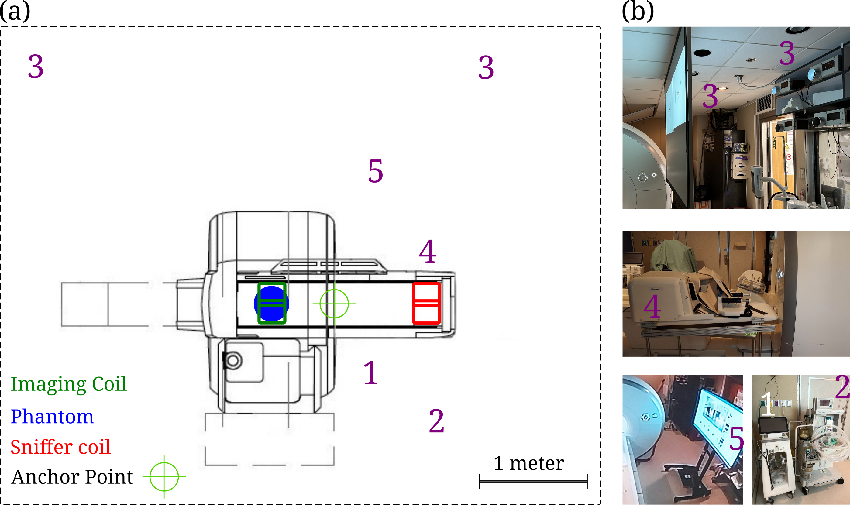
Figure 1: (a) Schematic showing the approximate positioning of coils and devices under test. Numbers 1 to 5 refers to the devices physiological monitor, ventilator, projectors, ergometer, and TV, respectively. (b) Images showing each of the devices.
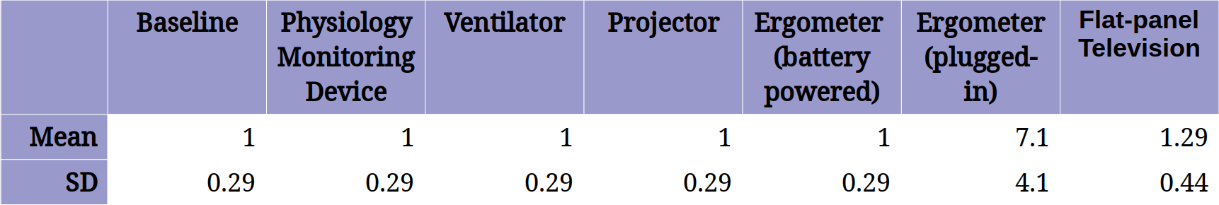
Table 1: Relative increase in the noise mean and standard deviation (SD) from the GRE0V images, normalized to baseline (no-device) GRE0V image mean. Significant elevation in both mean and SD was observed by ergometer, when plugged into a wall outlet, and the TV. No increase in the noise SD and mean was observed for the physiological monitor, projector and the ergometer when it is battery powered.
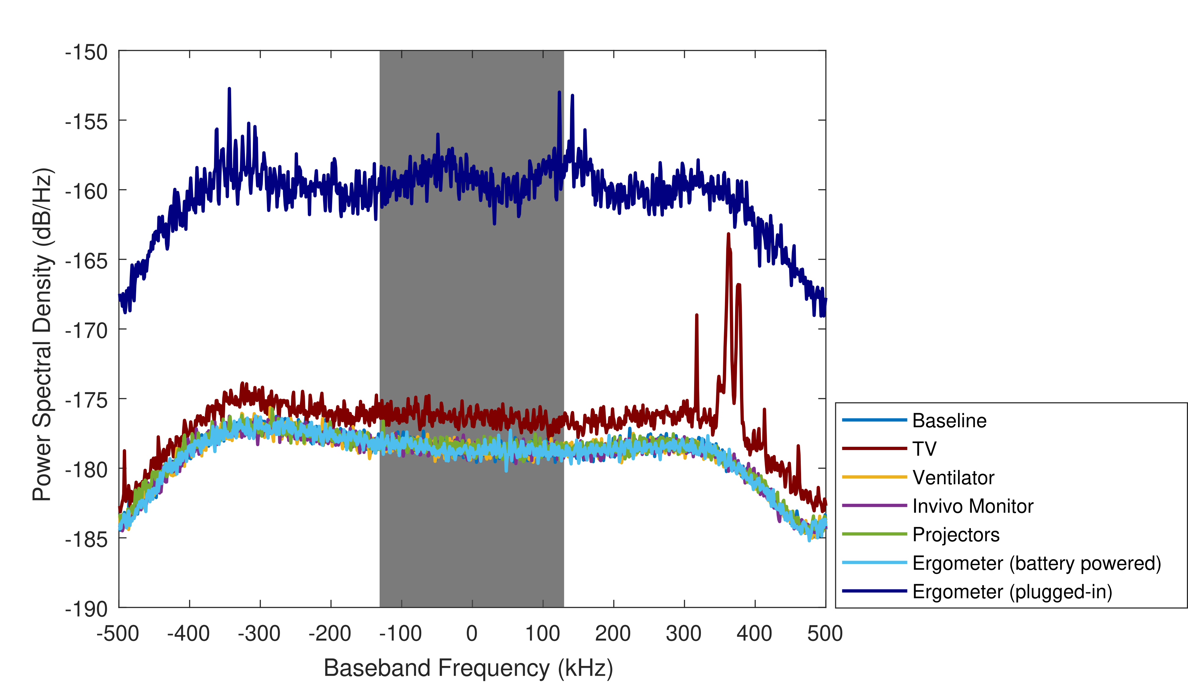
Figure 2: Power spectral densities of each spectrum measurement. Elevated noise floor and interference patterns are clearly visible for ergometer and TV. No elevation of the noise or interference was observed for the other devices. Note that, apart from the broadband interference, there are also narrow-band peaks for the offending devices. However, majority of the peaks are outside of the typical imaging bandwidth (250kHz or 4μs, gray shaded area) and does not have impact on the images.
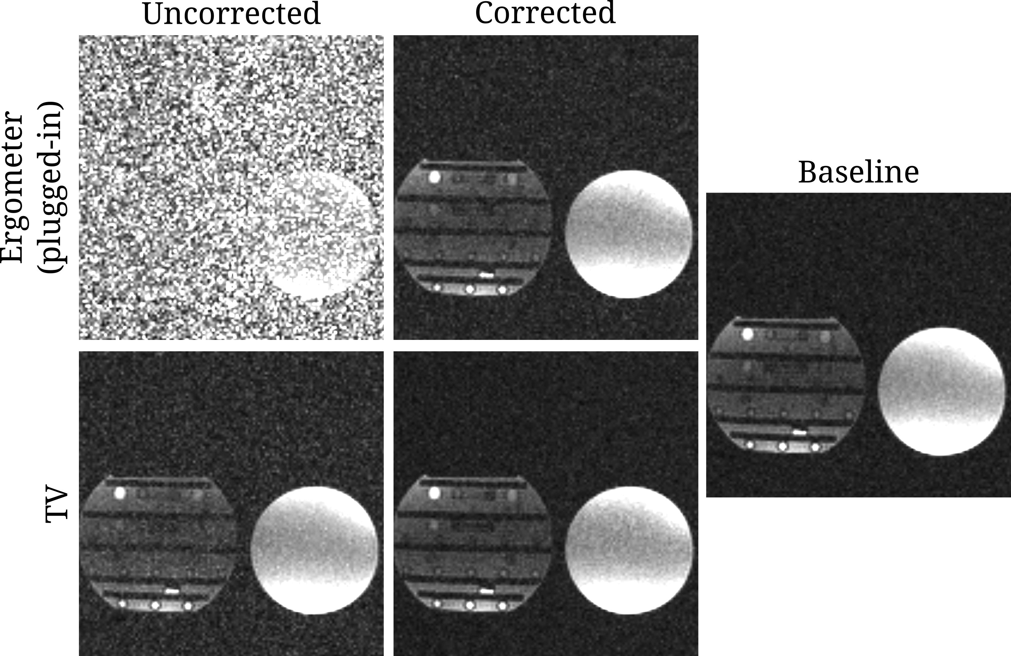
Figure 3: Comparison of uncorrected and EDITER corrected MRI images when offending devices are present. A baseline image without interference is also given for comparison. Significant improvement of image quality was observed with EDITER correction.
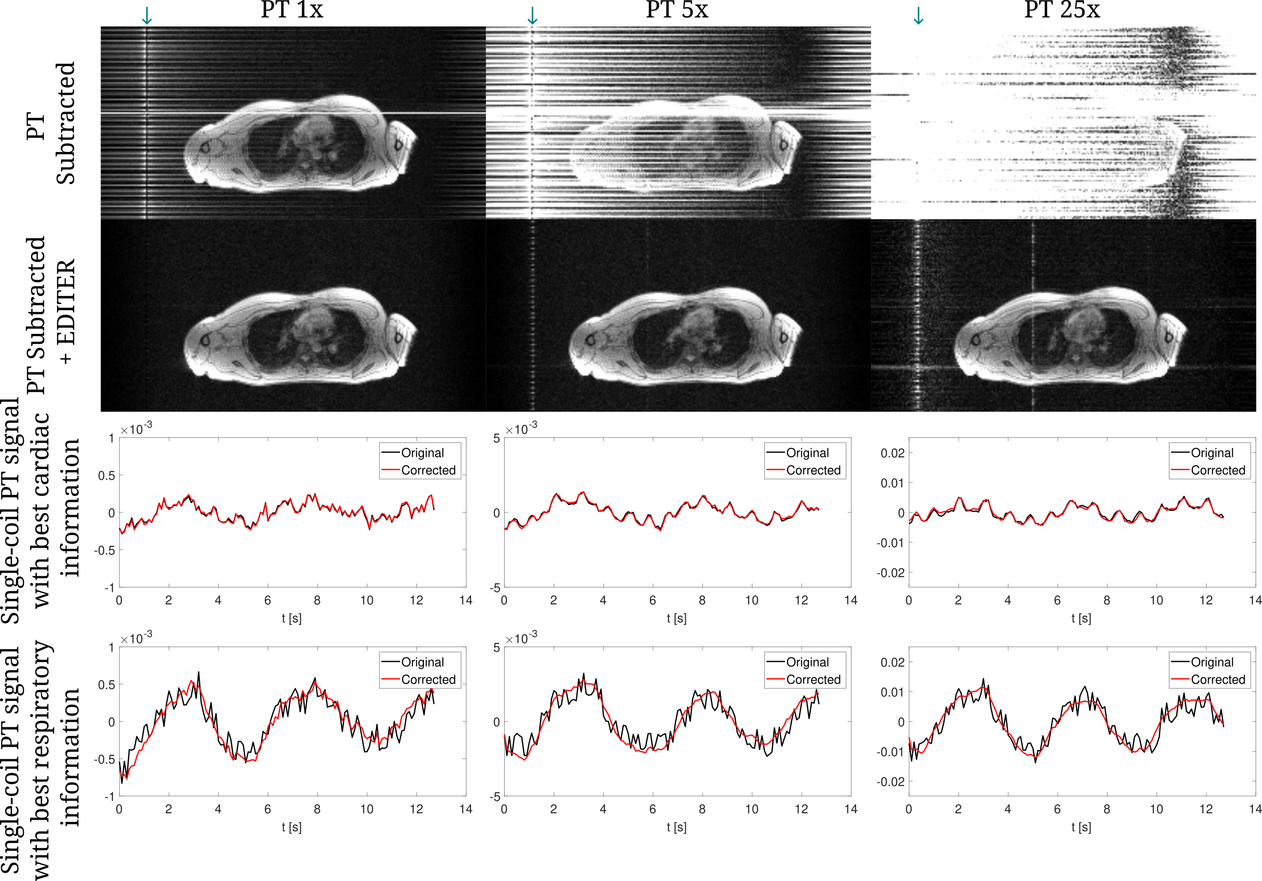
Figure 4: Removal of PT-induced artifacts. Images show an image corrected by PT model subtraction, and an EDITER corrected image, for 3 amplitudes of PT. Application of EDITER significantly reduces artifacts from imperfect PT. Even after EDITER, zipper artifacts remain for the 25x PT amplitude. Modulations due to cardiac and respiratory cycles are visible in PT signals. Imperfection removal using sniffer coils improved the respiratory PT. Increasing the PT amplitude improves the motion modulation on the PT.
DOI: https://doi.org/10.58530/2023/4428