4427
Comparison of universal phase shims determined from simulated and measured B1+ maps in the prostate and heart at 7T
Saskia Wildenberg1, Nico Egger2, Sophia Nagelstraßer2, Ralph Kimmlingen3, Titus Lanz4, Armin M. Nagel2,5, and Andreas K. Bitz1
1Electrical Engineering and Information Technology, University of Applied Sciences - FH Aachen, Aachen, Germany, Aachen, Germany, 2Institute of Radiology, Friedrich-Alexander Universität Erlangen-Nürnberg (FAU), Erlangen, Germany, Erlangen, Germany, 3Siemens Healthcare GmbH, Erlangen, Germany, Erlangen, Germany, 4Rapid Biomedical GmbH, Rimpar, Germany, Rimpar, Germany, 5Division of Medical Physics in Radiology, German Cancer Research Center (DKFZ), Heidelberg, Germany, Heidelberg, Germany
1Electrical Engineering and Information Technology, University of Applied Sciences - FH Aachen, Aachen, Germany, Aachen, Germany, 2Institute of Radiology, Friedrich-Alexander Universität Erlangen-Nürnberg (FAU), Erlangen, Germany, Erlangen, Germany, 3Siemens Healthcare GmbH, Erlangen, Germany, Erlangen, Germany, 4Rapid Biomedical GmbH, Rimpar, Germany, Rimpar, Germany, 5Division of Medical Physics in Radiology, German Cancer Research Center (DKFZ), Heidelberg, Germany, Heidelberg, Germany
Synopsis
Keywords: RF Pulse Design & Fields, Simulations
Universal RF shimming is a proven method to avoid the time-consuming process of calculating patient-specific RF shims for every individual subject. Nevertheless, this method requires numerous B1+ maps from in vivo measurements, which is a time-consuming process, especially in the context of product development. This work therefore investigates whether a universal RF shim based on simulated B1+ maps, which are needed for the safety assessment of the coil anyways, is a viable alternative compared to shim optimization based on in vivo data. The study was performed using a torso coil at 7T for the prostate and cardiac region.
Introduction
Parallel transmission (pTx) strategies, in particular RF Shimming1, allow an optimal patient-specific adaptation of the RF coil’s excitation profile to the anatomy of the subject, which, however, requires time-consuming calibration measurements and calculations for each individual subject. Also, for the more general approach of calculating a so called universal shim2, where the excitation profile is optimized over a representative set of subjects, numerous MRI measurements are necessary. Since safety assessments of RF coils in any case require a large number of simulations with anatomical human body models, the work presented here investigates whether a universal shim based on simulated B1+ maps is a valid alternative to the universal shim calculation from MRI data.Methods
The study was performed using an 8-channel torso array (RAPID Biomedical GmbH, Rimpar, Germany) that consisted of a ventral and dorsal array with 4 channels each. The corresponding setup for imaging the prostate region is shown for the human body model Duke3 in Figure 1. A database of B1+ maps was generated from thirty-five subject measurements for the universal phase shim based on MRI data (UPSMR), resulting in 23 maps for the prostate and 35 for the cardiac region. All volunteers except for two had an age between 22 and 34 years and a majority of 70% was of normal weight. 8% of the subjects were slightly underweight and 22% slightly overweight, the latter group includes 2 subjects whose age was 60 years. For the universal phase shim based on simulated data (UPSSIM) the B1+ maps calculated for the safety assessments of the RF coil (CST Studio Suite 2020, SIMULIA, Dassault Systèmes, France) were used. This database included 11 RF exposure models generated by scaling two male and two female human body models, aged between 26 and 42, with three scaling factors per model. With respect to the BMI, which was calculated from the height and tissue mass of the voxel models after scaling, except for two body models, which are slightly underweight, all models are of normal weight.For the calculation of the universal shims an in-house shimming tool was used, which optimizes the RF excitation profile across all subjects based on a predefined weighting of homogeneity (70%) and efficiency (30%) whereby the 3D B1+ field distributions within the region of interest (ROI) were passed as input data. For the prostate simulations also the field distributions within the female models were taken into account.
Results & Discussion
Figure 2 and Figure 3 show the phase distributions of the universal phase shims UPSMR and UPSSIM, as well as the individual phase shims for the cardiac (Fig. 2) and prostate region (Fig. 3). Tx phases of the individual shims are located within a limited variation range per channel and a common pattern as well as a symmetry between dorsal and ventral array can be observed. Furthermore, the universal shims show very good agreement with respect to the Tx phases for both regions, with a mean deviation of 37° for the prostate and 22° for the cardiac region.The performance of the universal phase shims UPSSIM and UPSMR is compared in terms of mean B1+, efficiency (magnitude of sums(B1+)/ sum of magnitudes(B1+)) and the coefficient of variation in both regions for the simulated (Figure 4) and measured B1+ maps (Figure 5). For the heart region a shim developed by the manufacturer (ShM) is evaluated additionally. As expected, the performance of UPSMR is slightly higher for the measured data, while UPSSIM performs a bit better for the simulated data; however, both universal RF shims are performing very good in the opposite dataset. Comparing the performance of UPSMR and UPSSIM averaged over all body models, volunteers and both regions, we observe the maximum deviation of 13% for the mean B1+, 12% for mean efficiency and 12%in the CoV. The SAR efficiency (B1+mean/√SAR10g,max) also agrees very well for both universal shims with 0.13 µT/√(W/kg) (UPSSIM) and 0.1 µT/√(W/kg) (UPSMR) for the prostate region and 0.074 µT/√(W/kg) (UPSSIM), 0.079 µT/√(W/kg) (UPSMR) and 0.065 µT/√(W/kg) (ShM) in the heart region. The universal shims even outperform the default shim (ShM) in SAR efficiency.
Conclusion
This work demonstrates that a universal phase shim determined from simulated B1+ maps in the prostate and cardiac regions can perform very good in comparison to universal phase shims calculated from in vivo measurements. Hence, universal RF phase shims can be determined directly during the simulation-based safety assessment of the RF coil without need for additional in vivo measurements. Further, the results show that in the present study a small set of anatomical human body models could be extended by applying moderate scaling factors to cover a wider range of body physiques in the simulation. It has to be noted that the database for the MRI measurements consist of a relatively homogeneous group of volunteers in terms of body physique. Therefore, additional volunteers with larger variation in anatomy will be included in the study.Acknowledgements
No acknowledgement found.References
[1] Collins CM et al., Magnetic Resonance in Medicine 2005;54(6):1327-1332. doi:10.1002/mrm.20729[2] Gras V, Boland M, Vignaud A, Ferrand G, Amadon A, Mauconduit F, et al. (2017) Homogeneous non-selective and slice-selective parallel-transmit excitations at 7 Tesla with universal pulses: A validation study on two commercial RF coils. PLoS ONE 12(8):e0183562.
[3] Christ et al. Physics Med Biol 2010;55(2):N23-38.
Figures
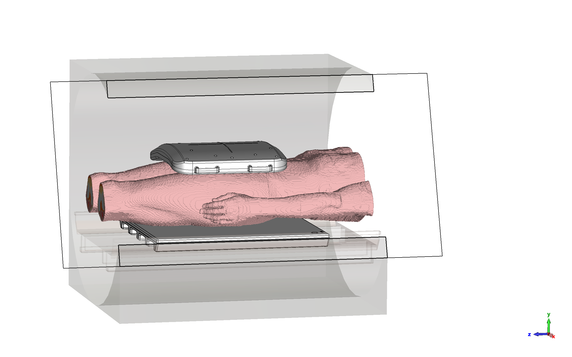
Figure 1: Exemplary exposure scenario for
imaging of the prostate region.
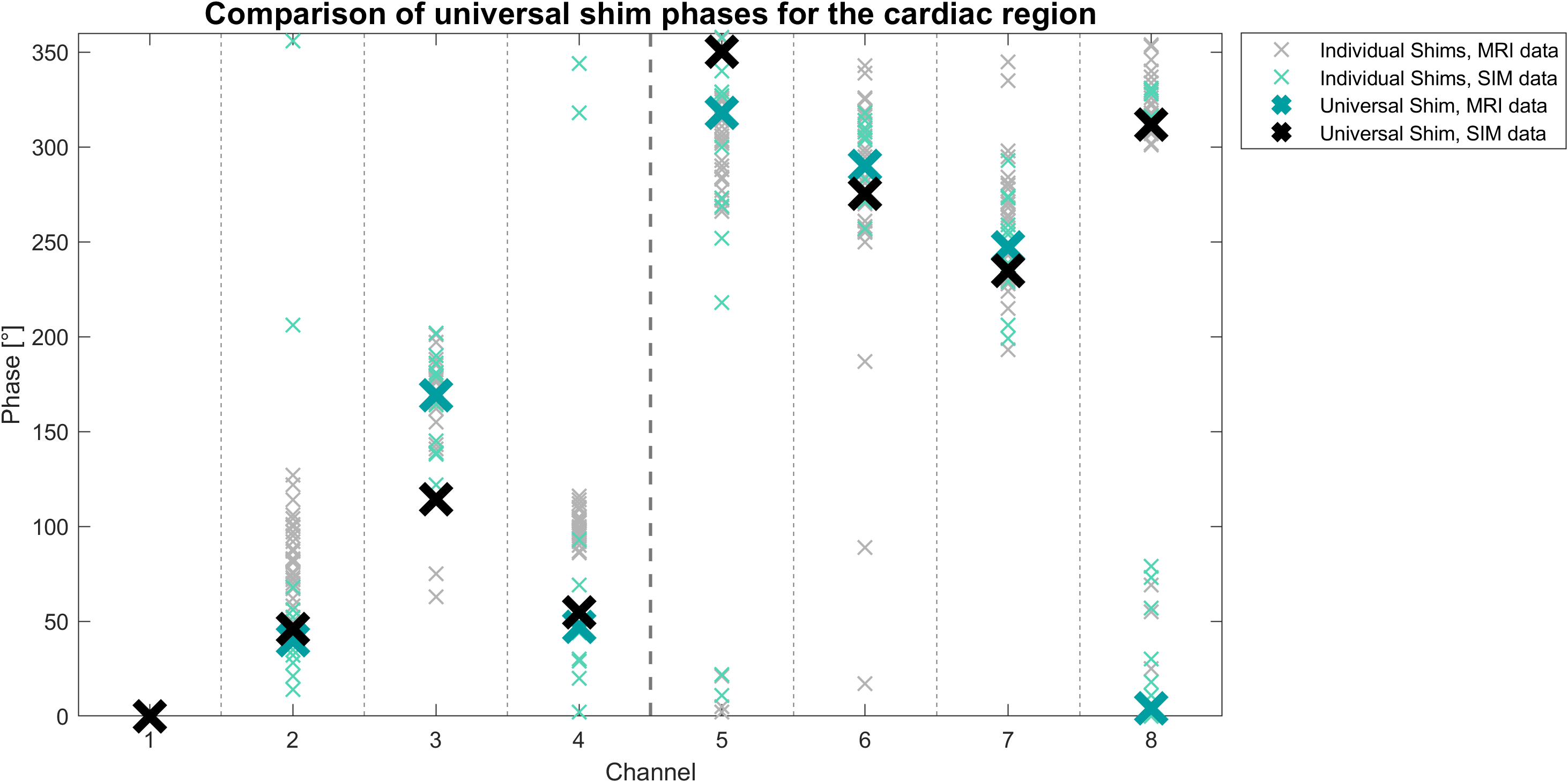
Figure 2: Comparison of the phase distribution of universal phase shims in the cardiac region. Also shown are the individual phase shims.
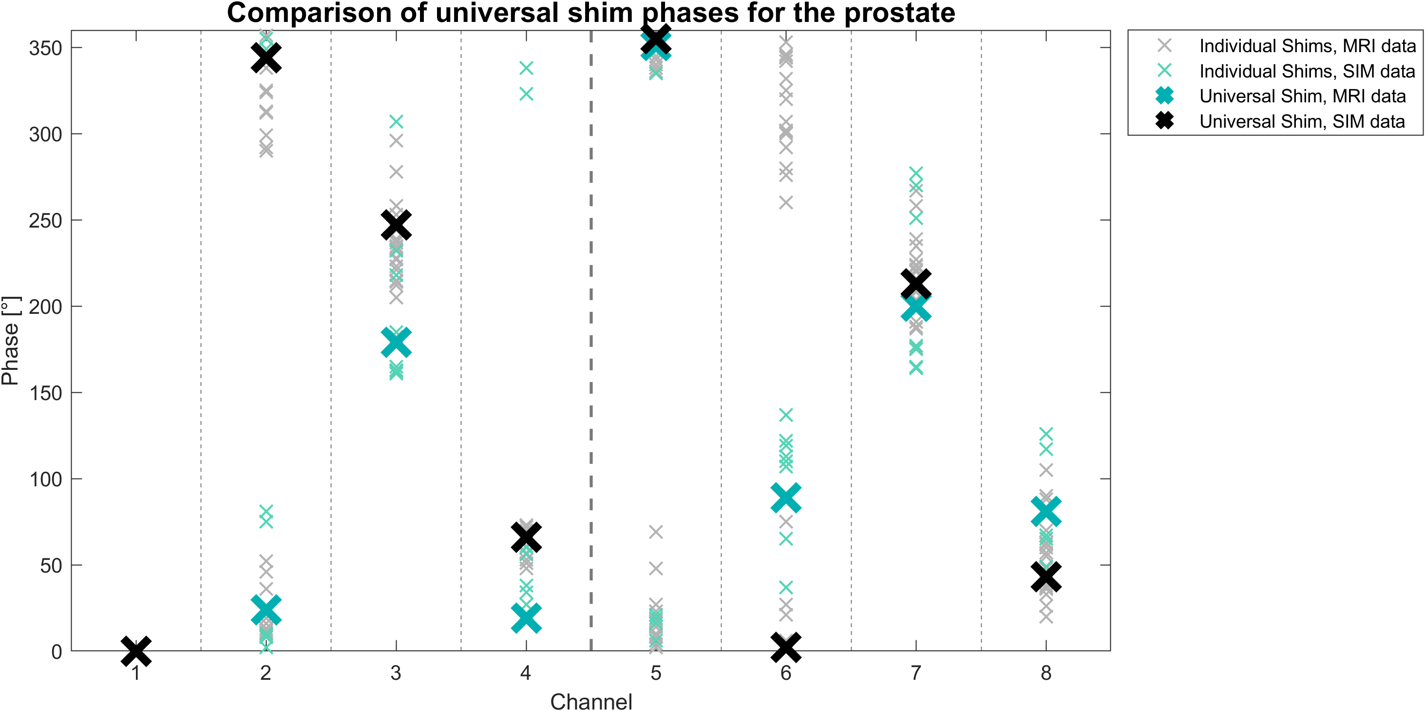
Figure 3: Comparison of the phase distribution of universal phase shims in the prostate region. Also shown are the individual phase shims.
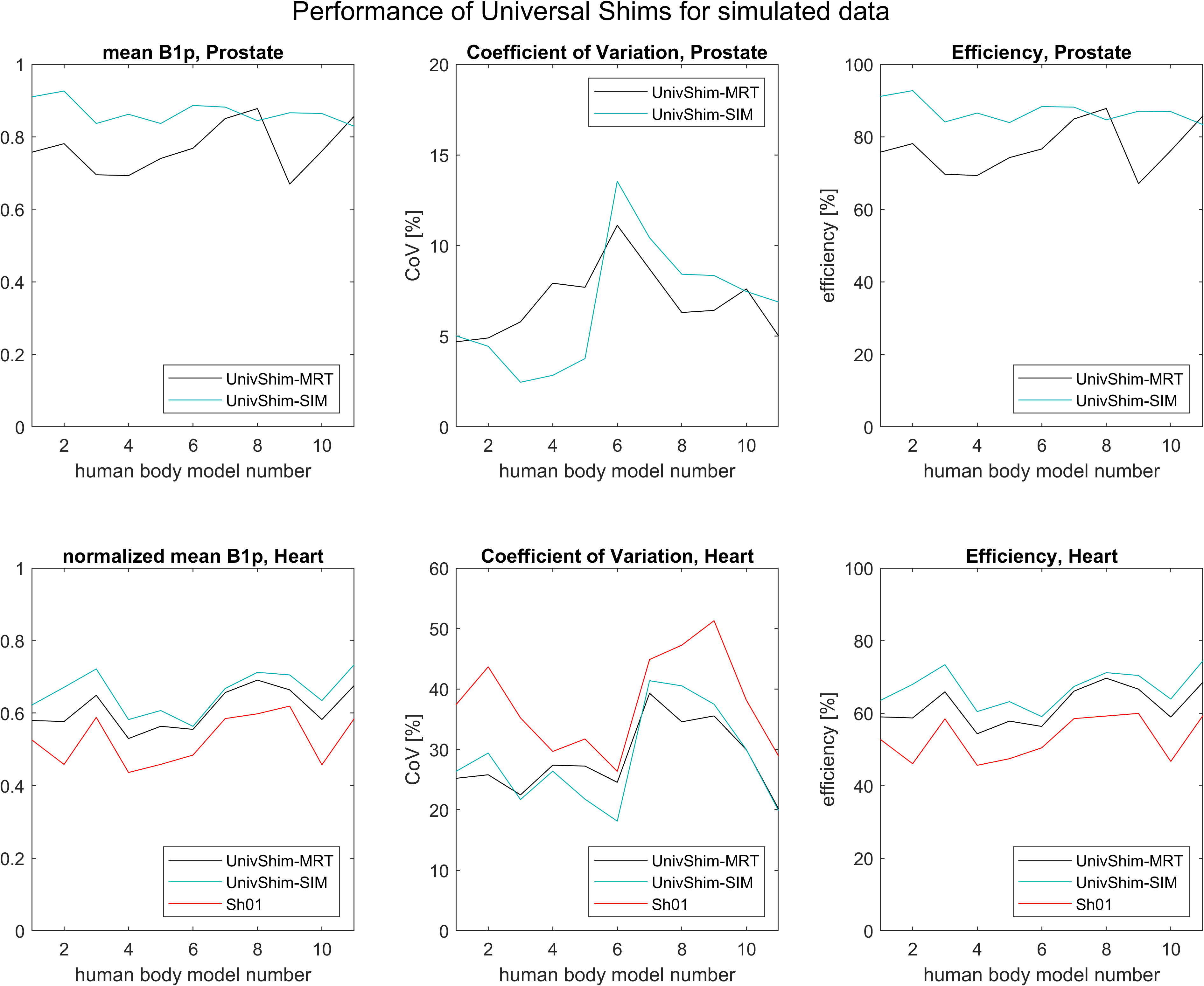
Figure 4: Performance of the universal phase shims for the simulated data set within the prostate and the cardiac region.
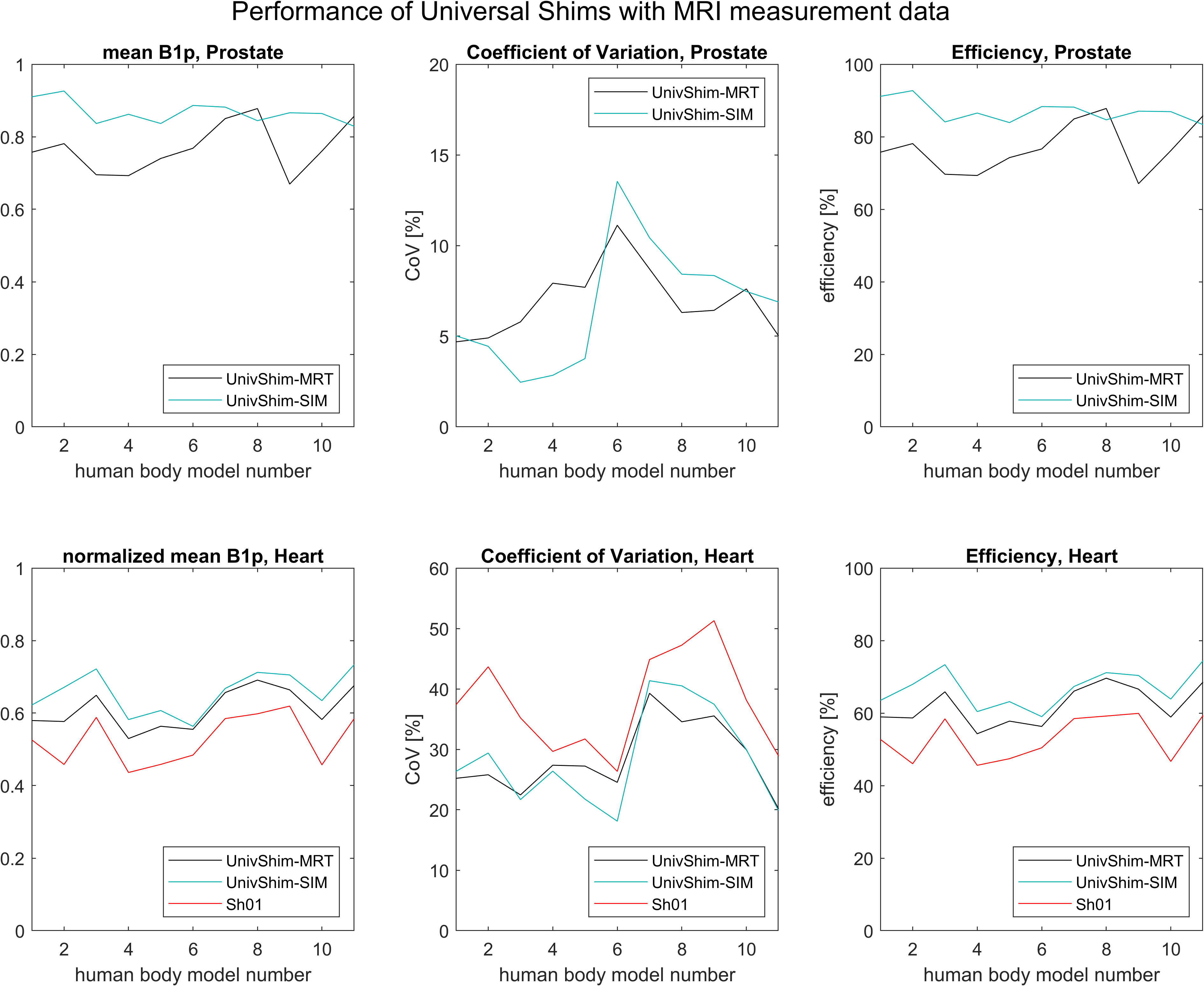
Figure 5: Performance of the universal phase shims for the measured data set within the prostate and the cardiac region.
DOI: https://doi.org/10.58530/2023/4427