4419
Correction of field drifting for single shot spiral diffusion imaging1Department of Radiology, Mayo Clinic, Rochester, MN, United States, 2MR R&D, Royal Philips, Rochester, MN, United States
Synopsis
Keywords: System Imperfections: Measurement & Correction, Diffusion Tensor Imaging
Diffusion MRI provides useful microstructural information of the soft tissue. Scans using a single-shot Spiral trajectory have demonstrated better geometric fidelity and SNR than those with a Cartesian EPI counterpart. This study shows that the field drifting during a diffusion scan can be a confounding factor to the image quality. A correction strategy exploiting the average off-resonance measurement before and after a scan was applied to mitigate the blurring artifact caused by field drifting.Introduction
Diffusion Weighted Imaging (DWI) is a useful tool to reveal microstructural information for clinical care and neurological research. Single-shot techniques such as Spiral imaging and EPI have been the principal sequences for this application due to their robustness in the presence of motion. Studies comparing diffusion scans using the Spiral and the Cartesian trajectories have suggested that the Spiral sequence can achieve better SNR 1,2 and geometry fidelity 3. A substantial challenge to Spiral MRI is that off-resonance leads to blurring; several studies have proposed methods to mitigate this artifact4,5.Spiral imaging and DWI both employ substantial gradient activity, which introduces system heating and can potentially lead to field drifting during the scan6,7. The effects of field drifting in Spiral MRI has been previously corrected by adding a frequency offset to the field map for improved reconstruction 8,9. The present work indicates similar but more substantial field drifting in DWI with Spiral encoding, leading to inconsistent blurring artifacts among the images. Therefore, a rapid frequency measuring sequence is employed before and after each scan to probe the frequency changes during the exam. The frequency offsets are then estimated and demodulated from the data to restore the diffusion weighted images for further processing.
Materials and Methods
Volunteer experiments were performed on a 3.0T scanner (Ingenia Elition X, Royal Philips) by a fat-suppressed twice-refocused diffusion sequence to minimize eddy current effects. The single shot Spiral encoding and the single shot Cartesian EPI were both utilized in acquisition for comparison. A field map is collected prior to the Spiral scan. Measurements of average frequency are included before and after the diffusion exam using a single-voxel FID sequence. Other image parameters are as follows: FOV = 23cm, Resolution = 1.4x1.4mm2, Slice Thickness = 6mm, Parallel imaging factor = 2, TR = 4s, TE = 90 ms(Spiral)/110 ms(EPI), b-values = 0 and 1000s/mm2, and 32 diffusion encoding directions with Nex = 3.Image reconstruction for the Spiral data starts with demodulating the frequency offset of each k-space data point according to:
$$ S_{corr}(k(t)) = S(k(t))\times e^{-j 2 \pi t {{\Delta f} \over {T}}}$$
where $$$\Delta f$$$ is the frequency difference before and after the exam, T is the total scan duration and t is the estimated time for the data being sampled according to the sequence timing and the sampling interval. The frequency demodulated data subsequently undergo an off-line conjugate gradient solver consisting of the field map and the sensitivity map to account for the local blurring and to remove aliasing artifacts. The EPI data were reconstructed by the scanner’s embedded method. The diffusion weighted images were then processed to produce DTI indices including the trace ADC map and the FA weighted orientation map (color FA).
Results
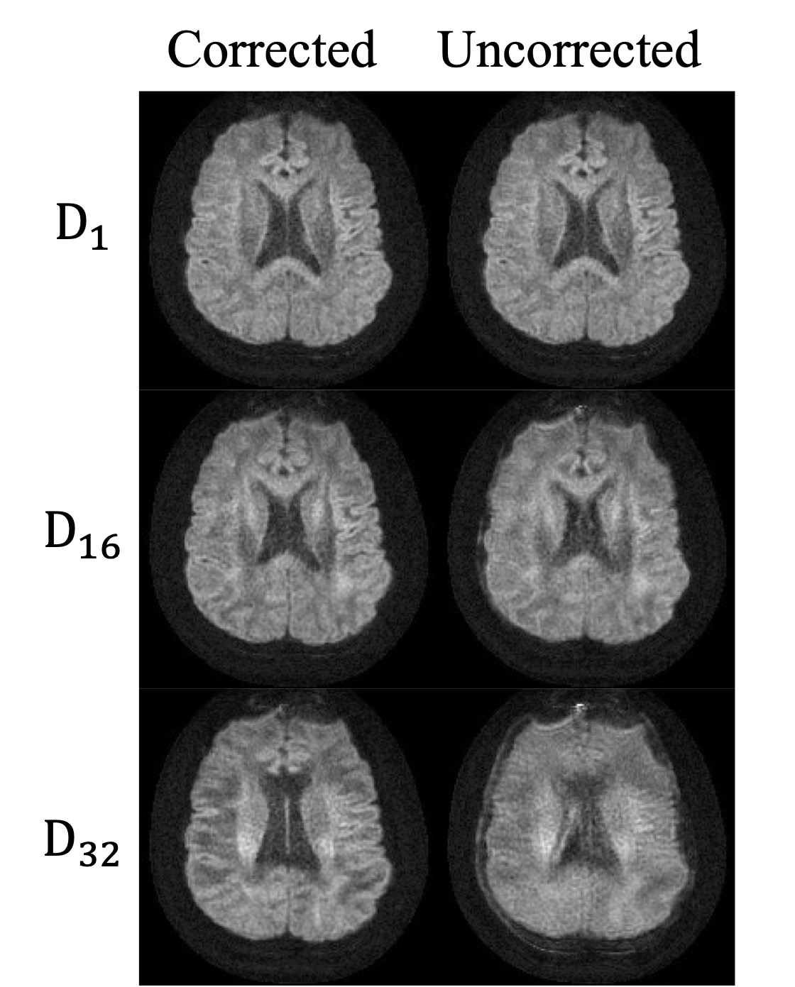
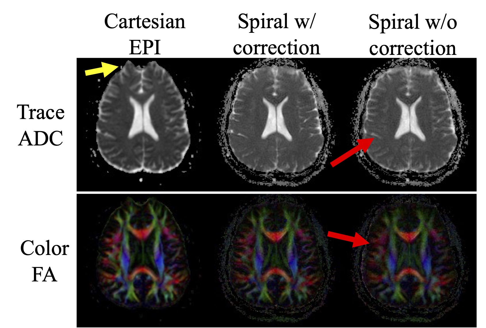
Fig 1. compares the Spiral DWI images with and without correction for frequency drifting. The difference in terms of blurring was subtle in the first diffusion encoding direction. As the scan proceeded, blurring in the diffusion encoded images became severer due to the main field drifting. The measured frequency change of the presented data was about 25Hz within a 6 min exam. The blurring artifact can then be mitigated with the frequency demodulation on the data by the proposed method.Fig 2. demonstrates the trace ADC maps and the color FA maps from the EPI and the Spiral data with and without correction. The trace ADC maps show little difference between the two spiral reconstructions except for noticeable blurring along the boundaries of CSF and parenchyma. However, the structural sharpness diminishes between different tissues in the color FA map if the frequency correction is not performed. Compared with the EPI image, the corrected spiral reconstruction shows better geometric fidelity due to the inclusion of the field map in the reconstruction.
Discussion
Field drifting caused by the gradient activity in a long scan is known to degrade image quality if the issue is not properly addressed. This study found that these frequency changes in a diffusion experiment induced substantial blurring in Spiral images. The introduction of a pre- and post-scan frequency measurement using a single voxel FID sequence only adds approximately six seconds to a scan. This extra frequency information is then critical to reduce the blurring artifact with the proposed frequency demodulation correction approach. The results suggest that the presented approach can be a useful addition to Spiral DWI to improve image quality for various applications.Conclusion
This study presents a strategy to demodulate the frequency drifting in the diffusion scans to improve image quality for the single shot Spiral imaging.Acknowledgements
This study was supported in part by Royal Philips.References
1. Lee Y, et al. On the signal-to-noise ratio benefit of spiral acquisition in diffusion MRI. Magn Reson Med. Apr 2021;85(4):1924-1937. doi:10.1002/mrm.28554
2. Holtrop JL, Sutton BP. High spatial resolution diffusion weighted imaging on clinical 3 T MRI scanners using multislab spiral acquisitions. J Med Imaging (Bellingham). Apr 2016;3(2):023501. doi:10.1117/1.JMI.3.2.023501
3. Glover GH. Spiral imaging in fMRI. Neuroimage. Aug 15 2012;62(2):706-12. doi:10.1016/j.neuroimage.2011.10.039
4. Wang D, Zwart NR, Pipe JG. Joint water-fat separation and deblurring for spiral imaging. Magn Reson Med. Jun 2018;79(6):3218-3228. doi:10.1002/mrm.26950
5. Noll DC, Meyer CH, Pauly JM, Nishimura DG, Macovski A. A homogeneity correction method for magnetic resonance imaging with time-varying gradients. IEEE Trans Med Imaging. 1991;10(4):629-37. doi:10.1109/42.108599
6. Lange T, Zaitsev M, Buechert M. Correction of frequency drifts induced by gradient heating in 1H spectra using interleaved reference spectroscopy. J Magn Reson Imaging. Mar 2011;33(3):748-54. doi:10.1002/jmri.22471
7. Hui SCN, et al. Frequency drift in MR spectroscopy at 3T. Neuroimage. Nov 1 2021;241:118430. doi:10.1016/j.neuroimage.2021.118430
8. Ooi MW, D; Anderson, AG; Li, Z; Zwart NR; Robison, RK; Pipe, JG. Spiral Deblurring Using B0 Maps with B0 Drift Correction. 2016:1760.
9. Anderson III AG, Robison RK, Wang D, Ooi MB, pipe JG. Intra-Scan Center Frequency Drift Correction for 3D Spiral Exams. 2017:1482.
Figures