4399
Evaluation of MRI performance and PET compatibility of a 32 channel coil for the Siemens Biograph mMR PET/MRI scanner
Harris Bidaman1, Katie Dinelle1, Reggie Taylor2,3, and Lauri Tuominen2,4
1Brain Imaging Centre, Royal Ottawa Mental Health Centre, Ottawa, ON, Canada, 2University of Ottawa Institute of Mental Health Research at The Royal, Ottawa, ON, Canada, 3Department of Physics, Carleton University, Ottawa, ON, Canada, 4Department of Psychiatry, University of Ottawa, Ottawa, ON, Canada
1Brain Imaging Centre, Royal Ottawa Mental Health Centre, Ottawa, ON, Canada, 2University of Ottawa Institute of Mental Health Research at The Royal, Ottawa, ON, Canada, 3Department of Physics, Carleton University, Ottawa, ON, Canada, 4Department of Psychiatry, University of Ottawa, Ottawa, ON, Canada
Synopsis
Keywords: PET/MR, Brain, Coil
We evaluate MRI and PET performance of the novel Siemens 32 channel head coil for the Siemens Biograph mMR. Results are compared to the system standard 12 channel coil. MRI performance (SNR) was improved with the 32 channel coil. PET image uniformity was not impacted. ROI analysis of the PET data indicated a 5% underestimation of activity concentration in the 32 channel coil, however this difference was mitigated by the use of an image derived reference region. Any small differences in PET performance introduced by the 32 channel coil are far outweighed by the superior MRI performance of this coil.Introduction
The Siemens Biograph mMR is a dual modality PET/MRI scanner1. For brain PET/MRI imaging this scanner was originally sold with a 12 channel head coil and a 16 channel head and neck coil. In order to fully exploit the ability to conduct simultaneous PET and MRI data, a 32 channel MRI coil for the Biograph mMR was recently released by Siemens. This coil is intended to be PET compatible. The greater number of channels in the 32 versus the 12 channel coil results in an increased amount of attenuation in the PET field of view. In this paper we compare the PET performance of the Biograph mMR using the 12 and 32 channel head coil for whole brain PET imaging.Methods
SNR was calculated by acquiring 50 repetitions of an EPI (TR = 2500 ms, TE = 26 ms, 64x64, FOV = 192 mm, ipat = 0) sequence of a phantom. A 0 volt image was also acquired. The mean signal and standard deviation of the noise were used to determine SNR.A 4L cylindrical Nalgene jug was filled with water and 36.8 MBq of F-18 was added. An accurate activity concentration measurement was acquired by measuring an aliquot of the radioactive phantom water on a gamma counter following completion of the scans.
Three phantom scans were acquired: (i) with the phantom supported on styrofoam in the centre of the scanner bore, (ii) with the phantom in the 12 channel coil and (iii) with the phantom in the 32 channel coil. Each scan acquisition ran until a total of 100 million counts were acquired.
Images were reconstructed using the Siemens offline reconstruction package E7-Tools (Siemens Healthineers, Knoxville, TN) with the following reconstruction parameters: OP-OSEM, 3 iterations, 21 subsets, 3mm Gaussian post-filter, with corrections for scattered and random events. Attenuation correction was applied using a mu-map generated from a CT scan of the phantom.
A circular ROI was centered on the phantom in the axial view and the activity in the ROI was averaged across the central 70 slices of the phantom. The average ROI activity in Bq/ml, and standard deviation within the ROI, for each scan was compared to the activity concentration measured using the gamma counter. To evaluate the uniformity of the phantom activity, vertical, horizontal and axial profiles were drawn on the central slice of the phantom for each scan. Decay corrected profiles were overlaid to compare uniformity in the presence of the different head coils. In addition, the standard deviation between 13 small circular ROIs distributed in-plane, on multiple slices, was used to compare variability on an ROI level.
Following a routine 18F-FDG brain scan (injected dose 186.1MBq, 40 minute uptake, 30 minutes scan) using the 12 channel coil, the participant was repositioned in the 32 channel coil and an additional 15 minutes of data were acquired. Data were reconstructed using E7-Tools with the same reconstruction parameters as the phantom. Only the first 15 minutes of the 12 channel coil data were reconstructed to match the scan length of the 32 channel dataset. The attenuation mu-map was calculated using the pseudoCT method2, which is a MR based (T1) technique to generate umaps.
The 12 and 32 channel images were co-registered and ROIs placed in the frontal lobe, occipital lobe, thalamus, cerebellum and pons. A direct comparison was made between ROI activity concentrations in each region between the two coils. Additionally, a comparison was made between ROI values normalized to the pons.
Results
A decrease in count rate of 33% was observed when the phantom was scanned in the 12 channel coil compared to the scan with no additional attenuating material in the FOV (e.g. scanner table, head coil). For the 32 channel coil the count rate was decreased by 54%. Using the large circular ROI averaged over 70 slices and decay correcting to the start of the first scan the results are (i) 1947(214) Bq/mL (ii) 2043(217) Bq/mL (iii) 2057(185) Bq/mL. From the smaller ROIs the average activity are (i) 1949(53) Bq/mL (ii) 2041(42) Bq/mL (iii) 2069(57) Bq/mL.A direct comparison of ROI activity concentrations, across all regions investigated, revealed that the 32 channel coil on average underestimated the 12 channel results by 5%. A comparison of the ROI values normalized to a reference region in the pons showed that the 32 channel coil on average underestimated the 12 channel results by 2%.
There was much better overall MRI signal in the 32 channel coil with a median SNR of 1068 in the 32ch compared to 691 in the 12 ch.
Conclusion
This study compared the impact on MRI and PET performance of the two available head coils (12 and 32 channel) manufactured by Siemens for the Biograph mMR (PET/MRI). Compared to the 12 channel coil, the 32 channel coil exhibited: improved MRI SNR, similar uniformity profiles in the PET phantom data, and minimal regional and global changes in PET ROI based phantom and human data. Our preliminary results indicate that for simultaneous PET/MRI studies the improved MRI performance of the 32 channel coil far outweigh any observed impacts on PET performance.Acknowledgements
We would like to acknowledge the Brain Imaging Centre at the Royal Ottawa Hospital.References
1. Delso G, Fürst S, Jakoby B, et al. Performance measurements of the Siemens mMR integrated whole-body PET/MR scanner. J Nucl Med. 2011 Dec;52(12):1914-22. doi: 10.2967/jnumed.111.092726. Epub 2011 Nov 11. Erratum in: J Nucl Med. 2012 Mar;53(3):507. PMID: 22080447.
2. Izquierdo-Garcia D, Hansen AE, Förster S, et al. An SPM8-based approach for attenuation correction combining segmentation and nonrigid template formation: application to simultaneous PET/MR brain imaging. J Nucl Med. 2014 Nov;55(11):1825-30. doi: 10.2967/jnumed.113.136341. Epub 2014 Oct 2. PMID: 25278515; PMCID: PMC4246705.
Figures
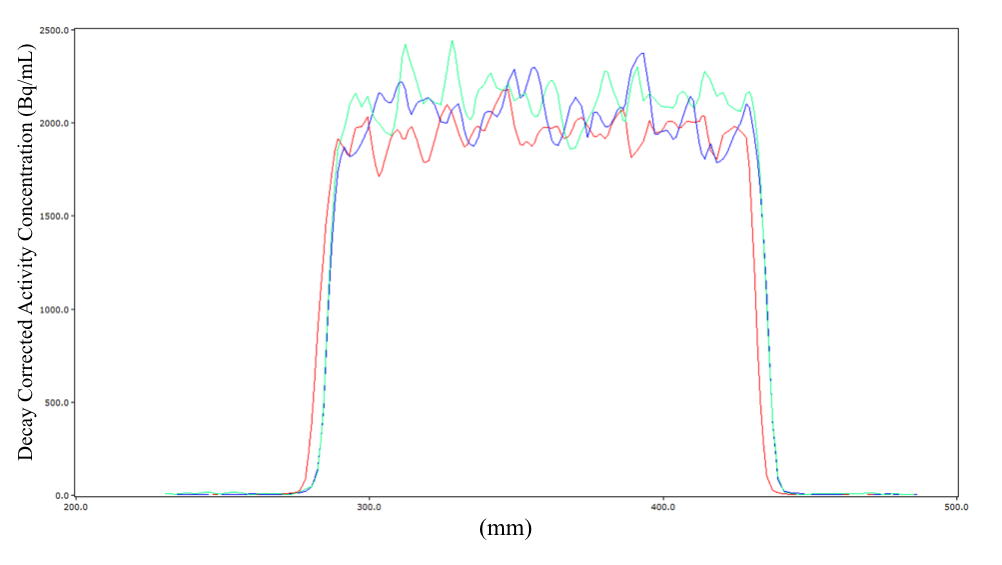
Profile of Decay Corrected Activity Concentration of phantom with no coil (red), phantom with 12 channel coil (blue) and phantom with 32 channel coil (green).
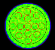
Example of defined regions of interest within the phantom.
DOI: https://doi.org/10.58530/2023/4399