4397
MRI compatibility study of a brain PET insert integrated with a quadrature birdcage transmit /47-channel receiver head coil assembly at 3 T1Paul C. Lauterbur Imaging Research Center, Shenzhen Institutes of Advanced Technology, Chinese Academy of Sciences, Shenzhen, China, 2Key Laboratory for Magnetic Resonance and Multimodality Imaging of Guangdong Province, Shenzhen, China, 3Institute of Computing Technology, Chinese Academy of Sciences, Beijing, China, 4University of Chinese Academy of Sciences, Beijing, China, 5Peng Cheng Laboratory, Shenzhen, China, 6Department of Biomedical Engineering, State University of New York at Buffalo, New York, NY, United States
Synopsis
Keywords: PET/MR, PET/MR
Simultaneous positron emission tomography/magnetic resonance imaging (PET/MRI) shows advantages in clinical and research applications for human brain. In this work, MRI compatibility of a brain PET insert integrated with a quadrature birdcage transmit /47-channel receiver head coil assembly was studied. The results of RF field (B1+) distribution, signal-to-noise ratio (SNR) and static field inhomogeneity (ΔB0) show MRI compatibility of the brain PET insert. Simultaneous PET/MRI was implemented, which demonstrated a high performance of the brain PET/MRI system.Introduction
Positron emission tomography/magnetic resonance imaging (PET/MRI) systems can be applied for simultaneous imaging of the anatomic, functional, and molecular states of human brain, showing advantages in clinical and research applications1,2. A dedicated brain PET device integrated with MRI systems is an economical choice3. Especially, both the PET insert and head coil assembly was carefully designed to avoid interferences. In our previous works, a brain PET scanner with dual-ended readout detectors4 and a dedicated multichannel head coil array5 for PET insert has been separately reported. In this work, we optimized the MRI shielding of the PET insert and the electronic components layout of the quadrature birdcage transmit/47-channel receiver head coil assembly for better compatibility. The RF field (B1+) distribution, SNR and static field inhomogeneity(ΔB0) were studied to demonstrated MRI compatibility of the brain PET insert. To evaluate the performance of brain PET/MRI system, simultaneous PET/MRI was implemented in a human subject.Methods
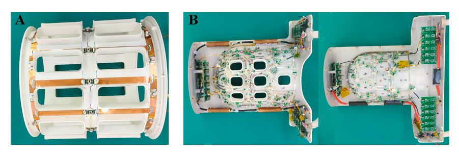
Figure 1 shows photograph of the optimized quadrature birdcage/47-channel receiver coil assembly on a 3 T MRI scanner (uMR 790, Shanghai United Imaging Healthcare, Shanghai, China). The high-pass birdcage coil with a 300mm diameter and a 390mm length was comprised of 12 rungs and was driven in circularly polarized mode. The coil shield has a 350mm diameter and a 390mm length. The receiver array was arranged on a close-fitting helmet former (A-P:238mm, L-R:206mm, I-S:265mm), of which 23 elements on the anterior and 24 elements on the posterior. The electronic components layout was optimized for PET compatibility. The brain PET insert with a 376.8mm detector ring diameter has 224 detector modules arranged in 8 detector rings with 28 detectors per ring. The axial field-of-view (FOV) is 329mm. For MRI shielding, carbon fiber tube and copper foil were used.A head-and-shoulder phantom filled with an aqueous solution of 3.75g NiSO4·6H2O and 5g NaCl per 1000g of demineralized water was used for MRI compatibility study. The measured B1+ field was characterized using the double-angle method (30o and 60o) with a 2D GRE sequence (TR/TE =2500ms/10ms, slice thickness=5mm, matrix=128×128, FOV=256mm×256mm, bandwidth=300Hz/pixel). For SNR calculation, the signal images were acquired using a 2D GRE sequence (TR/TE =300ms/10ms, flip angle=30o, slice thickness=5mm, matrix=128×128, FOV=256mm×256mm, bandwidth=300Hz/pixel). The noise images were obtained by setting the flip angle to zero. SNR maps were calculated using the sum-of-squares method. For the ΔB0 measurement, a 2D GRE with two echoes was applied with the parameters: TR=300ms, TE1/TE2=6.5ms/13ms, flip angle=30o, slice thickness=5mm, slice spacing=5mm, slices=12, matrix=128×128, FOV=256mm×256mm, bandwidth=300Hz/pixel.
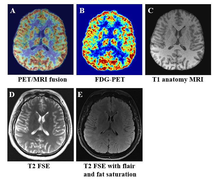
Before the human studies were initiated, informed consent was obtained from the volunteer. This study was approved by the institutional review boards of Shenzhen Institutes of Advanced Technology, Chinese Academy of Sciences. Human PET data from a volunteer with a 30-minute scan was acquired at 75 min after injection of [18F]FDG (6.9mCi injected activity). T1 anatomy images were acquired using a 3D GRE sequence at 1 mm isotropic resolution with following parameters: TR/TE=7.09ms/3.1ms, inversion time= 900ms, flip angle=10o, slices=176, echo train length=160, bandwidth=250Hz/pixel. T2 anatomy images were acquired using a 2D FSE sequence(TR/TE=5391ms/138.32ms, flip angle=80o, slices=19, matrix=667×768, FOV=200mm×230mm, echo train length=27, bandwidth=260 Hz/pixel) and a 2D FSE sequence with flair and fat saturation (TR/TE=8000ms/113.1ms, flip angle=150o, slices=19, matrix=396×456, FOV=200mm×230mm, echo train length=29, bandwidth=220Hz/pixel).
Results
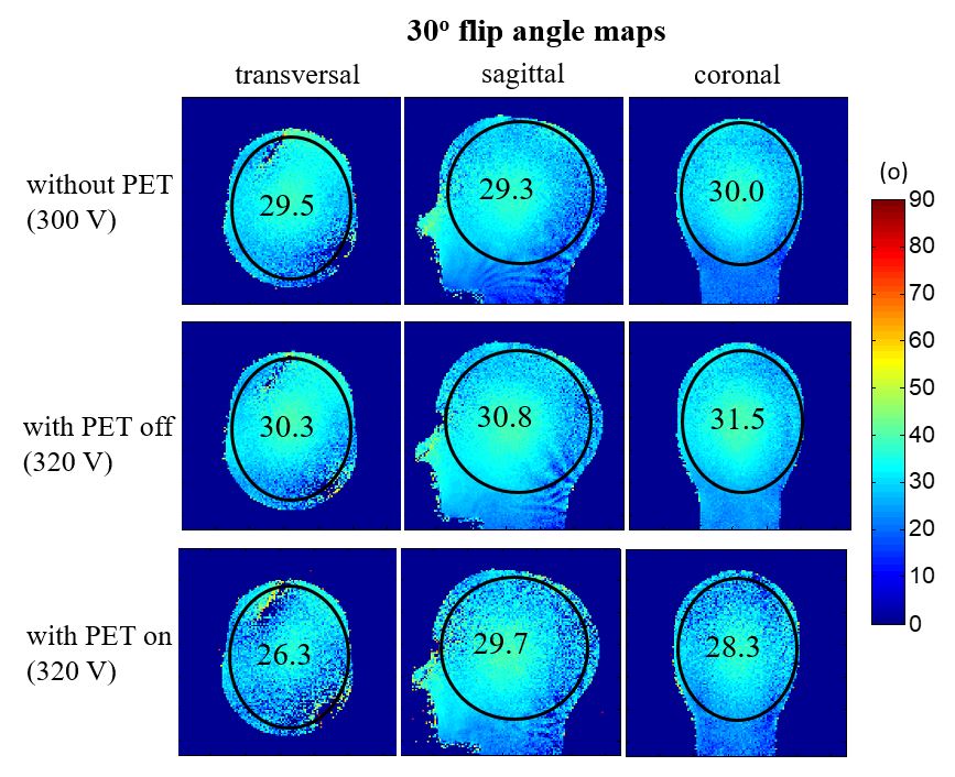
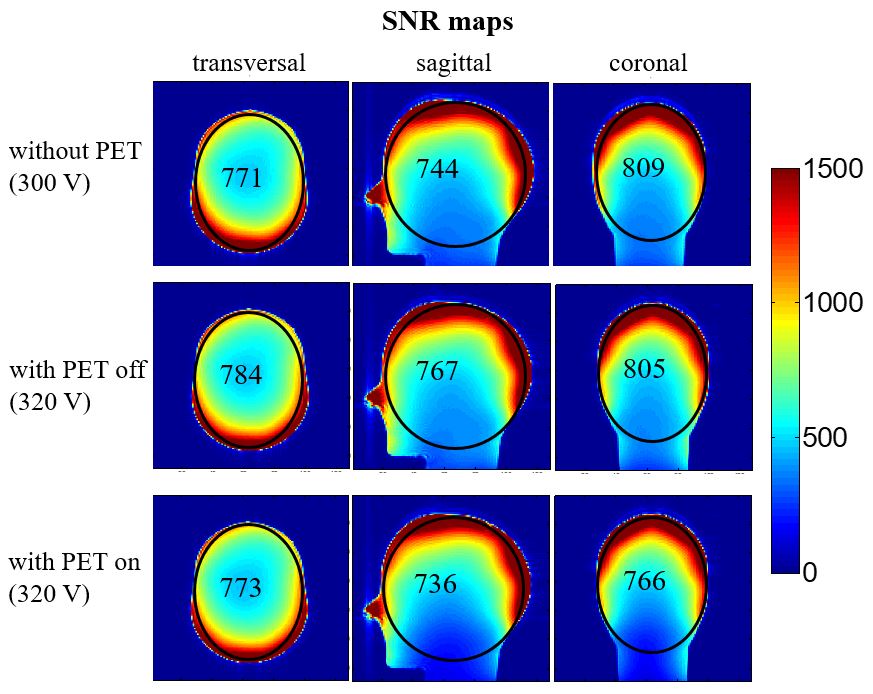
The measured B1+ field maps are shown in Figure 2, which were represented by 30o flip angle maps without PET using a 300V correction voltage, with PET off and with PET on using a 320V correction voltage. The mean values in the ROI were calculated and depicted in the maps. The B1+ field strength with PET on was reduced to about 90% and 95% of the B1+ field strength without PET in the transversal and coronal plane, respectively. It is not affected in the sagittal plane.The SNR maps are shown in Figure 3. The mean values in the ROI were calculated and depicted in the maps. Known from the results, SNR with PET on shows only a 5% loss in the coronal plane in comparison with the case of without PET. In the transversal and sagittal planes, SNR shows little difference.
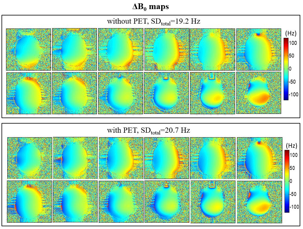
The measured ΔB0 field maps are shown in Figure 4. After using the B0 shimming in the MRI system for both cases (without PET and with PET), the B0 field homogeneity shows little difference, which was represented by the standard deviation (SD) value of the 12 slices.
Simultaneously acquired images from a volunteer using the brain PET/MRI system are shown in Figure 5A. The [18F]FDG-PET image and T1-weighted anatomy MR image are shown in Figure 5B and 5C, respectively. T2-weighted anatomy MR image are also respectively shown in Figure 5D and 5E. These results demonstrate a high performance of the brain PET/MRI system.
Discussions and Conclusions
The results of B1+ field, SNR and B0 field maps show MRI compatibility of the brain PET insert. Simultaneous PET/MRI demonstrated a high performance of the brain PET/MRI system. These show great significance in clinical and scientific research applications for brain imaging using the PET insert integrated with the quadrature birdcage transmit /47-channel receiver head coil assembly. Future work would include performance evaluation in PET compatibility of the head coil assembly and functional imaging.Acknowledgements
This work was supported in part by National Key R&D Program of China, 2021YFE0204400, 2021YFC2400203; the Strategic Priority Research Program of Chinese Academy of Sciences (Grant No. XDB25000000); National Natural Science Foundation of China, U22A20344; Youth Innovation Promotion Association of CAS No. Y2021098; Guangdong Province grants 2018B030333001; Key Laboratory Project of Guangdong Province, 2020B1212060051; Shenzhen city grant, RCYX20200714114735123.References
1. Catana C, Drzezga A, Heiss WD, Rosen BR. PET/MRI for neurologic applications. Journal of nuclear medicine, 2012, 53(12):1916-25.
2. Nensa F, Beiderwellen K, Heusch P, Wetter A. Clinical applications of PET/MRI: current status and future perspectives. Diagnostic and Interventional Radiology, 2014, 20(5):438-47.
3. Catana C. Development of Dedicated Brain PET Imaging Devices: Recent Advances and Future Perspectives. Journal of nuclear medicine, 2019, 60(8):1044-1052.
4. Kuang Z, Sang Z, Wang X, Ren N, Wu S, Zeng T, Niu M, Cong L, Mungai KS, Liu Z, Sun T, Hu Z, Yang Y. Development and initial performance of SIAT bPET: a high-resolution MRI compatible brain PET scanner with dual-ended readout detectors. 2022 IEEE Nuclear Science Symposium (NSS) and Medical Imaging Conference (MIC), 2022, p.1050.
5. Lee J, Sang Z, Yang Y, Zhang X, Li Y. A dedicated multichannel head coil array for PET insert on 3 T MRI. Proc. 29th Annual Meeting of ISMRM, online, 2021, p.4265.
Figures