4388
High permittivity material for catheter tracking in interventional imaging
Yunkun Zhao1 and Xiaoliang Zhang1
1Biomedical Engineering, State University of New York at Buffalo, Buffalo, NY, United States
1Biomedical Engineering, State University of New York at Buffalo, Buffalo, NY, United States
Synopsis
Keywords: Interventional Devices, MR-Guided Interventions
This study presents a technique of using high permittivity dielectric materials for catheter tracking in interventional endovascular imaging procedures. The design includes a high dielectric material cylinder at the tip of the catheter. Given the small size and limited space of the catheters, it is challenging to implement the current catheter tracking techniques. In the proposed approach, the high permittivity material mounted on tip of the catheter can amplify the local magnetic fields and makes the catheter tip visible. This method doesn’t need any feeding cables and is not sensitive to the frequency shift which makes it easy to implement.Introduction
The development of catheter tracking and guidance in catheterized interventions in the MR environment has increased rapidly in recent years. MRI has no ionizing radiation compared to conventional catheter tracking methods using X-ray, which is harmful to patients and physicians. Current applications include small RF (radio frequency) coils or antennas for active MR tracking. Both methods of active MR tracking require an extra power supply through the catheter, which adds complexity, instability, and safety concerns due to the potential SAR elevation caused by feeding cables and other associated conductors. Small RF coils and antennas also require some space inside the catheter for implementation, which may not fit a catheter used for neurosurgery with a diameter range from 1.5mm to 3mm. Studies show that high permittivity dielectric material can manipulate the B1 field and thus is widely studied today. New development in material engineering has produced high dielectric material with over 250000 relative permittivity material [1]. A critical property of high dielectric material is that it can tailor the electromagnetic fields within the human body. In this study, we proposed and investigated the use of high-dielectric material to achieve a simple and robust way of catheter tracking for interventional MR imaging. This design does not need any direct power supply through the catheter and can provide convenience for the tracking and guidance for catheterized interventional MR imaging.Methods
The proposed high dielectric tracker is designed on a 3-dimensional area, as shown in Fig.1. Based on the general size of the catheter used for neurosurgery. The high dielectric tracker is a cylinder with a 2.6mm diameter and 2.6mm length. It was placed at the tip of the catheter for tracking. The catheter outside the high dielectric tracker has a diameter of 2.64mm, and the material of it is PTFE which is commonly used for the catheter. There is also a cylindrical phantom outside the catheter to better show the magnetic field distribution around the catheter. A standard-designed birdcage coil generates the B1 field in this research operates at 298MHz (the Larmor Frequency of proton 1H at 7T) and has 190mm diameter and length for brain imaging. In order to investigate what kinds of dielectric material will provide the best tracking and guidance, nine dielectric materials with different permittivity from 5000 to 160000 have been tested. Because dielectric material with specific relative permittivity can turn into a resonator at a certain volume [2], a high dielectric tracker with 120300 relative permittivity, which is a resonator at 298MHz, has also been tested. The B1 field distribution plot evaluated the performance of the dielectric material in tracking. Numerical results of the proposed designs are obtained using electromagnetic simulation software CST Studio Suite (Dassault Systèmes, Paris, France). All result has been normalized to accepted power at the resonant frequency with 1Watt input power.Results
Fig.2 and Fig.3 show the simulated magnetic field distribution inside the birdcage coil. As shown in the figure, the birdcage generated a uniform magnetic field, and the magnetic field around the catheter significantly increased for the tracker with high relative permittivity. Fig.3 provides a closer look at the magnetic field distribution inside the phantom. The result indicates that there is no noticeable change for the tracker that has relative permittivity below 50000. However, after the relative permittivity increases higher than 50000, the magnetic field around the catheter can be easily identified. Results also show that when the relative permittivity reaches 120300, the tracker acts like a resonator with the highest magnetic field strength around the other cases. When the relative permittivity is higher than 120300, the resonator does not have the same magnetic field distribution as the distribution of trackers, which have relative permittivity lower than 120300 and affects less area around the tracker. Fig.4 shows the numerical result of magnetic field strength along the x-axis and y-axis, as shown in the schematic. Fig.5 also shows the percentage of increased or decreased magnetic field at the edge of the tracker compared with at the edge of the phantom. The percentage of magnetic field increase or decrease can be up to 3000% for resonator cases and up to 500% for non-resonator cases. Both figures show huge differences caused by the tailored effect from the high dielectric tracker.Conclusion
In this study, we developed a technique for catheter tracking using convenient high-permittivity materials for interventional MR imaging. With the use of an external RF coil, the miniature dielectric material can amplify and tailor the local magnetic field around the catheter and provide an enhanced MR signal, making the location of the catheter visible. The dielectric material, at a certain relative permittivity value, can turn into a resonator around which the B1 field is further augmented. This method thus can be possibly used for enhanced vascular imaging with improved resolution.Acknowledgements
This work is supported in part by the NIH under a BRP grant U01 EB023829, and by State University of New York (SUNY) under SUNY Empire Innovation Professorship Award.References
1. Guillemet-Fritsch S, Lebey T, Boulos M, et al. Dielectric properties of CaCu3Ti4O12 based multiphased ceramics[J]. Journal of the European Ceramic Society, 2006, 26(7): 1245-1257.
2. Wen H, Jaffer FA, Denison TJ, Duewell S, Chesnick AS, Balaban RS. The evaluation of dielectric resonators containing H2O or D2O as RF coils for high-field MR imaging and spectroscopy. J Magn Reson B. 1996 Feb;110(2):117-23. doi: 10.1006/jmrb.1996.0019.
Figures
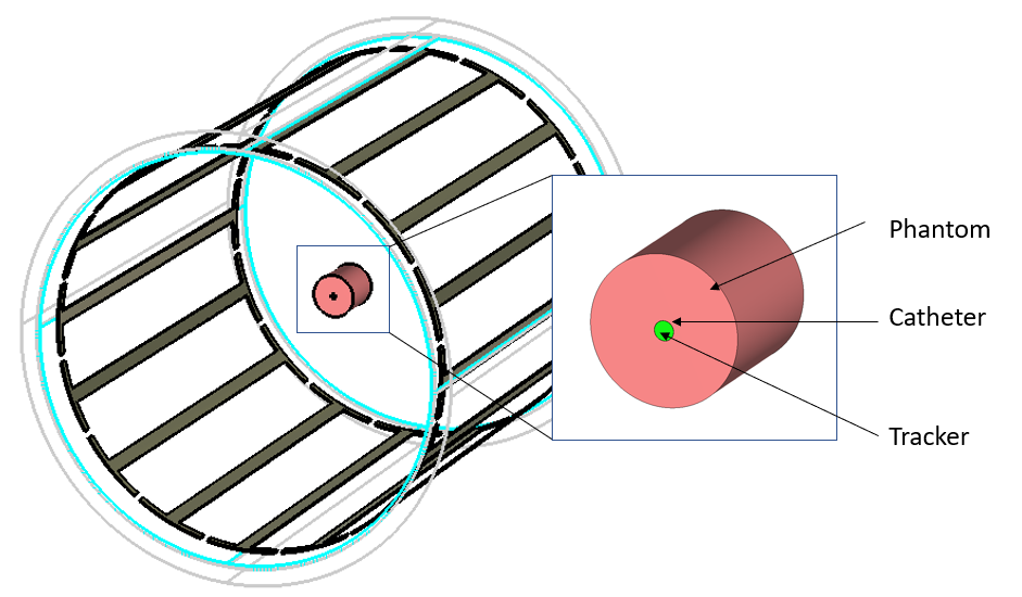
Fig. 1. Birdcage
coil, catheter, and tracker design
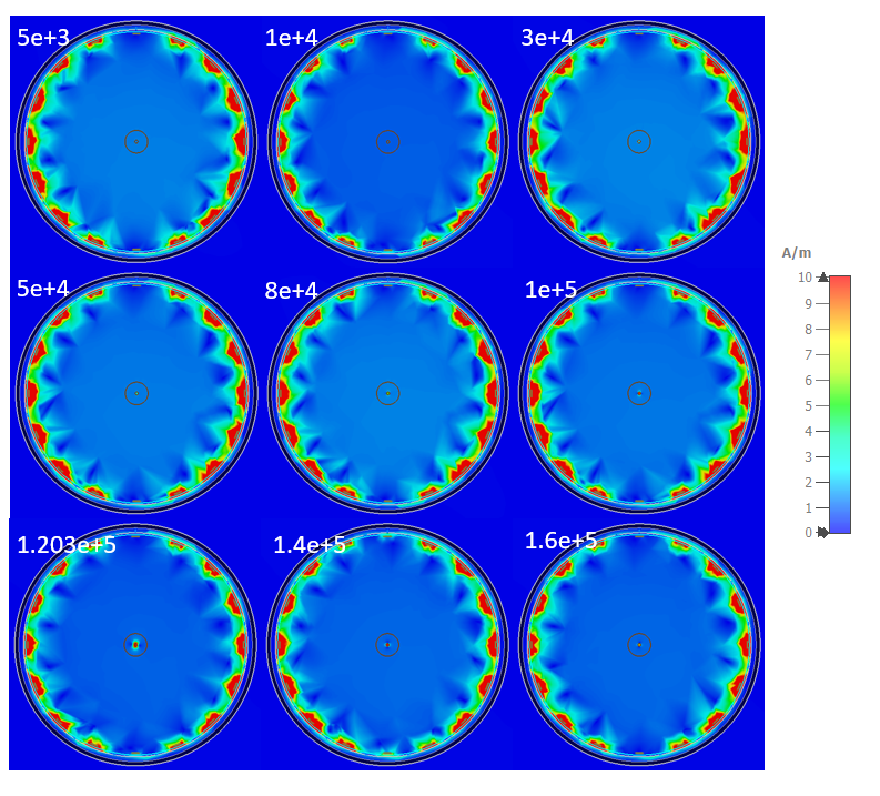
Fig. 2. Magnetic field distribution
for all trackers
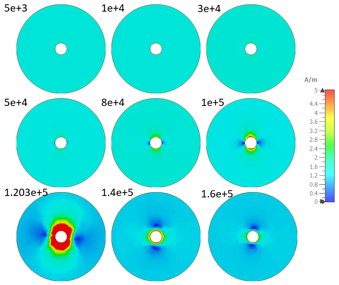
Fig. 3. Magnetic field distribution
inside phantom for all trackers
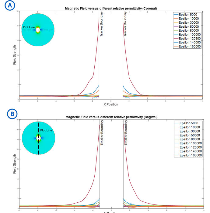
Fig. 4. Magnetic Field versus relative permittivity in
the phantom along the plot line (A) coronal
(B) sagittal.
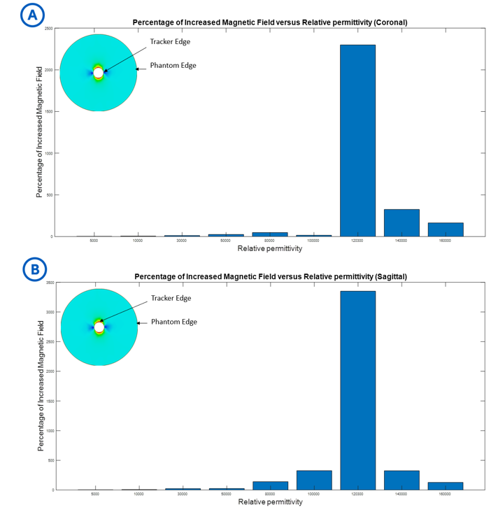
Fig. 5. The percentage increase in
the magnetic field at the edge of the tracker compared to the magnetic field at
the edge of the phantom at (A) coronal
(B) sagittal.
DOI: https://doi.org/10.58530/2023/4388