4380
Isotropic 3D MRI determined labral tear extent correlates with hyaline cartilage loss in hip dysplasia patients
Shuda Xia1, Avneesh Chhabra2, Gaurav Sharan2, Uma Thakur2, Holden Archer2, Yin Xi2, and Joel Wells2
1University of Texas Southwestern Medical Center, Plano, TX, United States, 2University of Texas Southwestern Medical Center, Dallas, TX, United States
1University of Texas Southwestern Medical Center, Plano, TX, United States, 2University of Texas Southwestern Medical Center, Dallas, TX, United States
Synopsis
Keywords: Joints, Joints
This study is a novel investigation into the clinical correlations between parameters measured through 3D MRI and outcomes for patients with hip dysplasia (HD). Although there were no statistically significant correlations seen between labral tear length and PROMs (p>0.05), a few non-significant correlation trends were observed. Furthermore, labral tear lengths directly positively correlated with worsening cartilage damage, the presence of subchondral bone cysts, para-labral cysts, and the number of labral tears seen.Introduction
Hip dysplasia (HD) is a developmental condition characterized by a shallow and upsloping acetabulum and sometimes accompanied by femoral head incongruency1. If left untreated, the joint's instability can lead to hyaline cartilage damage, labral tears, and premature hip osteoarthritis2. Radiographs are used for the initial imaging screening and assessment of HD and various validated measurements like Tonnis angle, lateral center edge angle and extrusion index have been described for grading the extent of HD3. Following radiographic assessment, multiplanar 2D (dimensional) MR imaging with or without arthrogram is routinely performed for outlining the health of labrum and hyaline cartilage4,5. Recently, 3D spin-echo MR imaging has been adopted for hip assessment. 3D MRI can be performed in an isotropic manner on high-field magnets (3 Tesla and newer 1.5T MRI scanners)6,7. Such volume imaging allows high-resolution assessment of labrum and cartilage lesions. In addition, it presents opportunities to measure the extent of labral tears with labrum-specific reconstructions8. Although the diagnosis of HD is established primarily based on a combination of clinical presentation, physical findings, and radiographic measurements, patient-reported outcome measures (PROMs) are vital in assessing the overall quality of life of the patient and how HD affects their joint health status. Common measurements included in PROMS include EqVas Health Rating, SF12, UCLA Activity Score, VAS Pain, HOS, HOOS, iHot 12, and HHS10-17. Each reported outcome measure provides a unique perspective on the patient’s health, which is essential in establishing an overall clinical picture of the patient and can influence treatment plans. This study aimed to correlate the extent of hyaline cartilage injury measured through 3D MRI with the presence, location, multiplicity, and length of labral tears in HD patients and with patient-reported outcome measures (PROMs).Methods
A total of 314 patient records with a final diagnosis of DDH were initially screened for inclusion in the study from a consecutive series of patients. Exclusion criteria included the presence of avascular necrosis of the hip (AVN), absence of 3D MR imaging, prior arthroplasty, and the presence of implants. Ultimately, 156 hips from 139 pre-operative patients with a final diagnosis of HD (ages 14-68 years, both genders) were included. 3D labrum-specific reconstructions were performed, and a multi-reader study was obtained. Inter-reader (ICC) analysis and Spearman correlations were calculated with the hypothesis that longer labral tears correlate with worsening cartilage injury and PROMs.Results
One hundred and twenty-two hips had intact cartilage, 10 had low-grade, 6 had high-grade, and 18 had full-thickness cartilage loss. The mean labral tear length was 15.38 mm with a standard deviation of 5.57 mm. An example of a hip with multifocal labral tears is included in Figure 1. There was a moderate inter-reader correlation (ICC = 0.66) on the lengths of labral tears. There were non-negligible correlations between the length of labral tears and various PROM parameters. There was a weak negative correlation between the length of the mean labral tear and the EqVas Health Rating (Spearman coefficient of -0.24 (95%CI: -0.40, -0.08)) and a weak positive correlation between the length of the mean labral tear and VAS worst pain (Spearman coefficient of 0.24 (95% CI: 0.07, 0.39)). However, none of the p values were significant after FDR adjustment. The mean labral tear size was directly positively correlated with worsening cartilage damage, the presence of subchondral bone cysts, para-labral cysts, and the number of labral tears.Discussion
HD is a common condition presenting in adult hip preservation practice. This work has shown that isotropic 3D MRI can be reliably used to measure labral tears, detect multifocality of tears and find correlations between the labral tears and hyaline cartilage loss. Labral tear lengths were directly correlated with worsening hyaline cartilage damage and the presence of subchondral and para-labral cysts. This suggests a possible protective role of the labrum on hyaline cartilage. Our first hypothesis was validated. However, we found no significant correlation between labral tear lengths and PROMs. This discrepancy is likely explained by confounding clinical factors, complex pathophysiology, and clinical presentation of HD along with the limitations of the study as discussed below. Existing literature suggests that features measured by 3D MRI are not correlated with PROMs in other conditions such as femoroacetabular impingement syndrome, but impaired physical functioning measured through PROMs was found to be associated with the severity of HD18,19. The correlation trends however somewhat support our hypothesis that worsening labral tear measurements correlate with poorer PROMs and a greater degree of hyaline cartilage damage.Conclusion
This novel study delves into the clinical correlations of 3D MRI measurements of a population of HD patients. Larger labral tears were positively correlated with deteriorating hyaline cartilage and the presence of subchondral and para-labral cysts, although worsening joint health was not directly correlated to hip symptomatology. Further studies could examine larger and more heterogenous cohorts of HD to determine the correlation trends among labral tear lengths, hyaline cartilage damage, and PROMs. Such studies can add to the existing literature regarding the usage of 3D MRI in diagnostic and treatment capacities, which can influence the practice of hip preservation.Acknowledgements
I would like to thank Dr. Avneesh Chhabra for mentoring and guiding me through this project. I would also like to acknowledge Dr. Uma Thakur, Dr. Gaurav Sharan, Dr. Joel Wells, Dr. Yin Xi, and Mr. Holden Archer for their contributions to this project.References
[1] S. Zhang, K.J. Doudoulakis, A. Khurwal, K.M. Sarraf, Developmental dysplasia of the hip, Br J Hosp Med (Lond) 81(7) (2020) 1-8. [2] S. Yang, N. Zusman, E. Lieberman, R.Y. Goldstein, Developmental Dysplasia of the Hip, Pediatrics 143(1) (2019). [3] K.S. McQuivey, E. Secretov, B.G. Domb, B.A. Levy, A.J. Krych, M. Neville, D.E. Hartigan, A Multicenter Study of Radiographic Measures Predicting Failure of Arthroscopy in Borderline Hip Dysplasia: Beware of the Tonnis Angle, Am J Sports Med 48(7) (2020) 1608-1615. [4] C.N. Anderson, G.M. Riley, G.E. Gold, M.R. Safran, Hip-femoral acetabular impingement, Clin Sports Med 32(3) (2013) 409-25. [5] D.G. Blankenbaker, M.J. Tuite, MR imaging of early hip joint degeneration, Magn Reson Imaging Clin N Am 19(2) (2011) 365-78. [6] M. Samim, 3D MRI Models of the Musculoskeletal System, Semin Musculoskelet Radiol 25(3) (2021) 388-396. [7] G. Zeng, F. Schmaranzer, C. Degonda, N. Gerber, K. Gerber, M. Tannast, J. Burger, K.A. Siebenrock, G. Zheng, T.D. Lerch, MRI-based 3D models of the hip joint enables radiation-free computer-assisted planning of periacetabular osteotomy for treatment of hip dysplasia using deep learning for automatic segmentation, Eur J Radiol Open 8 (2021) 100303. [8] O. Ashikyan, J. Wells, A. Chhabra, 3D MRI of the Hip Joint: Technical Considerations, Advantages, Applications, and Current Perspectives, Semin Musculoskelet Radiol 25(3) (2021) 488-500. [9] C.A. Barrera, S.A. Cohen, W.N. Sankar, V.M. Ho-Fung, R.W. Sze, J.C. Nguyen, Imaging of developmental dysplasia of the hip: ultrasound, radiography and magnetic resonance imaging, Pediatr Radiol 49(12) (2019) 1652-1668. [10] Y. Feng, D. Parkin, N.J. Devlin, Assessing the performance of the EQ-VAS in the NHS PROMs programme, Qual Life Res 23(3) (2014) 977-89. [11] D.R. Griffin, N. Parsons, N.G. Mohtadi, M.R. Safran, N. Multicenter Arthroscopy of the Hip Outcomes Research, A short version of the International Hip Outcome Tool (iHOT-12) for use in routine clinical practice, Arthroscopy 28(5) (2012) 611-6; quiz 616-8. [12] M.P. Jensen, C. Chen, A.M. Brugger, Interpretation of visual analog scale ratings and change scores: a reanalysis of two clinical trials of postoperative pain, J Pain 4(7) (2003) 407-14. [13] M. Kuhn, M. Harris-Hayes, K. Steger-May, G. Pashos, J.C. Clohisy, Total hip arthroplasty in patients 50 years or less: do we improve activity profiles?, J Arthroplasty 28(5) (2013) 872-6. [14] R.L. Martin, B.T. Kelly, M.J. Philippon, Evidence of validity for the hip outcome score, Arthroscopy 22(12) (2006) 1304-11. [15] F. Salaffi, G. Leardini, B. Canesi, A. Mannoni, A. Fioravanti, R. Caporali, G. Lapadula, L. Punzi, Gonorthrosis, A. Quality Of Life, Reliability and validity of the Western Ontario and McMaster Universities (WOMAC) Osteoarthritis Index in Italian patients with osteoarthritis of the knee, Osteoarthritis Cartilage 11(8) (2003) 551-60. [16] J. Ware, Jr., M. Kosinski, S.D. Keller, A 12-Item Short-Form Health Survey: construction of scales and preliminary tests of reliability and validity, Med Care 34(3) (1996) 220-33. [17] D.S. Yu, E.C. Yan, C.K. Chow, Interpreting SF-12 mental component score: an investigation of its convergent validity with CESD-10, Qual Life Res 24(9) (2015) 2209-17. [18] S. Okpara, P. Nakonezny, J. Wells, Do psychological factors or radiographic severity play a role in the age of onset in symptomatic developmental dysplasia of hip and femoroacetabular impingement syndrome?, BMC Musculoskelet Disord 20(1) (2019) 412. [19] T.L. Ratcliff, A. Chhabra, S.O. Okpara, P. Lawson, S. Kayfan, Y. Xi, E.P. Mulligan, J.E. Wells, Correlation of the Imaging Features of Femoroacetabular Impingement Syndrome With Clinical Findings and Patient Functional Scores, Orthopedics 44(4) (2021) e577-e582.Figures
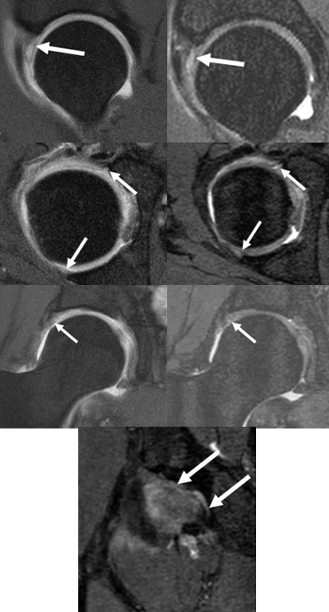
Figure 1. Multifocal labral tear seen in all quadrants in a
29-year-old male patient, with a 3D MRI image quality of 4. Images on the
left-hand side panel were taken from 2D MRI sequences, with tears outlined by
white arrows. Images on the right-hand side panel were taken from 3D MRI
sequence and correspond with the same viewing plane as the 2D MRI images. The
last image outlines the posteroinferior tear measured for the study (arrows).
DOI: https://doi.org/10.58530/2023/4380