4371
Improved identification of cartilage lesion in chondromalacia patellae (CMP) with 7.0 T MRI compared with 3.0T MRI: a preliminary study
Yan Wang1, Jiayu Huang1, Jianxun Qu2, Xiangbing Bian1, Caohui Duan1, Chunbao Li3, Jing Zhang1, and Xin Lou1
1Radiology, PLA general hospital, Beijing, China, 2MR Collaboration, Siemens Healthineers Ltd, Beijing, China, 3Orthopaedics, PLA general hospital, Beijing, China
1Radiology, PLA general hospital, Beijing, China, 2MR Collaboration, Siemens Healthineers Ltd, Beijing, China, 3Orthopaedics, PLA general hospital, Beijing, China
Synopsis
Keywords: Cartilage, Cartilage
To compare the ability in revealing subtle changes of patellar cartilage in suspected CMP between 7.0T and 3.0T MRI. We documented the total number of identified cartilage lesions by two radiologists and compared the number of cartilage lesions identified between 3.0T and 7.0T MRI. The result showed good consistency between readers. The mean number of cartilage lesions identified on 3.0T images was significantly less than 7.0T images. Considering the preliminary and small-sample feature of the study, we conclude conservatively that compared with 3.0T MRI, 7.0T MR images reveal more lesion of the patellar cartilage in CMP patients.Background
Chondromalacia patellae (CMP) is commonly seen among young people. While the disease process is reversible in the early stage, it can progress to osteoarthritis, affecting the quality of life and leading to high prevalence of physical disability [1]. Therefore, early diagnosis is crucial important for the best prognosis. Traditionally, 3.0T MRI is accepted as the most sensitive and non-invasive method to detect CMP [2]. With clinical application of 7T MR in recent years, the visualization of knee cartilage is supposed to be improved and earlier identification of CMP should be achieved.Purpose
To compare the ability in revealing subtle changes of patellar cartilage in suspected CMP between 7.0T and 3.0T MRI.Material and Method
With ethics board approval, three untreated patients with clinically suspicious CMP were recruited in this study. The study was performed on a 3.0T whole-body scanner (MAGNETOM Skyra, Siemens Healthcare, Erlangen, Germany) and a 7.0T whole-body scanner (MAGNETOM Terra, Siemens Healthcare, Erlangen, Germany). Each patient received fat-suppressed proton density weighted (fs-PDw) imaging and other routine protocols on the scanners within the same day. At 3T, the protocol parameters for fs-PDw are TR 4145ms, TE 42ms, slice thickness 4mm, slice gap 0.8mm, and FOV 16 * 16 cm2, the in-plane resolution is 0.4 * 0.4 mm2,and the acquisition time is 1 minute and 35 seconds. At 7T, the parameters are TR 4500ms, TE 35ms, slice thickness 2.5mm, slice gap 0.8mm, FOV 16 * 16 cm2, the in-plane resolution is 0.2 * 0.2 mm2, and the acquisition time is 3 minutes and 59 seconds.Two experienced radiologists in the musculoskeletal specialty independently read all the 3.0T and 7.0T images. Based on the MRI grading system for chondromalacia patellae [3], the total number of identified cartilage lesions was documented by two radiologists, respectively. Based on the number of lesions identified in each patient, the intraclass correlation coefficient (ICC) was calculated to reveal the consistency between readers. Then the number of cartilage lesions identified was compared between 3.0T and 7.0T MRI with matched samples t-test. All the statistical analysis were performed on SPSS 17.0, and p <0.05 was set as a significant level.
Results
All the 3 patients completed the 3.0T and 7.0T scan successfully (Figure 1). The ICC for two radiologist was 0.93 (CI: 0.911~0.975, p<0.001) on 3.0T images and 0.94 (CI: 0.90~0.97) on 7.0T images, indicating a very good consistency between readers both on 3.0T and 7.0T scan. The mean number of cartilage lesions identified on 3.0T images was significantly less than 7.0T images (1.7 vs 4.0 p<0.001), respectively indicating the superiority of 7.0T to 3.0T MRI on the identification of CMP-related cartilage lesion.Discussion and Conclusion
Compared with 3.0T MRI, the 7.0T MR can reveal more lesion of the patellar cartilage in CMP patients and might be a promising method for the earlier detection of CMP-related cartilage lesion (Figure 2). Owing to the preliminary feature, the final result might be weakened by the lack of arthroscopy and pathology-imaging correlation. The sample size is relatively small and need to be enlarged. Further study is ongoing in our institute to prove the finding of the present study.Acknowledgements
No acknowledgement found.References
1. Chondromalacia patellae: current options and emerging cell therapies. Weitao Zheng, Hanluo Li , Kanghong Hu, et al. Stem Cell Res Ther, 2021, 12(1):412.
2. MRI of the knee at 7.0 Tesla. O Kraff, J M Theysohn, S Maderwald, et al. Rofo, 2007, 179(12):1231-5.
3. A new MRI grading system for chondromalacia patellae. Ali Özgen, Neslihan Taşdelen and Zeynep Fırat. Acta Radiol. 2017, 58(4): 456–463.
Figures
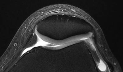
Figure 1. FS PD
images of a normal patellae on 7T. Patellar cartilage showed uniform signal
intensity. Periphery of the cartilage might show slightly hyper-signal near the
junction of patellar cartilage and patellar retinaculum.
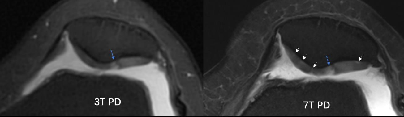
Figure 2. FS PD images of a CMP patient on 3T and 7T. On axial 3T image (A), 1 lesion was detected (blue arrow head). On axial 7T image (B), another 4 lesions were detected obviously (white arrow heads) beside the lesion that detected on 3T (blue dashed arrow) due to higher resolution of the image.
DOI: https://doi.org/10.58530/2023/4371