4369
Assessment of articular cartilage in response to an impact injury using µMRI
Amanveer Singh1, Hannah Mantebea2, Farid Badar2, Syeda Batool2, Gabrielle Abdelmessih3, Talia Sebastian3, Michael Newton4, and Yang Xia2
1Department of Physics and Center for Biomedical Research, Oakland University, Rochester, MI, United States, 2Department of Physics and Center for Biomedical Research, Oakland University, Rochestor, MI, United States, 3Department of Chemistry, Oakland University, Rochestor, MI, United States, 4Beaumont Hospital, Royal Oak, MI, United States
1Department of Physics and Center for Biomedical Research, Oakland University, Rochester, MI, United States, 2Department of Physics and Center for Biomedical Research, Oakland University, Rochestor, MI, United States, 3Department of Chemistry, Oakland University, Rochestor, MI, United States, 4Beaumont Hospital, Royal Oak, MI, United States
Synopsis
Keywords: Cartilage, Osteoarthritis, Osteoarthritis, Cartilage, Impact injury
This project aimed to study the properties of articular cartilage in response to mechanical injury in rabbit knees using μMRI. Femoral cartilage bone plugs were excised 2 and 14 weeks after impact and imaged using μMRI at a resolution of 11.7 μm/pixel. Higher T2 relaxation values in affected specimens illustrated deterioration in the cartilage after impact. Furthermore, the higher T2 values in impacted samples of the 14-week batch compared to the 2-week batch indicate progressive cartilage degradation over time.INTRODUCTION
Trauma to a joint can result in post-traumatic osteoarthritis, contributing to approximately 12% of all osteoarthritis (OA) cases, leading to joint pain and disability1. Articular cartilage (AC) injury does not heal well on its own due to its limited repair ability and the lack of blood supply to the tissue, ultimately leading to the degeneration of AC in OA. Therefore, it is crucial to understand the biological changes initiated by the injury and how the degradation progresses over time. This study aimed to investigate the consequences of a mechanical injury to the knee at two different time points after the impact in a rabbit model using microscopic magnetic resonance imaging (µMRI) at high resolution.METHODS
The AC of the right knee of 12 adult female NZW rabbits was impacted one time by the surgeon using a limb clamp with a specially designed impact device on the operating table with a pressure of approximately 30 MPa. Animals were allowed ad libitum activity until reaching their assigned euthanizing time points (2 and 14-week post-impact). AC was extracted from both the knee joints and quantified using μMRI-T2 images at 11.7 μm/pixel resolution at 0° and 55° sample orientations to B0 in a 7T magnet. In addition, cartilage tissues were harvested from the anterior and posterior portions of the femur; the water (H2O) content in the specimens was determined by weighting/drying analysis. Bonferroni tests were performed in statistical analysis to compare mean T2 values with a significance level of p<0.05.RESULTS
T2 relaxation values from µMRI images with the cartilage oriented at both 0˚ and 55˚ to the external magnetic field B0 showed similar trends, values for the impacted right knee (IRK) were greater than the non-impacted left knee (NILK) in both the 2-week and 14-week samples, this increment in the T2 values of the IRK was observed throughout all the cartilage zones. Furthermore, it was observed that the T2 values were higher for 14-week IRK than for the 2-week IRK. Biochemistry analysis showed that H2O% mean value for IRK was higher than the NILK for both 2 and 14-week samples.DISCUSSION
This study evaluated the effects of impact injury contributing to OA through progressive cartilage degeneration in knee joints at 2 and 14 weeks after the surgery. Evaluation of the T2 data from µMRI depicted cartilage degradation in the impacted knees with higher values due to elevated water content. Also, greater degradation in affected knees at 14 weeks than at 2 weeks indicates progressive cartilage deterioration over time. The µMRI results were in accord with the biochemistry analysis with higher H2O% in the IRK compared to the NILK, indicating structural degradation in the AC. Other complementary analyses are in progress.Acknowledgements
This research was funded by NIH R01 (AR 69047, PI: Xia). Dr. Kevin Baker (Henry Ford Hospital, Detroit, MI) for technical assistance and expertise with animal protocols.References
1. Punzi L, Galozzi P, Luisetto R, Favero M, Ramonda R, Oliviero F, Scanu A. Post-traumatic arthritis: overview on pathogenic mechanisms and role of inflammation. RMD Open. 2016 Sep 6;2(2):e000279. doi: 10.1136/rmdopen-2016-000279.Figures
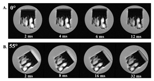
T2 weighted
intensity images of articular cartilage at different echo times with pixel
resolution of 11.7 μm. The orientation of the specimen was 0° (A) and 55° (B), angles were defined as the
angle between the normal axis of the articular surface and the direction of the
magnetic field (B0).
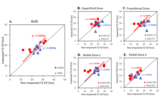
A
representation of T2 data at 55° for (2 and 14-week) non-impacted and
impacted specimens with linear regression illustrating Bulk (A), Superficial Zone
and Transitional Zone (B,C), and Radial Zone1 and Radial Zone2 (D,E). The
dotted black line in the center has a slope of 1. The slopes of linear fit more
than 1 indicate an increment in T2 values after the impact.
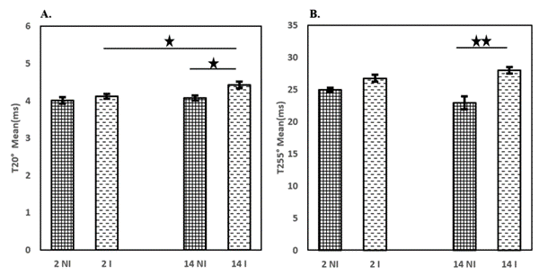
Statistical comparison of T2
values between non-impacted (NI) left knee and impacted (I) right knee for 2 and 14-week articular cartilage specimens
at 0° (A) and 55° (B). The statistical significances are denoted
as * *(P<0.01) and *(P<0.05).
DOI: https://doi.org/10.58530/2023/4369