4366
Deep-learning based fast spin echo magnetic resonance imaging improved clarity of nasal cartilage1renmin hospital of wuhan university, wuhan, China, 2GE Healthcare, MR Research China, beijing, China
Synopsis
Keywords: Cartilage, Cartilage
In this study, a comparative study of the original images (T1WI, T2WI) and ARDL images based on deep learning reconstruction (T1WI-DL, T2WI-DL) was conducted for magnetic resonance imaging of nasal cartilage. We aim to evaluate the feasibility and performance of a novel deep learning based MRI reconstruction method to show the structures of nasal cartilage, especially lower lateral cartilage. The results showed that applying ARDL to nasal cartilage MRI showed significant improvement in both SNR and CNR. we conclusion that the DL-based reconstruction algorithm was feasible to be applied to nasal cartilage MRI and improve the image quality.Synopsis
In this study, a comparative study of the original images (T1WI, T2WI) and ARDL images based on deep learning reconstruction (T1WI-DL, T2WI-DL) was conducted for magnetic resonance imaging of nasal cartilage. We aimed to evaluate the feasibility and performance of a novel deep learning based MRI reconstruction method to show the structures of nasal cartilage, especially lower lateral cartilage. The results showed that ARDL nasal cartilage MRI had significant improvement in both SNR and CNR. Overall, the DL-based reconstruction algorithm was feasible to improve the image quality of nasal cartilage MRI and shorten scan time without SNR loss.Introduction and purpose
The nose, as a unique facial landmark, is basically composed of cartilage. Lower lateral cartilage is a thin cartilage plate curved in pairs like a horseshoe, located on both sides of the nasal tip, which constitutes the main scaffold of the nose and is divided into medial and lateral pedicles. Lower lateral cartilage is the main influencing factor of the aesthetic appearance of the nose. Nasal cartilage defects and deformities caused by trauma, congenital diseases (e.g. congenital nasal deformity, cleft lip and palate, etc.), nasal tumors (e.g. Nasal basal cell carcinomas), etc., it is essential to restore its physiological function and appearance throug surgical reconstruction or implantation. Proper preoperative observation of these anatomical structures can improve surgical outcomes and increase postoperative patient satisfaction [1]. Nowadays, Rhinoplasty and surgical reconstruction of nasal cartilage structures remains a great challenge, and the limitations of most rhinoplasty procedures stem from the low diagnostic and imaging capabilities of the region in the preoperative phase[2]. Traditional preoperative evaluation methods for rhinoplasty include direct anthropometric measurements, 3D facial scanners and subjective patient/doctor evaluations, and measurement tools and measurement practices are difficult to standardize. The key aspects of rhinoplasty are preoperative imaging and placement selection [3].MRI can help to achieve the preoperative imaging needs of rhinoplasty[4], and the combination of imaging techniques with biomedicine is useful for the study of nasal support structures. Conventional MRI sequences are usually a compromise between scan time and image quality, a novel deep learning based MRI reconstruction method (hereafter referred to as “ARDL”) can achieve improved image quality with reduced scan time. In this study, for nasal cartilage MRI images, a comparative study of origin images (T1WI, T2WI) and ARDL images (T1WI-DL, T2WI-DL) was conducted to evaluate the feasibility of applying ARDL to display the anatomical structures of nasal cartilage, especially lower lateral cartilage.Materials and methods
38 patients consulting rhinoplasty underwent an MRI exam on 3.0T scanner (Signa Architect, GE Healthcare) using a 19-channel head and neck coil. All MR data was reconstructed with and without DLR. A total of 4 sets of images including reconstructed and original images were obtained using ARDL (AIR™ Recon DL, GE healthcare) . For qualitative image quality assessment , Two radiologists scored the images’ quality and the display quality of the nasal cartilage structures (including the septal cartilage, lateral nasal cartilage and lower lateral cartilage) using 5-point scoring system. Signal-to-noise ratio (SNR) and contrast-to-noise ratio (CNR) were measured to evaluate image quality.Result
Thirty-eight volunteers (age: 25.5 ± 2.3 years) were included in this study. For overall image quality, the differences in subjective scores between reconstructed images(T1WI-DL and T2WI-DL) were not statistically significant (p > 0.05), but all were significantly improved over the original images, respectively (p values < 0.001);for nasal cartilage anatomical structure scores, nasal septal cartilage, lateral nasal cartilage and lower lateral cartilage were better displayed by T2WI-DL (p < 0.01). By Kappa consistency test, κ = 0.823, p < 0.001, the 2 doctors had good agreement in the diagnosis. The SNR and CNR of reconstructed images were better than the original sequences ( p<0.001); for the display of lateral nasal cartilage, the image quality of T2WI-DL group was better than that of T1WI-DL. p<0.001, the difference was statistically significant.Conclusion and Discussion
In this study, a comparative study of ARDL images based on deep learning reconstruction (T1WI-DL, T2WI-DL) with the original images (T1WI, T2WI) was conducted for magnetic resonance imaging of nasal cartilage to evaluate the feasibility of this novel method to display the anatomical structures of nasal cartilage, especially lower lateral cartilage. The results showed that the reconstructed images showed significant improvement in SNR and CNR on the T1WI and T2WI sequences compared to the original images. For the display of the lower lateral cartilage, the image quality of the T2WI-DL group was optimal, which led to the conclusion that ARDL images based on deep learning reconstruction was feasible to be applied to MRI to display the nasal cartilage structure and improve the image quality.Acknowledgements
No acknowledgement found.References
[1] Nguyen C, Nicolai E, He J J, et al. 3D surface imaging technology for objective automated assessment of facial interventions: A systematic review[J]. J Plast Reconstr Aesthet Surg,2022.
[2] Chuang J, Barnes C, Wong B. Overview of Facial Plastic Surgery and Current Developments[J]. Surg J (N Y),2016,2(1):e17-e28.
[3] Xie K, Sun X, Wang L, et al. One-Stage Repair of Alveolar Cleft and Nasal Deformities Using Grafts From Nasal Septum: Application of Vomer, Ethmoid, and Septal Cartilage[J]. J Craniofac Surg,2022,33(6):1869-1874.
[4] Recht M P, Goodwin D W, Winalski C S, et al. MRI of articular cartilage: revisiting current status and future directions[J]. AJR Am J Roentgenol,2005,185(4):899-914.
Figures
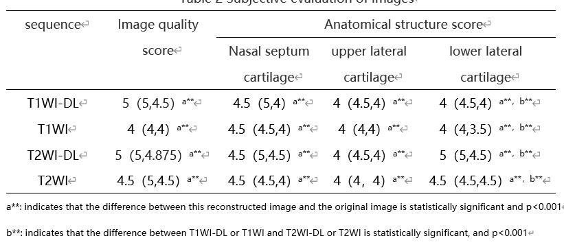
Table 2 Subjective evaluation of images
a**: indicates that the difference between this reconstructed image and the original image is statistically significant and p<0.001
b**: indicates that the difference between T1WI-DL or T1WI and T2WI-DL or T2WI is statistically significant, and p<0.001
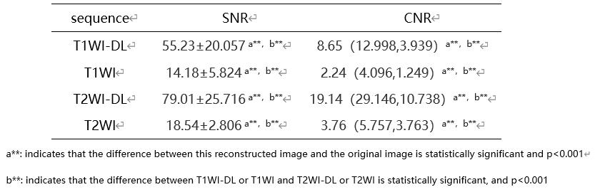
Table3 Objective evaluation of images
a**: indicates that the difference between this reconstructed image and the original image is statistically significant and p<0.001
b**: indicates that the difference between T1WI-DL or T1WI and T2WI-DL or T2WI is statistically significant, and p<0.001
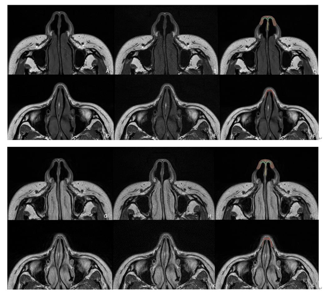
Figure 1 Transverse axial image of nasal cartilage
1) A, D and G, J are T1WI and T2WI ARDL reconstructed images respectively, the nasal cartilage structure is clearly displayed without motion artifacts;
2)B, E and H, K are T1WI and T2WI original images respectively, the image noise is large and affects the display of nasal cartilage structure.
3)C,F, I, L are schematic diagrams of nasal cartilage structure, green represents nasal wing large cartilage, yellow represents nasal septum cartilage, red represents lateral nasal cartilage