4365
Comparing the effect of body weight, BMI and physical activity intensity on calcified and uncalcified cartilage using quantitative UTE MRI1Ruijin Hospital, School of Medicine, Shanghai Jiao Tong University, Shanghai, China, 2MR Collaborations, Siemens Healthineers Ltd., Shanghai, China, Shanghai, China, 3Siemens Healthineers International AG, Zurich, Switzerland, Zurich, Switzerland, 4Swiss Center for Musculoskeletal Imaging (SCMI), Balgrist Campus, Zurich, Switzerland, Zurich, Switzerland, 5Advanced Clinical Imaging Technology (ACIT), Siemens Healthineers International AG, Lausanne, Switzerland, Lausanne, Switzerland
Synopsis
Keywords: Cartilage, Cartilage
UTE technique was utilized to quantitatively assess calcified cartilage (CC) and uncalcified cartilage (UC). T2* values of UC in patella, medial tibia and lateral femur positively moderately correlated with body weight, while T2* values of CC showed no significant correlation. T2* values of UC showed no significant correlation with BMI, while T2* values of CC in medial femur and lateral tibia showed negatively strong and positively moderate correlation, respectively. T2* values of CC and UC showed no significant correlation with physical activity level. The mechanical loading effect performs different between CC and UC, indicating their different bio-mechanical properties.Background and Purpose
Articular cartilage can be divided into calcified cartilage (CC) and overlying uncalcified cartilage (UC) [1]. The composition and state of chondrocytes in CC and UC are different [2]. The mechanical property and the effect of mechanical loading in CC may be different with UC, which may play an important role in abnormal mechanical stress induced OA. Conventional sequences failed to show CC clearly because of its short T2 relaxing time. Ultrashort echo time (UTE) sequences use hard-pulse excitations and radial center-out k-space sampling trajectory to make it able to show tissues with short T2 relaxing time [3]. Therefore, we intend to investigate the effect of body weight, BMI and physical activity level on CC and UC using UTE sequences in this study.Methods
Following informed consent, thirty-one healthy young adults (14 males [45.2%] and 17 females [54.8%], median age, 23 years [range, 20-34 years]) were enrolled and underwent left knee MR imaging using a prototype UTE sequence. Four images at different echo times were acquired with the following parameters on a 3T MRI system (MAGNETOM Prisma, Siemens Healthcare, Erlangen, Germany): FA = 8°, TR = 180 ms (containing segmented fat saturation module, 1 fat saturation per 20 radial spokes), total radial spokes = 59840; dual echo acquisition were performed twice under TEs (four echoes) = 0.1, 0.5, 2.8 and 4.0 ms; resolution = 0.6 mm3 isotropic. Quantitative T2* values were generated offline using MATLAB (v2017A, Natick, MA, USA). UTE subtraction images (TE1 = 0.1 ms, TE2 = 2.8 ms) were used to identify CC and UC. Cartilage was manually divided into 5 regions (patella, medial, and lateral femur and tibia). Regions of interests (ROIs) were delineated on 5 regions using UTE subtraction images and copied to the associated quantitative images to calculate the T2* values. The data of height and body weight were collected, and body mass index (BMI) was calculated. Participants took the short form of the International Physical Activity Questionnaire (IPAQ) to evaluate physical activity performed in the previous 7 days. The physical activity level (insufficiently active, sufficiently active, and highly active) was defined according to the IPAQ score. Pearson correlation and Spearman correlation were used to evaluate the correlation of T2* value of CC and UC with body weight and BMI, according to whether the data conforming to normal distribution. Spearman correlation was used to evaluate the correlation of T2* value of CC and UC with and physical activity level. Correlation coefficients of 0.30-0.50 were considered as moderate correlation and above 0.50 as strong correlation [4].Results
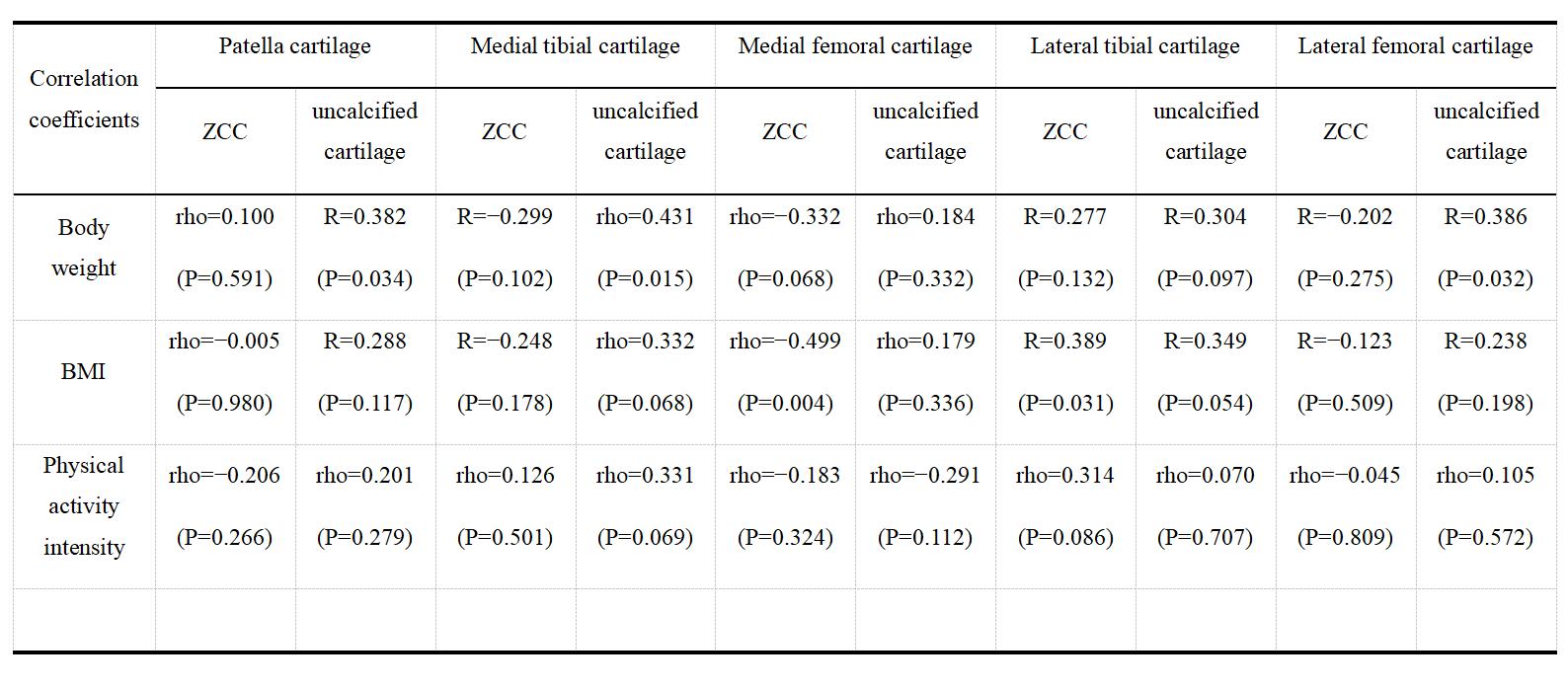
T2* values of UC in patella (R=0.382, P=0.034), medial tibia (rho=0.431, P=0.015) and lateral femur (R=0.386, P=0.032) positively moderately correlated with higher body weight, while T2* values of CC in 5 regions showed no significant correlation with body weight. For BMI, T2* values of UC in 5 regions showed no significant correlation. While T2* values of CC showed negatively moderate correlation with higher BMI in medial femur (rho=−0.499, P=0.004), and positively moderate correlation in lateral tibia (R=0.389, P=0.031). T2* values of CC and UC in all 5 regions showed no significant correlation with physical activity level (Table 1).Discussion
Using UTE sequences to measure CC and UC, our study showed that the effect of body weight and BMI was different for CC and UC, indicating different bio-mechanical properties in CC and UC. It may be due to different populations of chondrocytes in CC and UC and their different mechanosensitive signaling pathways [5]. CC contains quiescent chondrocytes, while UC contains chondrocytes which maintain an equilibrium between anabolic and catabolic metabolism [6, 7]. In contrast to previous MRI quantitative study demonstrating that physical activity is associated with cartilage microstructure and composition [8-11], we found physical activity seems to have limited effect on T2* value of CC and UC, which may need further investigation.This study has some limitations. First, it is a single-centered prospective study with small sample size and narrow age range, which may limit the statistical conclusions one can draw from this work. Second, the border of fluid and UC was sometimes difficult to identify on the UTE subtraction images which may impact the T2* values of the interested regions. Third, as the CC layer is very thin, ROIs may be delineated beyond the exact border of CC sometimes, which could cause measurement error in results.
Conclusion
Accessed by quantitative UTE, CC and UC show different mechanical effects to the body weight and BMI, potentially due to their different bio-mechanical properties.Acknowledgements
References
1. Pearle AD, Warren RF, Rodeo SA. Basic science of articular cartilage and osteoarthritis. Clin Sports Med. 2005 Jan;24(1):1-12.
2. Hoemann CD, Lafantaisie-Favreau CH, Lascau-Coman V, et al. The cartilage-bone interface. J Knee Surg. 2012 May;25(2):85-97.
3. Chang EY, Du J, Chung CB. UTE imaging in the musculoskeletal system. J Magn Reson Imaging. 2015 Apr;41(4):870-83.
4. Alodaibi FA, Fritz JM, Thackeray A, Koppenhaver SL, Hebert JJ. The Fear Avoidance Model predicts short-term pain and disability following lumbar disc surgery. PLoS One. 2018 Mar 5;13(3):e0193566.
5. Ji Q, Zheng Y, Zhang G, Hu Y, Fan X, Hou Y, Wen L, Li L, Xu Y, Wang Y, Tang F. Single-cell RNA-seq analysis reveals the progression of human osteoarthritis. Ann Rheum Dis. 2019 Jan;78(1):100-110.
6. Hoemann CD, Lafantaisie-Favreau CH, Lascau-Coman V, et al. The cartilage-bone interface. J Knee Surg. 2012 May;25(2):85-97.
7. 6.Goldring SR, Goldring MB. Changes in the osteochondral unit during osteoarthritis: structure, function and cartilage-bone crosstalk. Nat Rev Rheumatol. 2016 Nov;12(11):632-644.
8. Kumar D, Souza RB, Singh J, Calixto NE, Nardo L, Link TM, Li X, Majumdar S. Physical activity and spatial differences in medial knee T1rho and t2 relaxation times in knee osteoarthritis. J Orthop Sports Phys Ther. 2014 Dec;44(12):964-72.
9. Hovis KK, Stehling C, Souza RB, Haughom BD, Baum T, Nevitt M, McCulloch C, Lynch JA, Link TM. Physical activity is associated with magnetic resonance imaging-based knee cartilage T2 measurements in asymptomatic subjects with and those without osteoarthritis risk factors. Arthritis Rheum. 2011 Aug;63(8):2248-56.
10. Halilaj E, Hastie TJ, Gold GE, Delp SL. Physical activity is associated with changes in knee cartilage microstructure. Osteoarthritis Cartilage. 2018 Jun;26(6):770-774.
11. Wellsandt E, Emory J, Golightly YM, Dudley AT, Michaud K, Tao MA, Manzer MN, Sajja BR. Individual and cumulative measures of knee joint load associate with T2 relaxation times of knee cartilage in young, uninjured individuals: A pilot study. Knee. 2021 Oct;32:19-29.
Figures
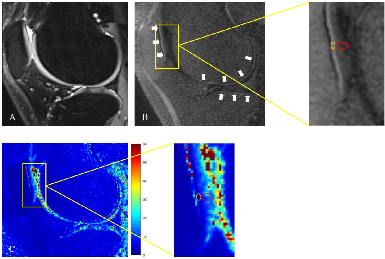
Figure1. PD-FS image (A), UTE subtraction image (B, TE1=0.1 ms, TE2=2.8 ms) and UTE-T2*maps (C) of a 23 year-old male. The linear high signal intensity (white arrows) on B indicates CC. Using A as a reference to ensure the boundary of cartilage, ROIs were delineated on CC (orange) and UC layers (red) of patella cartilage on B and copied to C to measure the T2* values (3.51 ms for CC and 14.80 ms for UC).