4360
Deep learning Based Automatic Segmentation of Brain Magnetic Resonance angiography Images1Radiology, The First Hospital of Jilin University, Changchun, China, 2Shukun (Beijing) Technology Co., Ltd, Beijing, China
Synopsis
Keywords: Machine Learning/Artificial Intelligence, Vessels
Accurate cerebrovascular segmentation plays an important role in clinical diagnosis and the related research. Magnetic Resonance angiography (MRA) postprocessing manually recognized by technologists is extremely labor intensive and error prone. However, automatic segmentation of brain vessels remains challenging because of the variable vessel shape and high complex of vessel geometry. We propose an artificial intelligence brain vessel segmentation system supported by an optimized physiological anatomical-based 3D convolutional neural network that can automatically achieve brain MRA vessel segmentaion in healthcare services. The overall segmentation accuracy of the independent testing dataset is 0.931.Introduction
MRA is a widespread, non-invasive, and cost-efficient imaging modality that is employed in routine clinical diagnoses of head and neck vessels, especially in cerebrovascular disease, which represents one of the leading causes of severe disability and mortality worldwide1. To visually analyze the vasculature of the head and neck more efficiently, vessel segmentation is usually performed by experienced computed tomography technologists. However, as the number of requests for MRA examinations have increased, the postprocessing staff cohort has become overwhelmed due to the time-comsuming manual process2. Considering that vessel imaging segmentation is required in clinical settings, an automatic segmentation system can be easily integrated into the clinical workflow if processed segmentations are available.Methods
Deep learning-based segmentation approaches have drawn increasing interest due to their self-learning and generalization abilites from large data columes3. A major technological challenge of this study is accurate vessel segmentation without interruption of the blood vessels which is difficult because the vessels are tortuous and branched. In this paper ,we uesd a method to segment vessel which has been used in CTA image4. As is shown in Fig.1, the 3D-CNN model was trained using a modified U-net with the addition of bottleneck-ResNet(BR) which automatically achieved optimized model parameter selection. The core design of the segmentation system was the deep learning model, Unet has been widely used to segment various medical images since it was proposed5. Generally, Unet consists of a constriction path and a symmetrical expansion path, and skip connections are used between the paths for feature fusion. The constriction path contains successive down-sampling layers to capture context, while the symmetrical expansion path contains a series of up-sampling layers to recover localization. In our modified Unet, inception modules and dilated convolutions are inserted into adjacent down-sampling layers in the constriction path.Result
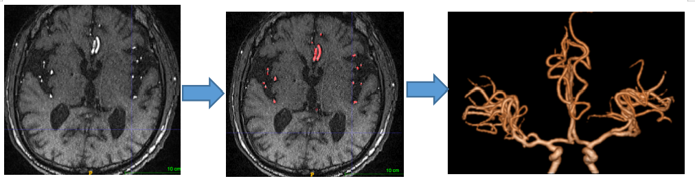
This system is trained and tested with 365 head 1.5T and 3T time-of-flight (TOF) MRA datas collected between 2021.10.1 and 2022.1.16. One case of brain vessel segmentation result is shown in Fig.2. The vessel segmentation attained dice similarity coefficient(DSC) of 0.975 and 0.944, weighted vessel score(V-scores) of -0.975 and -0.929, and recall of 0.961 and 0.933 for the training and validation sets, respectively.Discussion
In this study, we validated a deep learning algorithm through physiological anatomical-based 3D CNN for MRA head image postprocessing. The model was developed with an optimized network by the distribution of BRs, and we proposed the CGPM to revise vascular segmentation errors and avoid partially missing vasculature. our results indicated that a 3D-CNN deep learning algorithm can be trained to complete vessel segmentation automatically with high sensitivity and specificity in a wide variety of enhanced MRA scnas.Conclusion
In conclusion, we found a time-saving and subjectivity-independent method compared to currently available techniques to segment brain vessel of MRA images, saving costs, and increasing efficiency. It is clinically applicable due to its consistency with manually processed images. Thus, the system facilitaes clinical workflows and provides an opportunity for clinical technologists to improve humanistic patient care.Acknowledgements
No acknowledgement found.References
[1] Xu, G, Ma, M, Liu, X, et al. Is there a stroke belt in China and why? STROKE. 2013; 44 (7): 1775-83.
[2] Hilkewich, MW. Written Observations as a Part of Computed Tomography Angiography Post Processing by Medical Radiation Technologists: A Pilot Project. J MED IMAGING RADIAT. 2014; 45 (1): 31-36.e1.
[3] McBee, MP, Awan, OA, Colucci, AT, et al. Deep Learning in Radiology. ACAD RADIOL. 2018; 25 (11): 1472-1480.
[4] Fu, F, Wei, J, Zhang, M, et al. Rapid vessel segmentation and reconstruction of head and neck angiograms using 3D convolutional neural network. Nat Commun. 2020; 11 (1): 4829.
[5] Ronneberger, O., Fischer, P. & Brox, T. U-Net: convolutional networks for biomedical image segmentation. MICCAI. 2015; 9315: 34–241.
Figures