4321
Evolution of Microstructural Changes in a Mouse Model of Mild Traumatic Brain Injury1Robarts Research Institute, London, ON, Canada, 2Medical College of Wisconsin, Milwaukee, WI, United States
Synopsis
Keywords: Diffusion/other diffusion imaging techniques, Microstructure, Brain Injury
Single mild concussive impacts remain sparsely explored by in vivo neuroimaging techniques. Multimodal microstructural MRI has shown increased sensitivity and specificity to microstructural changes in various disease and injury models. In this work, we apply oscillating gradient spin-echo (OGSE) diffusion MRI, microscopic anisotropy (µA) diffusion MRI, and magnetization transfer (MT) MRI longitudinally to increase sensitivity to smaller spatial scales, disentangle fiber orientation dispersion from true microstructural changes, and acquire myelin sensitivity, respectively. We demonstrate that multimodal microstructural MRI provides sensitivity to evolving changes following a mild impact in both acute and chronic regimes, with OGSE demonstrating higher sensitivity than µA.Introduction
Multimodal microstructural MRI has shown increased sensitivity and specificity to microstructural changes in various disease and injury models. Oscillating gradient spin echo (OGSE) diffusion MRI (dMRI)1 and microscopic fractional anisotropy (µFA) dMRI2 may provide additional insight by increasing sensitivity to smaller spatial scales and disentangling fiber orientation dispersion from true microstructural changes, respectively, compared to conventional diffusion tensor imaging. Here, we evaluate mean diffusivity difference between OGSE frequencies (ΔMD), microscopic fractional anisotropy (µFA), traditional dMRI metrics, and magnetization transfer ratio (MTR) longitudinally in vivo in sham and injured mice, following a single mild traumatic brain injury (mTBI).Methods
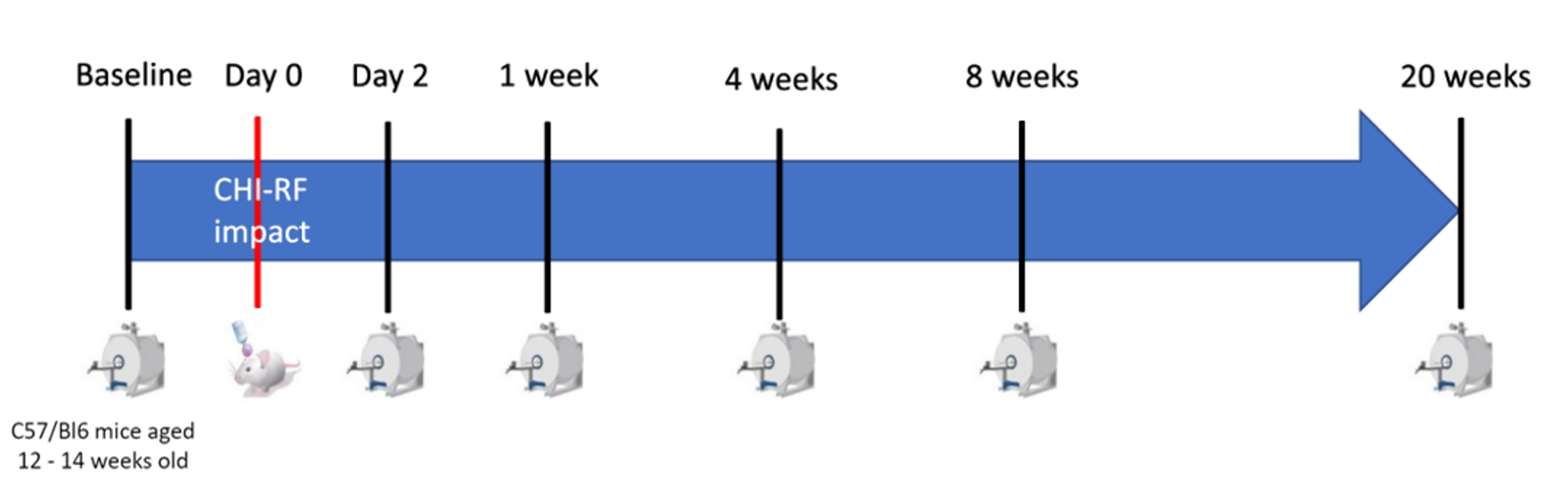
AnimalsThe sham and concussed cohort each consisted of six female and six male C57Bl/6 mice, aged 10-12 weeks at the start of the study. Longitudinal imaging was performed on the sham and concussed cohorts at the timepoints shown in Fig. 1.
Imaging
Imaging was performed at 9.4T with a 1 T/m gradient insert using single-shot EPI with an in-plane resolution of 0.175mm x 0.2mm, 0.5mm slice thickness, and a total scan time of 2 hours. The OGSE sequence was implemented with b=800s/mm, TE=37ms, 10 directions and OGSE frequencies of 0, 50, 100, 145, and 190 Hz. The µA sequence was implemented using a single diffusion encoding (SDE) scheme with linear and spherical tensor encodings at b=2000s/mm (30 directions) and b=1000s/mm (12 directions)3. The MT protocol comprised two FLASH-3D scans, with MT-weighting achieved by applying an off- resonance Gaussian-shaped RF pulse (12-ms duration, 385° nominal flip angle, 3.5 kHz frequency offset, 5 μT RF peak amplitude) prior to the excitation. Post processing included PCA denoising4 and eddy current correction with FSL5. The protocols have been described in detail in earlier work6,7.
Statistical Analysis
Parameters were measured in the corpus callosum (CC), prefrontal cortex (PFC), and fornix. The mean values in each ROI were averaged across all subjects in each cohort, and the change in the averages between each timepoint and the baseline was calculated. A repeated measures ANOVA was used for each metric to determine if there were statistically significant interaction effects (p < 0.05) between sham and concussed mice over time, and post-hoc analysis (with Tukey-Kramer multiple comparison correction) was used to determine if the groups differed within each timepoint post-mTBI.
Results
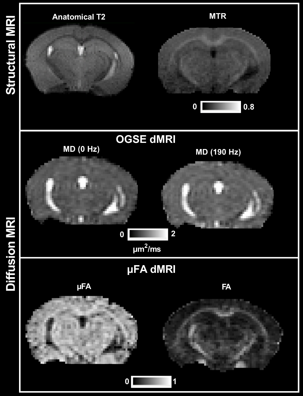
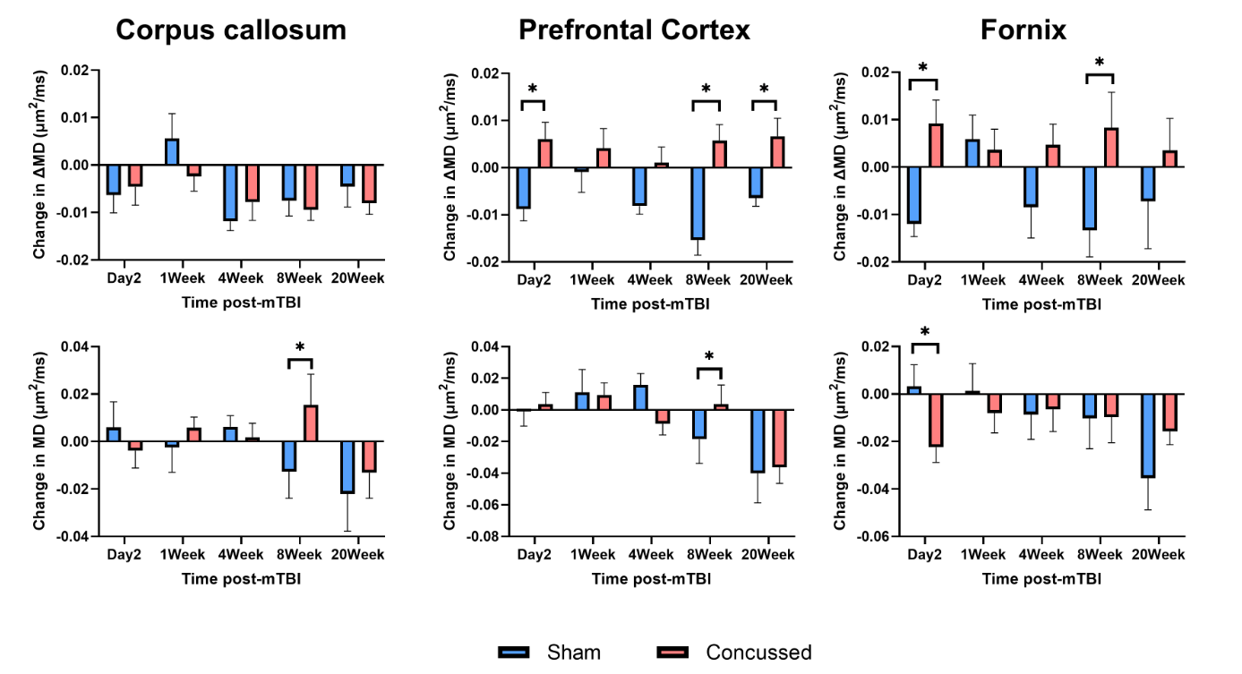
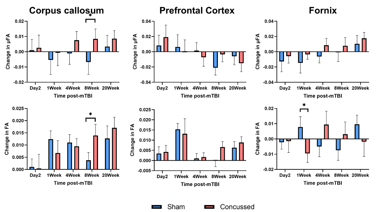
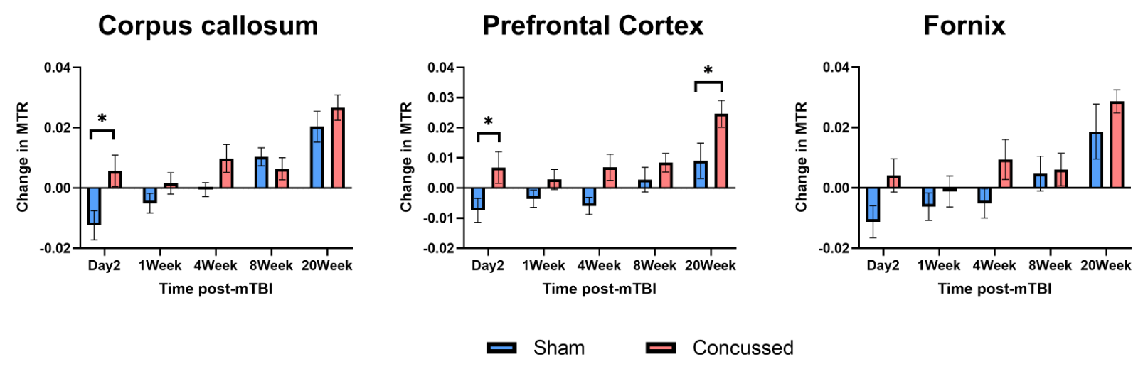
Notably, the single mild impact resulted in no observable symptoms, seizures, skull fractures, hemorrhages or qualitative MRI differences. Representative parameter maps at baseline are shown in Fig. 2. Figures 3, 4, and 5 show the changes in ΔMD (difference in MD(190Hz) and MD(0Hz)), µFA, and MTR, respectively, between each timepoint post-mTBI and the baseline for each cohort. In Figure 3 and 4, the changes in MD and FA between each timepoint post-mTBI and the baseline are also shown, for reference.Discussion
The net increases in ΔMD in the concussed cohort observed in both acute and chronic timepoints in the PFC and fornix are consistent with neurite beading8,9. In the concussed cohort, in both PFC and fornix, there is a trend of acute net increased ΔMD, pseudo-normalization of ΔMD, and chronic net increased ΔMD. This is consistent with the multi-phase concussion recovery10, in which we hypothesize neurite beading is contributing to ΔMD increases in the acute stage, and other mechanisms, such as tissue remodeling11, may be contributing to ΔMD increases in the chronic stage.The significant changes in ΔMD observed in the fornix, but not CC, may indicate that the fornix is more susceptible to microstructural changes caused by rotational acceleration, due to the superior-inferior orientation of fibers. Interestingly, rotational acceleration and shear strain were recently reported to significantly impact MD in only the fornix (out of 49 brain regions investigated)12. This is also supported by the acute net decrease in MD and FA in only the fornix, which is consistent with other DTI mTBI studies13,14.
µFA was not sensitive to changes following this mTBI model, which suggests little axon degeneration. The more subtle changes in MD and FA reported here, compared to mild multi-hit models15,16, reinforces the notion that multiple hits are more damaging.
The trend of a net increase in MTR in both cohorts, which is compatible with the net increases in FA and net decreases in MD, may be related to ongoing mouse brain maturation, as FA variations with age in the mouse brain have been related to ongoing tissue organization processes17, and this motivates further investigation of the sham cohort. The trend of a greater net increase in MTR at the final time interval (significant only in PFC), in concussed compared to sham, may be an indication of excessive myelin in the chronic stage due to remyelination or aberrant myelin synthesis18. The consistent net decrease in MTR in the sham cohort at acute timepoints may indicate anesthetic effects on microstructure19,20, as all mice were anesthetized for seven hours in the span of four days from baseline to the 2Day timepoint.
Conclusion
Overall, our data shows that ΔMD provides greater sensitivity than MD alone, and that µFA is not as sensitive to changes post-mTBI. In conclusion, we demonstrate that multimodal microstructural MRI provides sensitivity to evolving neurobiological changes following mTBI at both acute and chronic stages.Acknowledgements
No acknowledgement found.References
1. Baron, C. A. & Beaulieu, C. Oscillating gradient spin-echo (OGSE) diffusion tensor imaging of the human brain. Magn. Reson. Med. 72, 726–736 (2014).
2. Lasič, S., Szczepankiewicz, F., Eriksson, S., Nilsson, M. & Topgaard, D. Microanisotropy imaging: Quantification of microscopic diffusion anisotropy and orientational order parameter by diffusion MRI with magic-angle spinning of the q-vector. Front. Phys. 2, 1–14 (2014).
3. Arezza, N. J. J., Tse, D. H. Y. & Baron, C. A. Rapid microscopic fractional anisotropy imaging via an optimized linear regression formulation. Magn. Reson. Imaging 80, 132–143 (2021).
4. Veraart, J., Novikov, D. S., Christiaens, D., Ades-aron, B., Sijbers, J. & Fieremans, E. Denoising of diffusion MRI using random matrix theory. Neuroimage 142, 394–406 (2016).
5. Andersson, J. L. R. & Sotiropoulos, S. N. An integrated approach to correction for off-resonance effects and subject movement in diffusion MR imaging. Neuroimage 125, 1063–1078 (2016).
6. Rahman, N., Xu, K., Omer, M., Budde, M. D., Brown, A. & Baron, C. A. Test-retest reproducibility of in vivo oscillating gradient and microscopic anisotropy diffusion MRI in mice at 9.4 Tesla. PLoS One 16, e0255711 (2021).
7. Rahman, N., Ramnarine, J., Xu, K., Brown, A. & Baron, C. Test-retest reproducibility of in vivo magnetization transfer ratio and saturation index in mice at 9.4 Tesla. J Magn Reson Imaging 1–11 (2022) doi:10.1002/jmri.28106.
8. Budde, M. D. & Frank, J. A. Neurite beading is sufficient to decrease the apparent diffusion coefficient after ischemic stroke. Proc. Natl. Acad. Sci. U. S. A. 107, 14472–14477 (2010).
9. Baron, C. A., Kate, M., Gioia, L., et al. Reduction of Diffusion-Weighted Imaging Contrast of Acute Ischemic Stroke at Short Diffusion Times. Stroke. 46, 2136–2141 (2015).
10. To, X. V. & Nasrallah, F. A. A roadmap of brain recovery in a mouse model of concussion: insights from neuroimaging. Acta Neuropathol. Commun. 9, 1–20 (2021).
11. Romeu-Mejia, R., Giza, C. C. & Goldman, J. T. CONCUSSION Concussion Pathophysiology and Injury Biomechanics. Curr. Rev. Musculoskelet. Med. 105–116 (2019).
12. Post, A., Hashim, E., Hoshizaki, T. B., Gilchrist, M. D. & Cusimano, M. D. A preliminary examination of the relationship between biomechanical measures and structural changes in the brain. Trauma (United Kingdom) 23, 24–32 (2021).
13. Mac Donald, C. L., Dikranian, K., Bayly, P., Holtzman, D. & Brody, D. Diffusion tensor imaging reliably detects experimental traumatic axonal injury and indicates approximate time of injury. J. Neurosci. 27, 11869–11876 (2007).
14. Tu, T. W., Williams, R. A., Lescher, J. D., Jikaria, N., Turtzo, L. C. & Frank, J. A. Radiological-pathological correlation of diffusion tensor and magnetization transfer imaging in a closed head traumatic brain injury model. Ann. Neurol. 79, 907–920 (2016).
15. Wortman, R. C., Meconi, A., Neale, K. J., et al. Diffusion MRI abnormalities in adolescent rats given repeated mild traumatic brain injury. Ann. Clin. Transl. Neurol. 5, 1588–1598 (2018).
16. Haber, M., Hutchinson, E. B., Sadeghi, N., et al. Defining an analytic framework to evaluate quantitative MRI markers of traumatic axonal injury: Preliminary results in a mouse closed head injury model. eNeuro 4, 1–15 (2017).
17. Hammelrath, L., Škokić, S., Khmelinskii, A., et al. Morphological maturation of the mouse brain: An in vivo MRI and histology investigation. Neuroimage 125, 144–152 (2016).
18. Mierzwa, A. J., Marion, C. M., Sullivan, G. M., McDaniel, D. P. & Armstrong, R. C. Components of myelin damage and repair in the progression of white matter pathology after mild traumatic brain injury. J. Neuropathol. Exp. Neurol. 74, 218–232 (2015).
19. Li, Q., Mathena, R. P., Xu, J., Eregha, O. N., Wen, J. & Mintz, C. D. Early postnatal exposure to isoflurane disrupts oligodendrocyte development and myelin formation in the mouse hippocampus. Anesthesiology 131, 1077–1091 (2019).
20. Zhang, L., Xue, Z., Liu, Q., et al. Disrupted folate metabolism with anesthesia leads to myelination deficits mediated by epigenetic regulation of ERMN. EBioMedicine 43, 473–486 (2019).
Figures