4290
Second-order motion compensated single-shot spin echo planar imaging sequence using Compressed SENSE: a pilot study1Radiology, Guangdong Provincial People’s Hospital, Guangzhou, China, 2Philips Healthcare China, Shenzhen, China
Synopsis
Keywords: Heart, Diffusion/other diffusion imaging techniques
Cardiac diffusion weighted imaging (DWI) is a novel tool that could provide non-invasive in-vivo microstructural assessment. However, cardiac DWI remains a challenging task due to low signal-to-noise-ratio (SNR) and motion artifacts. The second-order motion compensated (M2C) could be used to decrease the motion artifacts, and Compressed SENSE could improve the SNR of the echo planar imaging (EPI) based sequences. Hence, the combination of M2C DWI and Compressed SENSE was firstly proposed in the present study. We found that M2C DWI using Compressed SENSE showed better overall image quality and higher SNR.Summary of Main Findings
The combination of the second-order motion compensated DWI and Compressed SENSE was firstly proposed in the present study. The method we proposed showed better overall image quality and higher SNR with the similar scan time than the M2C DWI.Purpose
The goal of this work is to provide a new solution which combines M2C DWI and Compressed SENSE to improve the image quality of cardiac DWI. A pilot study of CS M2C DWI was explored.Introduction
Cardiac DWI is a novel tool that could provide noninvasive in-vivo microstructural assessment [1]. It could offer the information of the cardiomyocyte organization, myocardial fibrosis, intramyocardial iron, edema, etc [1-4]. However, the cardiac DWI is mainly limited by the relatively low SNR [5]. M2C is considered optimal compared with a stimulated echo acquisition (STEAM) based technique [5]. However, the echo time (TE) is increased when using M2C DWI [6], and it will decrease the SNR dramatically. Recently, it has been reported that the Compressed SENSE framework applied to EPI sequence could reduce noise-like artifacts and improve the overall image quality of EPI based DWI [7]. In the clinical application of the abdomen, using Compressed SENSE could improve the image quality of DWI [8,9]. Hence, it is reasonable to assume that Compressed SENSE probably improves the image quality of M2C DWI. To our knowledge, there is no studies that explored the combination of cardiac DWI and Compressed SENSE, especially for M2C DWI.Methods
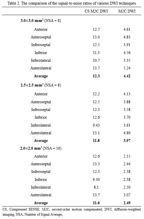
Figure 1 illustrated the sequence diagram for the CS M2C DWI in the present study. The CS M2C DWI and M2C DWI were implemented on a 3.0 Tesla Philips Ingenia CX system (Philips Healthcare, Best, The Netherlands). The signal was received with a 32-channel torso and spine coil. Written informed consent was obtained from healthy volunteers before scanning.The DWI data was acquired with healthy volunteers without history of cardiac disease. The DWI sequences were ECG-triggered to mid-systole systole (65% of peak systole), and the mid-ventricular in short-axis view was acquired. The DWI acquisition was performed with a b-value of 0 and 450 s/mm2 during free breathing with respiratory navigator-based slice tracking. The CS M2C DWI and M2C DWI were acquired at the in-plane resolution of 3.0×3.0, 2.5×2.5, 2.0×2.0 mm2, respectively. The other imaging parameters were listed as Table 1. The SNR of the cardiac DWI was determined for the b-values of 450 s/mm2. Figure 2 illustrated how to measure SNR in the present study, and the SNR of DWI image was defined as the Eq. (1).
SNR = SImyo / SIair (1)
where the SImyo was defined as the mean intensity in the myocardium of left ventricular, while SIair defined as the mean intensity of the air within the lung. The image qualities of the CS M2C DWI and M2C DWI were compared in the present study.
Results
Figure 3 showed the DWI images with a b-value of 450 s/mm2 using various in-plane resolution. It turned out that the overall image quality of CS M2C DWI was better than those of M2C DWI with in-plane resolution of 3.0×3.0 mm2, 2.5×2.5 mm2, 2.0×2.0 mm2, respectively. Table 2 illustrated that the average SNR of CS M2C DWI with the in-plane resolution of 3.0×3.0 mm2, 2.5×2.5 mm2, 2.0×2.0 mm2 were 12.3, 11.8, 11.6, respectively. The SNRs of CS M2C DWI in every segment of the mid-ventricular with different in-plane resolution were higher than that of M2C DWI.Discussion
In the present study, the new method we proposed was to combine the M2C DWI and Compressed SENSE. Compared with the M2C DWI, CS M2C DWI showed better overall image quality and higher SNR. These results were similar with Kaga et al and Tamada et al [8,9], and the Compressed SENSE seemed to increase the image quality of M2C DWI. Therefore, the proposed method may be a useful solution to address the relatively lower SNR of M2C DWI and STEAM DWI.Recently, similar studies were concentrated to improve the SNR of cardiac DWI [6,10-12]. The reduced field-of-view (FOV) technique improve the image quality of cardiac DWI [10-12], but it might be unsuitable for the patients with significantly enlarged cardiac size. In addition, the combination of M2C DWI and spiral sequences has been proposed to reduce the TE and increase the SNR [6]. However, the repetitive breath-holding was needed, and the patients hard to hold their breath might not be benefit from it [6].
Conclusion
The combination of the M2C DWI and Compressed SENSE was proposed in the present study. The results showed that CS M2C DWI has better overall image quality and higher SNR with the similar scan time.Acknowledgements
No acknowledgement.References
1. Khalique Z, Ferreira PF, Scott AD, Nielles-Vallespin S, Firmin DN, Pennell DJ (2020) Diffusion Tensor Cardiovascular Magnetic Resonance Imaging: A Clinical Perspective. J Am Coll Cardiol Img 13 (5):1235-1255.
2. Moulin K, Viallon M, Romero W, Chazot A, Mewton N, Isaaz K, Croisille P (2020) MRI of Reperfused Acute Myocardial Infarction Edema: ADC Quantification versus T1 and T2 Mapping. Radiology 295 (3):542-549.
3. Das A, Kelly C, Teh I, Sharrack N, Stoeck CT, Kozerke S, Schneider JE, Plein S, Dall'Armellina E (2022) Detection of Intramyocardial Iron in Patients Following ST-Elevation Myocardial Infarction Using Cardiac Diffusion Tensor Imaging. Journal of magnetic resonance imaging : JMRI doi:10.1002/jmri.28063.
4. Nguyen C, Lu M, Fan Z, Bi X, Kellman P, Zhao S, Li D (2015) Contrast-free detection of myocardial fibrosis in hypertrophic cardiomyopathy patients with diffusion-weighted cardiovascular magnetic resonance. Journal of cardiovascular magnetic resonance : official journal of the Society for Cardiovascular Magnetic Resonance 17 (1):107.
5. von Deuster C, Stoeck CT, Genet M, Atkinson D, Kozerke S (2016) Spin echo versus stimulated echo diffusion tensor imaging of the in vivo human heart. Magnetic resonance in medicine 76 (3):862-872.
6. van Gorkum RJH, Guenthner C, Koethe A, Stoeck CT, Kozerke S (2022) Characterization and correction of diffusion gradient-induced eddy currents in second-order motion-compensated echo-planar and spiral cardiac DTI. Magnetic resonance in medicine 88 (6):2378-2394.
7. Yoneyama M, Morita K, Peeters J, Nakaura T, Van Cauteren M Noise reduction in prostate single-shot DW-EPI utilizing compressed SENSE framework. In: Proceedings of the 27th annual meeting of ISMRM, Montreal, 2019.
8. Kaga T, Noda Y, Mori T, Kawai N, Takano H, Kajita K, Yoneyama M, Akamine Y, Kato H, Hyodo F, Matsuo M (2021) Diffusion-weighted imaging of the abdomen using echo planar imaging with compressed SENSE: Feasibility, image quality, and ADC value evaluation. Eur J Radiol 142:109889.
9. Tamada T, Ueda Y, Kido A, Yoneyama M, Takeuchi M, Sanai H, Ono K, Yamamoto A, Sone T (2022) Clinical application of single-shot echo-planar diffusion-weighted imaging with compressed SENSE in prostate MRI at 3T: preliminary experience. Magma doi:10.1007/s10334-022-01010-w:1-8.
10. Gotschy A, von Deuster C, van Gorkum RJH, Gastl M, Vintschger E, Schwotzer R, Flammer AJ, Manka R, Stoeck CT, Kozerke S (2019) Characterizing cardiac involvement in amyloidosis using cardiovascular magnetic resonance diffusion tensor imaging. Journal of cardiovascular magnetic resonance : official journal of the Society for Cardiovascular Magnetic Resonance 21 (1):56.
11. Ma S, Nguyen CT, Christodoulou AG, Luthringer D, Kobashigawa J, Lee SE, Chang HJ, Li D (2018) Accelerated Cardiac Diffusion Tensor Imaging Using Joint Low-Rank and Sparsity Constraints. IEEE transactions on bio-medical engineering 65 (10):2219-2230.
12. Wu Z, Zhang Y, Hu X, Zhang J, Zeng F, Xu X, Jiang G, Zhao Y, Gilbert G, Wang J (2022) iZoom with 2 nd order flow compensated diffusion for Improving cardiac diffusion imaging: a preliminary study. Proceedings of the 30th annual meeting of ISMRM, London.
Figures
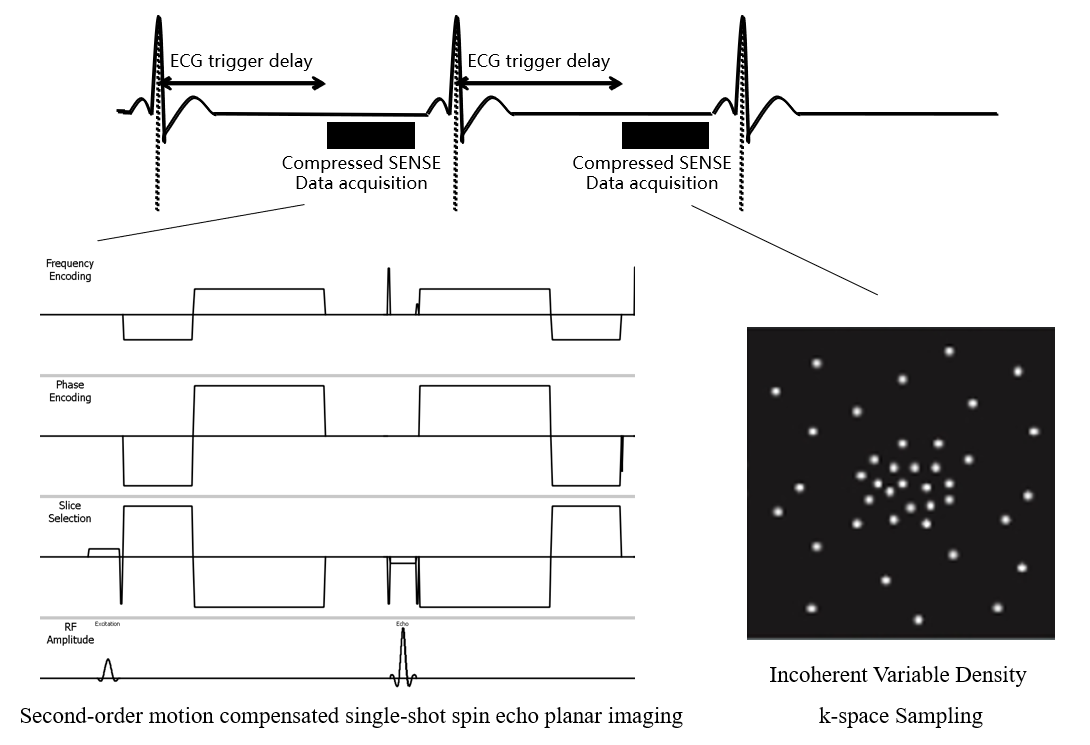

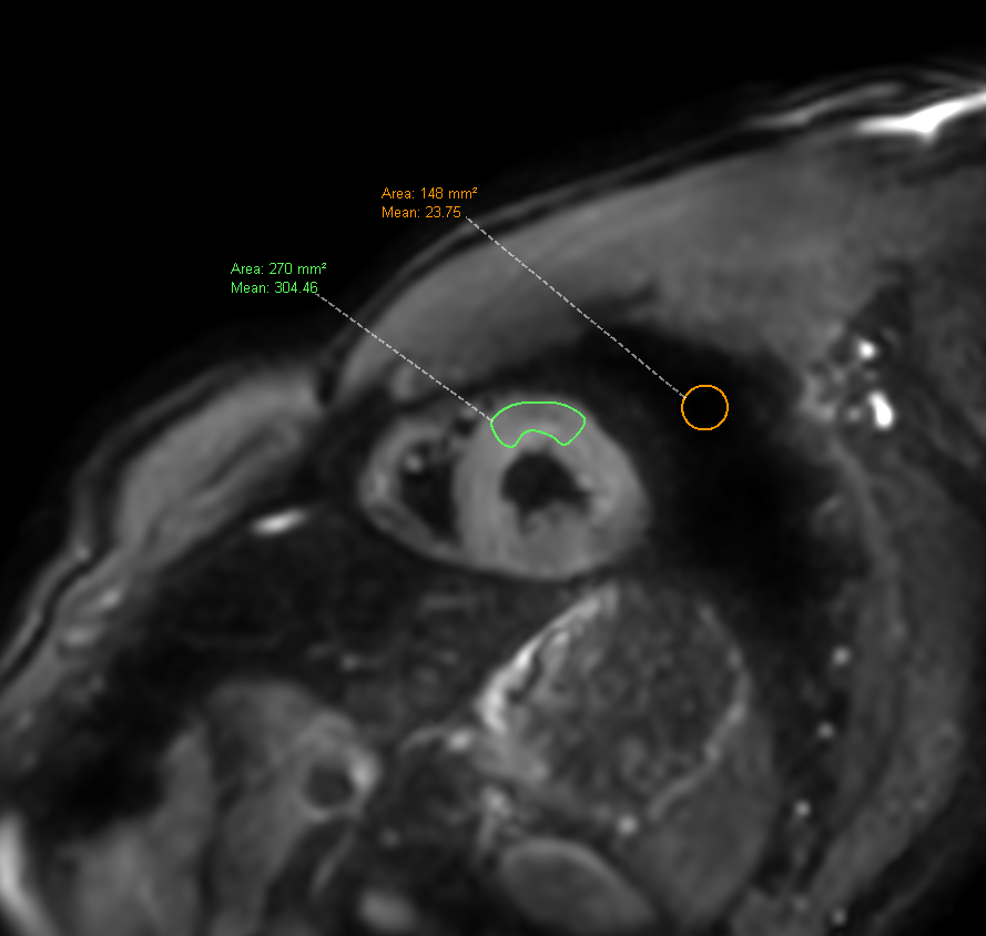
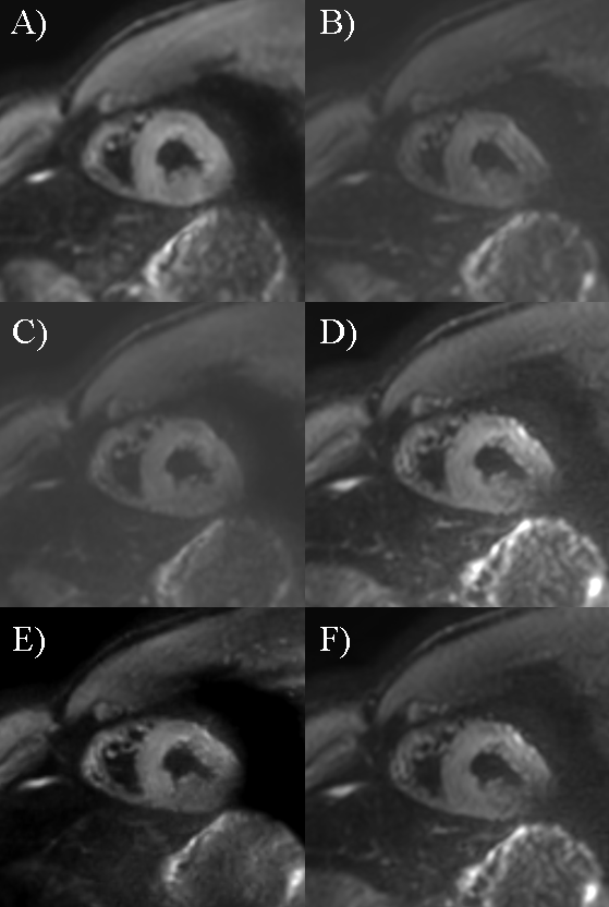
Figure 3. Diffusion weighted images with a b-value of 450 s/mm2 using various in-plane resolution.
(A) the CS M2C DWI with in-plane resolution of 3.0×3.0 mm2; (B) the M2C DWI with in-plane resolution of 3.0×3.0 mm2; (C) the CS M2C DWI with in-plane resolution of 2.5×2.5 mm2; (D) the M2C DWI with in-plane resolution of 2.5×2.5 mm2; (E) the CS M2C DWI with in-plane resolution of 2.0×2.0 mm2; (F) the M2C DWI with in-plane resolution of 2.0×2.0 mm2;
CS, Compressed SENSE; M2C, second-order motion compensated; DWI, diffusion-weighted imaging;