4282
An add-on local external shim array to improve the main magnetic field homogeneity in the prostate
Carlijn Tenbergen1, Angeliki Stamatelatou1, Sahar Nassirpour2, Paul Chang2, and Tom Scheenen1,3
1Department of Medical Imaging, Radboud University Medical Center, Nijmegen, Netherlands, 2MR Shim, Reutlingen, Germany, 3Erwin L. Hahn Institute, Essen, Germany
1Department of Medical Imaging, Radboud University Medical Center, Nijmegen, Netherlands, 2MR Shim, Reutlingen, Germany, 3Erwin L. Hahn Institute, Essen, Germany
Synopsis
Keywords: Prostate, Shims, Diffusion, Spectroscopy
B0-field inhomogeneities can influence (spectroscopic) image quality of the prostate, causing geometric distortion in diffusion weighted EPI images as well as line broadening in MRSI voxels. We added a 16-element external local shim array around the pelvis to improve B0-field homogeneity within the prostate and evaluated its effects compared to standard shimming in 9 healthy volunteers. Resulting B0-maps showed a comparable or reduced frequency variation across subjects, with a significant decrease of MRSI citrate line widths. DWI did not show a robust effect over the group of subjects.Introduction
Homogeneity of the B0-field within the prostate is essential for high quality MR (spectroscopic) imaging, but bowel gas and motion can influence the B0-field and therefore image quality, mostly at the posterior part of the prostate1. With EPI-based DWI and MRSI sensitive to B0 inhomogeneities, off-resonance effects can cause geometric distortions in DWI, and spectral line broadening and frequency shifts in MRSI affecting water suppression and causing lipid contamination of spectra within the prostate1,2. One way to partly prevent possible susceptibility-induced artifacts of this air is by patient preparation using antispasmodic agents and a rectum catheter3. An alternative method as proposed in the brain is to improve the B0-shim quality locally using advanced shimming methods such as a local shim array4. We present initial results of the first add-on local shim array for the prostate and analyse its effect on B0-maps, EPI-based DWI and MRSI.Method
A 16-channel external array of local abdominal shim coils (named “Dia”, MR Shim GmbH, Reutlingen, Germany) was used in a 3T MR-system (Siemens Magnetom Prisma), consisting of 8 shim coils housed in two anterior modules, positioned onto a 18-channel body receive RF coil (Siemens), and 8 shim coils in the posterior modules (Figure 1), positioned on the spine-array bed.Two shimming methods were compared: 1) The scanner’s prostate shimming routine with additional manual refining using the interactive adjustments of the scanner until a magnitude FWHM of around 30Hz of the shim volume around the prostate was achieved. 2) Dia shimming, with the “Dia”-system, in which a reference field map was acquired using a dual-echo gradient-echo sequence, and shim values were calculated non-iteratively using the provided software tool with a constrained convex optimization algorithm.
Nine healthy volunteers (mean age 40y, written informed consent) were scanned without any bowel preparation. After anatomical T2w scans in three orientations (transversal: TR/TE 5660/92ms, FOV 256x240mm, resolution 0.3×0.3×3.0mm), field maps were acquired with both shim methods with a dual gradient echo sequence and compared by the standard deviation of the off-resonance frequencies in the prostate region.
For DWI analysis, EPI images with b-value of 50s/mm2 were acquired with both shim methods (TR/TE 3200/63ms, FOV 256x240mm, resolution 2.0x2.0x3.0mm, 3 averages). The two different b50-maps were registered to the transversal T2w scan as undistorted anatomical reference, and a whole gland prostate outlining was performed per slice for all three series by a single reader. Both b50-maps were compared to the anatomical images by a Dice similarity coefficient (DSC) and Hausdorff distance (HD) measure5,6 and tested for significant differences by Wilcoxon Signed-Ranks tests.
Finally, 3D MRSI data were acquired with a GOIA semi-LASER pulse sequence with MEGA lipid and water suppression7 (TR/TE 750/88ms, FOV 77x56x56mm3, matrix 8x8x11, Hamming-filtered and interpolated to 16x16x16). Citrate (Cit) linewidth maps were generated for multiple slices of all subjects for both shim methods, and a paired samples t-test was performed in 236 selected independent voxels from the full cohort of acquired datasets.
Results
Examples of 2 representative slices of B0-maps show an improved homogeneity of the B0-field for the Dia shim (Figure 2). Overall the off-resonance frequencies of the Dia shim showed a comparable or reduced frequency variation (Table 1).EPI b50-images showed some geometric distortions (Figure 3). The corresponding quantitative measures in Table 1, showed no significant overall effect for DWI with or without the Dia shim. On subject-level, the mean DSC was significantly decreased for the Dia shim in two volunteers, indicating a better standard shim (mean DSC 0.81±0.03 and 0.71±0.07 with Dia shim vs 0.85±0.04 and 0.86±0.03 with standard shim). In one volunteer the Dia shim performed better, as shown by a significantly improved HD with the Dia shim (16.3±5.2mm vs 27.1±5.7mm).
Figure 4 presents the effect of both shimming methods on MRSI, with representative examples showing a quantitative (Cit linewidth maps) and qualitative (spectra) comparison. Analysis of Cit linewidths within the prostate over the full cohort, showed 66% of all standard shim voxels reached linewidths below 15Hz, while this result was met in 82% of the voxels with Dia shim. The mean Cit linewidth over all selected voxels showed a significant improvement (p<0.01) for the Dia shim (12.8±2.5Hz) compared to the scanner shim (14.3±4.1Hz). The mean Cit linewidths per subject are presented in Table 1.
Conclusion and Discussion
This study shows the potential of an add-on local shim array to improve the shim quality for prostate imaging in healthy volunteers. For DWI, it was shown that for some slices, geometric distortion was decreased resulting in a lower DSC and HD, although in other subjects, where the scanner shim performed well without distortions, the Dia shim did not result in any improvements. For MRSI, the Dia shimming approach succeeded in overall significantly narrower Cit linewidths than the conventional scanner shim, leading to an improved spectral quality, possibly improving the accuracy of spectral quantification.In patients with generally larger prostates and bowel-motion-suppressing medication the effect of additional shimming remains to be seen in following work. In this older population metallic implants can be present with severe influence on local field homogeneity that could strongly benefit from additional shimming flexibility.
Acknowledgements
No acknowledgement found.References
1. Shrestha Kakkar L, Usman M, Arridge S, Kirkham A, Atkinson D. Characterization of B0-field fluctuations in prostate MRI. Phys Med Biol. 2020 Nov 5;65(21):21NT01. doi: 10.1088/1361-6560/abbc7f. PMID: 32992306; PMCID: PMC8528180.
2. Stamatelatou A, Scheenen TWJ, Heerschap A. Developments in proton MR spectroscopic imaging of prostate cancer. MAGMA. 2022 Aug;35(4):645-665. doi: 10.1007/s10334-022-01011-9. Epub 2022 Apr 20. PMID: 35445307; PMCID: PMC9363347.
3. Engels RRM, Israël B, Padhani AR, Barentsz JO. Multiparametric Magnetic Resonance Imaging for the Detection of Clinically Significant Prostate Cancer: What Urologists Need to Know. Part 1: Acquisition. Eur Urol. 2020 Apr;77(4):457-468. doi: 10.1016/j.eururo.2019.09.021. Epub 2019 Sep 30. PMID: 31582290.
4. Juchem C, Nixon TW, McIntyre S, Boer VO, Rothman DL, de Graaf RA. Dynamic multi-coil shimming of the human brain at 7 T. J Magn Reson. 2011 Oct;212(2):280-8. doi: 10.1016/j.jmr.2011.07.005. Epub 2011 Jul 23. PMID: 21824794; PMCID: PMC3183127.
5. Dice LR. Measures of the amount of ecologic association between species. Ecology 1945; 26(3): 297–302. doi: 10.2307/1932409.
6. Chen X, Zhang Y, Cao Y, Sun R, Huang P, Xu Y, Wang W, Feng Q, Xiao J, Yi J, Li Y, Dai J. A feasible study on using multiplexed sensitivity-encoding to reduce geometric distortion in diffusion-weighted echo planar imaging. Magn Reson Imaging. 2018 Dec;54:153-159. doi: 10.1016/j.mri.2018.08.022. Epub 2018 Aug 29. PMID: 30171998.
7. Steinseifer IK, van Asten JJ, Weiland E, Scheenen TW, Maas MC, Heerschap A. Improved volume selective (1) H MR spectroscopic imaging of the prostate with gradient offset independent adiabaticity pulses at 3 tesla. Magn Reson Med. 2015 Oct;74(4):915-24. doi: 10.1002/mrm.25476. Epub 2014 Sep 29. PMID: 25264976.
Figures
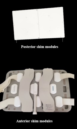
Figure 1: 16-ch local array of shim coils (“Dia”) consisting of two posterior modules (top) which are placed on the spine array coil in the patient bed and two anterior modules (bottom) which are positioned on the RF receive array as shown in the figure.
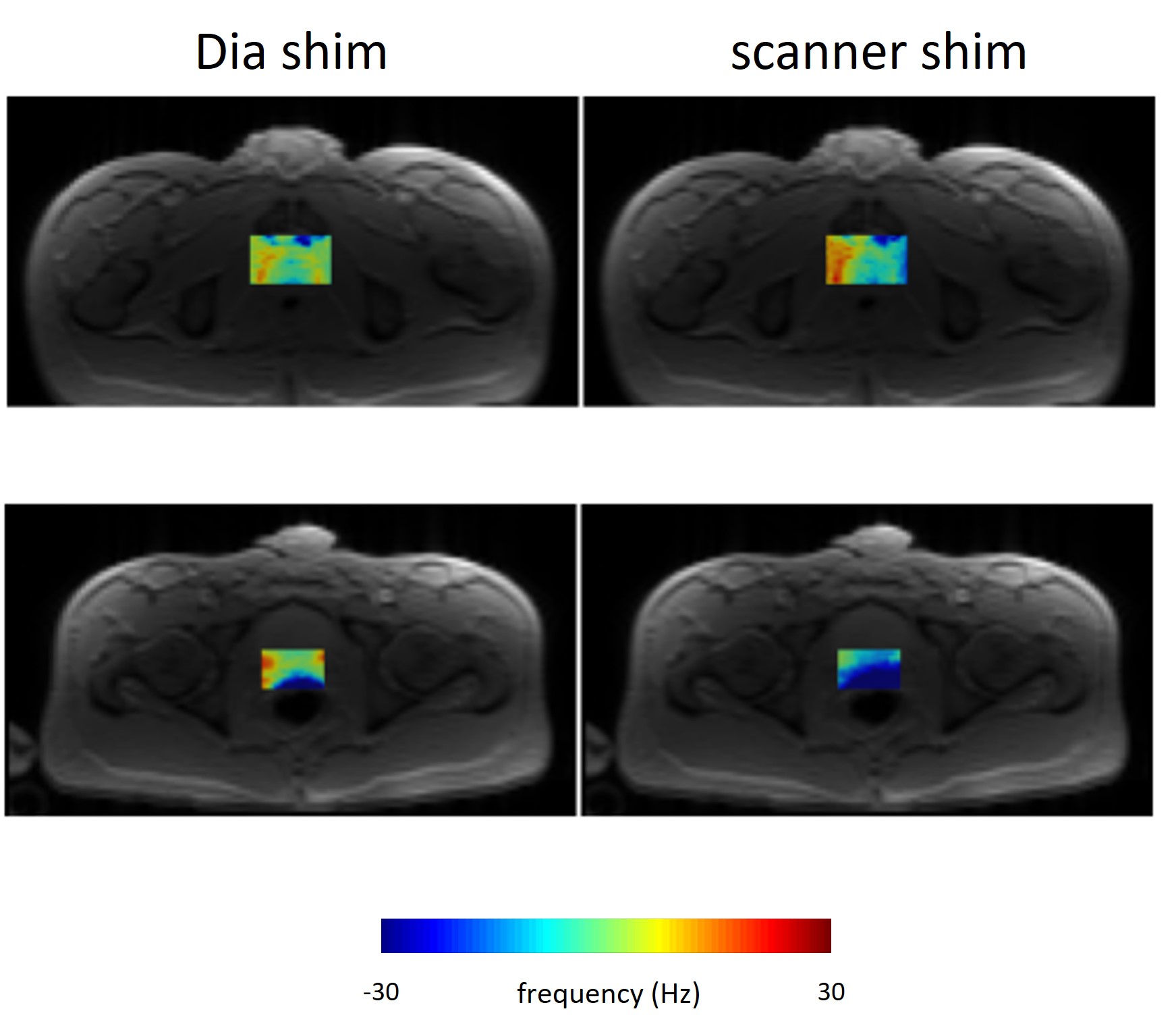
Figure 2: Examples of single slices of B0-maps for two subjects, volunteer 5 (top row) and 8 (bottom row), in which the Dia shim presents a more homogeneous B0-field (left) compared to the scanner shim (right).
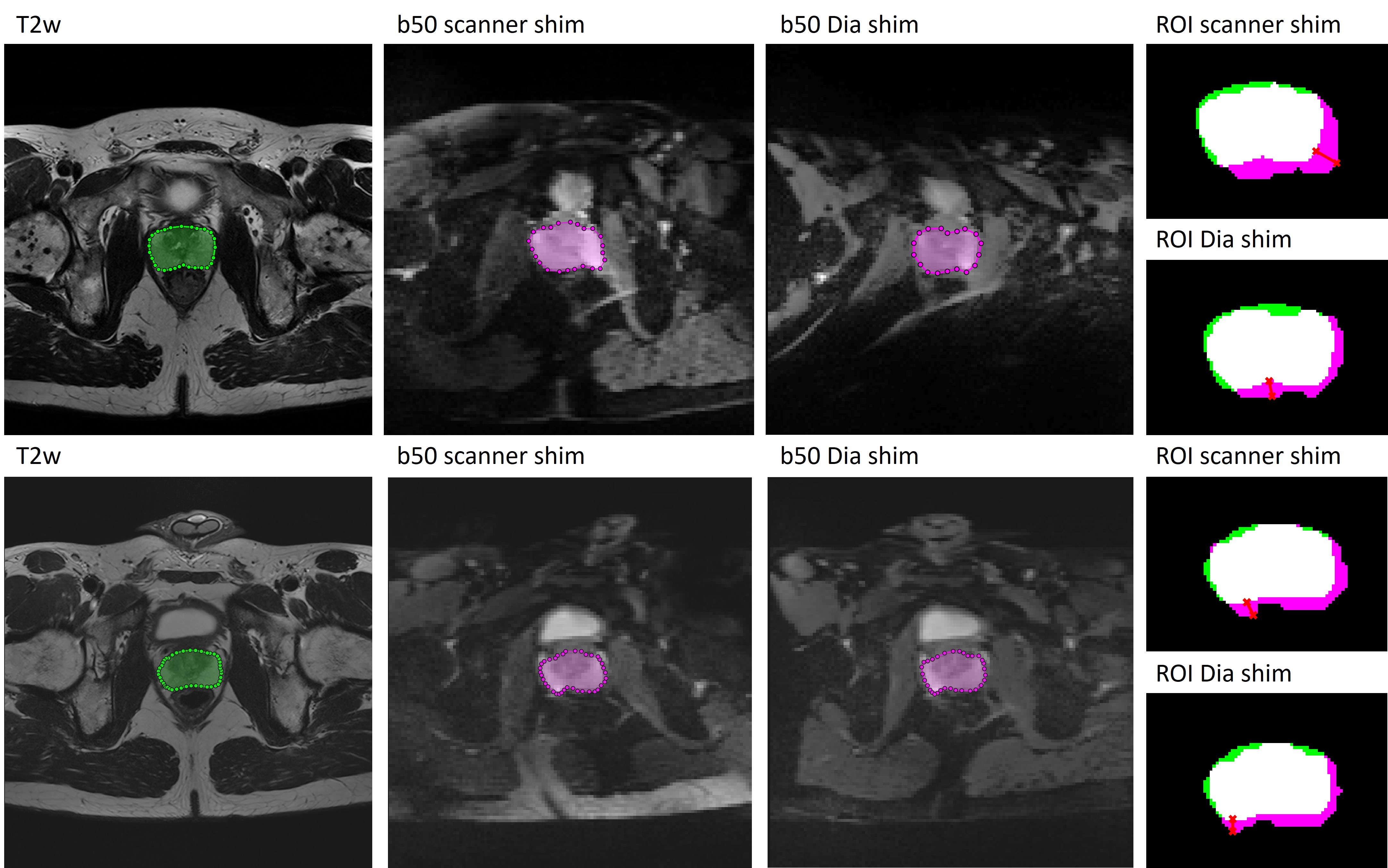
Figure 3: DWI analysis of example slices in 2 subjects, with transversal T2w image as an anatomical reference (left), b50 EPI images for scanner and Dia shim (middle), and resulting overlaid prostate segmentations (right panels, HD with red line). Top row (volunteer 7): minimized distortion of the prostate in Dia shim, with DSC of 0.88 for Dia shim and 0.84 for scanner shim. Bottom row (volunteer 8): a typical example with visible differences in prostate distortion, though resulting in a similar DSC and HD for both shim methods.
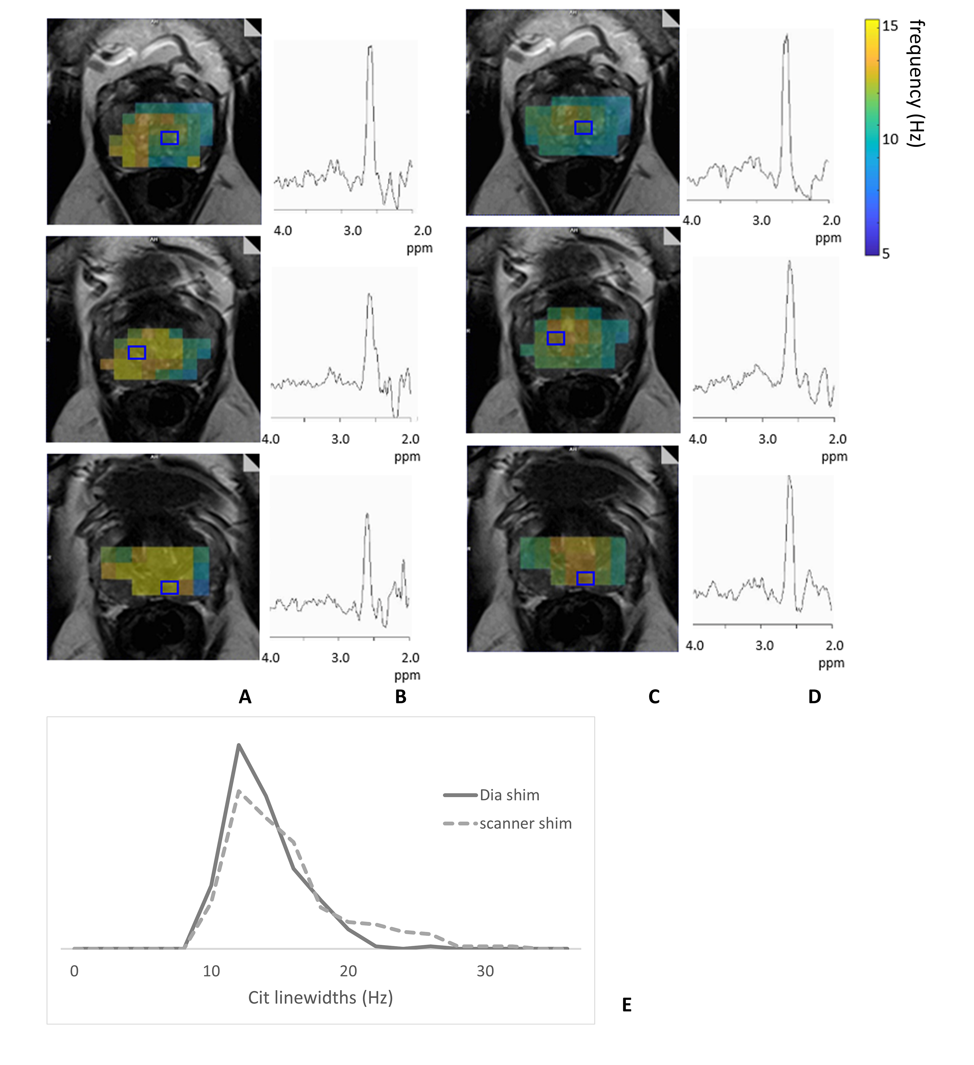
Figure 4: Shimming comparison in MRSI with: (A) Cit linewidth maps (3 slices from volunteer 9) with scanner shim (B) spectra from the indicated voxels on A (blue) (C) Cit linewidth maps (same 3 slices) of MRSI voxels with Dia shim (D) spectra from indicated voxels on B (blue) (E) Cit linewidth histograms N=236 voxels from the full cohort, with Dia shim (line, median 12.9Hz, width 18Hz) and scanner shim (dotted, median 14.3Hz, width 24Hz)
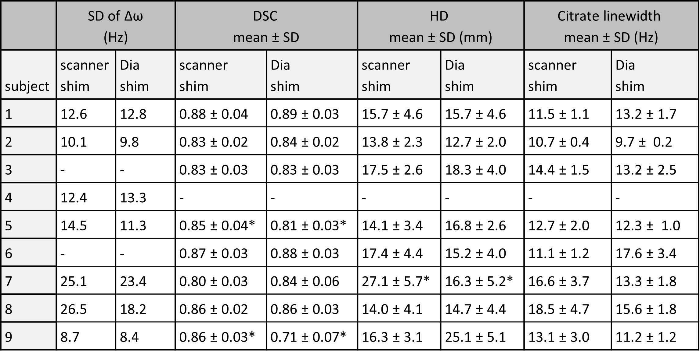
Table 1: Overview of results on B0-map, DWI and MRSI for all volunteers for scanner and Dia shim methods. In volunteer 3 and 6, B0-maps were not representative as reshimming was needed due to air passing by in the shim area or severe phase wrapping. In volunteer 4, there was a mismatch in FOV positioning between the transversal T2w and DWI series. *significant differences with p<0.05, Δω: off-resonance frequencies, DSC: Dice similarity coefficient, HD: Hausdorff distance, SD: standard deviation
DOI: https://doi.org/10.58530/2023/4282