4281
Amide proton transfer values in testicular spermatogenic function evaluation: a preliminary study1The First Affiliated Hospital, Sun Yat-Sen University, Guangzhou, China, 2Philips Healthcare, Guangzhou, China
Synopsis
Keywords: Urogenital, fMRI
In this study, we determined APT of normal testes possible variations with age, and to assess the feasibility of APT in testicular spermatogenic function evaluation. Our results showed that APT of normal testicular tissue decrease with advancing age, and could be promising in evaluation of male infertility.Introduction
The advantages of magnetic resonance imaging (MRI) of scrotum can provide adequate anatomic information, satisfactory tissue contrast, and functional information [1]. Functional MRI, such as DWI and MTI can be used to evaluate male infertility [2]. Amide proton transfer (APT) imaging has been introduced as a new endogenous contrast mechanism for MR imaging by means of detecting low concentration solutes such as mobile proteins and peptides in tissues or tumors that contain abundant amide chemical constituents [3, 4]. The clinical utility of APT imaging for estimating the tumor grade of brain tumors, lung cancers, and prostate cancers has been reported [5-8]. We hypothesized that APT imaging might also be useful to estimate testicular spermatogenic function. In this study, we aimed to determine APT of normal testes possible variations with age, and to assess the feasibility of APT in testicular spermatogenic function evaluation.Methods and Materials
12 men with orchitis (Group A) and 37 male volunteers (control group B, age range: 20–80 years) including three subgroups (Group Byoung, 20–39 years, n=12; Group Bmiddle, 40–69 years, n=13,) and Group Bolder, older than 69 years, n=11) were included. All subjects underwent conventional MRI and APT examination on a 3.0 T MR scanner (Ingenia CX, Philips Healthcare, Best, The Netherlands). APT was performed with a three-dimensional TSE-mDixon sequence. And parameters of the APT sequence were as follows: saturation power= 1.5 µT, saturation duration= 1 s, frequency offsets= ±3.5, ±3.42, ±3.58, −1540 ppm, repetition time (TR)/echo time (TE), 4000/8.8 ms; field of view (FOV), 250*346 mm2; sampling resolution, 2.02*2mm3; slice thickness, 4 mm; sensitivity encoding (SENSE) factor, 1.6; total scan time, 3:40 minutes. APT signal intensity (APT SI) of testes were measured in each group. The differences of APT SI between two groups were compared using the independent two-samples t-test or Mann–Whitney U test. P < 0.05 was considered statistically significant.Results
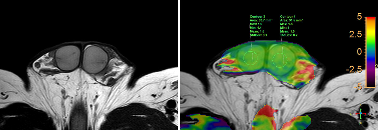
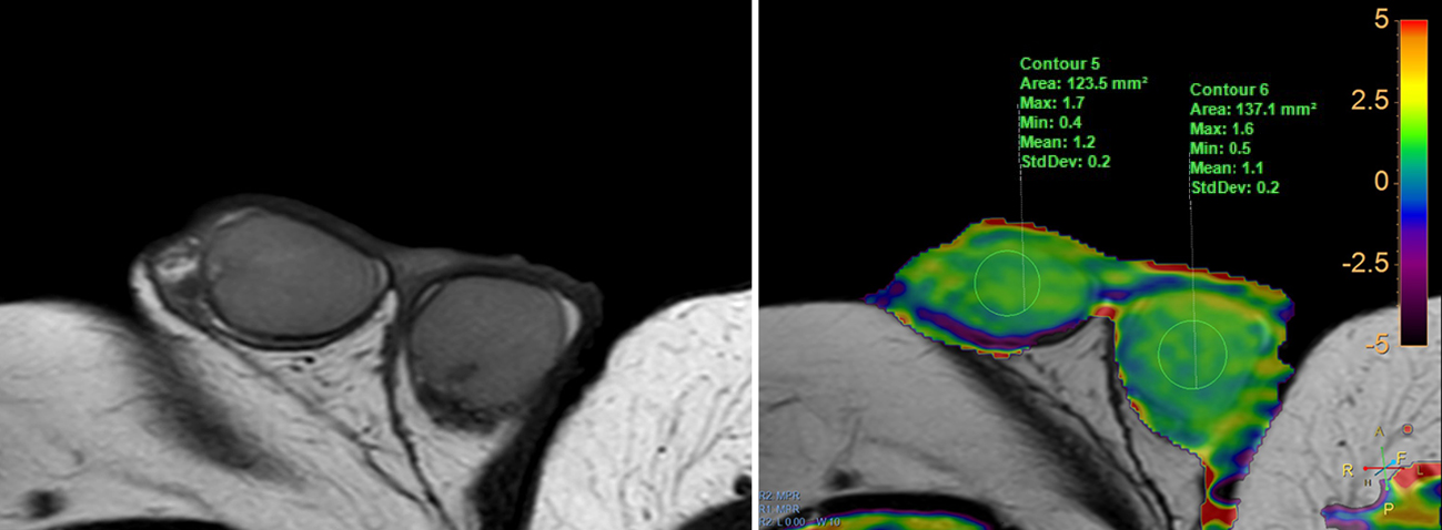
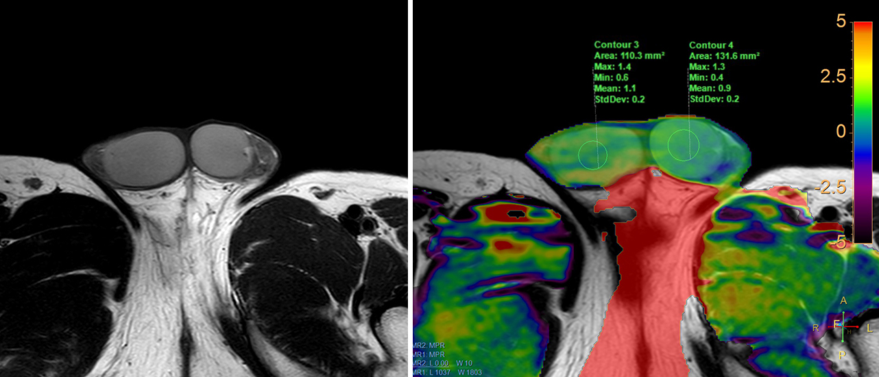
APT SI of group Bolder (1.05 ±0.14) were significantly lower than that of group Byoung (1.40±0.20, P<0.001) and group Bmiddle (1.29±0.13, P=0.02) (Figure 1 and 2). The APT SI in group A (1.15±0.13) were significantly lower than that of control group B (1.30±0.17, P=0.001), group Byoung (P<0.001), and group Bmiddle (P=0.01) (Figure 3), whereas there were no significantly difference between group A and group Bolder.Conclusions
APT SI of normal testes decrease with advancing age. The decrease of APT SI of testes may indirectly reflect the testicular spermatogenesis hypofunction.Acknowledgements
None.References
[1] Tsili AC, Ntorkou A, Baltogiannis D, et al. Magnetization transfer imaging of normal and abnormal testis: preliminary results. Eur Radiol, 2016;26:613–621.
[2] Wang H, Jian Guan, Lin JH, et al. Diffusion weighted and magnetization transfer imaging in testicular spermatogenic function evaluation: preliminary results. Journal of Magnetic Resonance Imaging. 2018,47(1):186-190.
[3] Zhou J, Payen JF, Wilson DA, et al. Using the amide proton signals of intracellular proteins and peptides to detect pH effects in MRI. Nat Med 2003;9(8):1085–1090.
[4] Zhao X, Wen W, Huang F, et al. Saturation power dependence of amide proton transfer image contrasts in human brain tumors and strokes at 3T. Magn Reson Med, 2011, 66: 1033-1041.
[5] Wen Z, Hu S, Huang F,et al. MR imaging of high-grade brain tumors using endogenous protein and peptide-based contrast. Neuro-Image,2010, 51:616–622.
[6] Togao O, Yoshiura T, Keupp J, et al. Amide proton transfer imaging of adult diffuse gliomas: correlation with histopathological grades. Neuro-oncol 2014;16(3):441–448.
[7] Ohno Y, Yui M, Koyama H, et al. Chemical exchange saturation transfer MR imaging: preliminary results for differentiation of malignant and benign thoracic lesions. Radiology 2016;279(2):578–589.
[8] Takayama Y, Nishie A, Sugimoto M, et al. Amide proton transfer (APT) magnetic resonance imaging of prostate cancer: comparison with Gleason scores. MAGMA 2016;29(4):671–679.
Figures