4277
Effect of Compressed SENSE on 3D T2-weighted sequence for Rectum Imaging with a deep learning constrained Compressed SENSE Reconstruction
Ying Qiu1, Yi Zhu2, Dandan Guo1, and Ke Jiang2
1Department of radiology, First Hospital of Jilin University, ChangChun, China, 2Philips Healthcare, BeiJing, China
1Department of radiology, First Hospital of Jilin University, ChangChun, China, 2Philips Healthcare, BeiJing, China
Synopsis
Keywords: Urogenital, Pelvis
Rectal MRI examination requires high resolution three-dimensional (3D) sequences to observe the morphology, structure and the location of the lesions. However, 3D sequences may lead to longer scan time ,patient discomfort and image quality problem. In this study, we investigated the use of a deep learning-based reconstruction algorithm (CS-AI) to highly accelerate 3D rectum MRI. The result showed that CS-AI reconstruction can use the same scan time with sufficient image quality compared to SENSE and might be clinically useful in assessment of rectum cancer.Introduction
High resolution MRI is the best imaging method to evaluate the efficacy of rectum radiotherapy and chemotherapy before operation. Due to its high spatial resolution and soft tissue resolution, MR Images can clearly show the depth of tumor invasion and the presence or absence of extramural venous infiltration. It also has high accuracy in evaluating the status of mesangial fascia and the presence or absence of lymph node infiltration1-3. However, this always takes a very long scan time and causes patient discomfort. Recently, integrating arterial intelligence (AI) into MRI reconstruction has attracted much attention. This allows to further minimize scan time. At the 2019 fastMRI challenge, a novel deep neural network was introduced as Adaptive-CS-Net and showed superior performance for reconstructing knee images from highly undersampled k-space data4-7. The Adaptive-CS-Net was expanded to multiple contrasts and anatomical areas and is presented here as Compressed SENSE AI (CS-AI) reconstruction. It is hypothesized that the acquisition time for rectum MRI may be significantly shortened while maintaining image quality by using the CS-AI reconstruction algorithm. The purpose of this study was to acquire highly accelerated rectum MRI using the CS-AI reconstruction and compare the image quality with the SENSE, and compressed-SENSE (CS).Method
The study was approved by the local IRB, and written informed consent was obtained from all subjects. A total of 23 patients were examined on a 3.0 T system (Ingenia Elition , Philips Healthcare) using a Torso coil , including 14 men and 9 women(mean age 57.35±6.15 years, range: 52-69 years).Preparation before examination: All patients fasted 6h before the examination, underwent clean enema 30 minutes before examination, and used glycerine to clean the intestine. All patients received three different acceleration methods SENSE、CS and CS-AI of 3D PelvisVIEW_T2 sequence with the same acceleration factor of 9. The following parameters were common to all examinations: voxel size=1*1*1mm3, FOV=400*400*100mm3, 200 slices, TR=1250ms, TE=157ms,TSE factor=120, NSA = 2. The scan results were exported to Philips IntelliSpace Portal (ISP, Philips Healthcare).The total scan time for SENSE and CS/CS-AI was 2:23 and 2:39. Quantitative image analysis was performed by a radiologist with more than 4 years of experience. Signal to noise ratio (SNR) and contrast to noise ratio (CNR) were calculated by setting the rectal wall as the region of interest (ROI) and setting the peri-bowels fat tissue as a contrast. The image quality of SENSE、CS and CS-AI was visually compared, including anatomical structures, diagnostic certainty and artifacts scored on a five-point-scale. The paired Wilcoxon test was performed to test for an influence of the different sequences. P values< 0.05 were considered significant.Results and Discussions
For quantitative analysis of image quality, the SNR and CNR values of CS-AI were all significantly higher than SENSE (P<0.05)and Compressed SENSE, and the CS-AI sequence was rated highest for SNR and CNR value with an average of 9.20±2.91 and 7.47±4.32. On the other hand, all the CS-AI sequences had significantly higher CNR values than the CS sequences with the same acceleration factor( P<0.05). The mean SNR,CNR and subject scores for all sequences were listed in the Table1.For subjective analysis, CS-AI sequences had higher scores for artifacts compared to others (P<0.05), and CS-AI had higher scores for anatomical structures and diagnostic certainty than others(P<0.05). Overall, the CS-AI images demonstrated high denoising performance. In all sequences, the CS-AI showed image quality comparable to the SENSE method, which took about the same scan time as the accelerated scans. Figure 1 shows the comparison of the SENSE, C-SENSE, and CS-AI for 3D PelvisVIEW_T2w images. The Compressed SENSE and CS-AI showed less noise than SENSE method. The CS-AI produced visually sharper images compared to SENSE and C-SENSE. Both the SENSE and CS-AI showed good image quality, but the CS-AI demonstrated a better rectum of small structures such as mesorectal fascia (arrow). This suggests that the scan acceleration with noise reduction by CS-AI reconstruction may be useful for getting a high-quality image.Conclusion
In this study, we compared the effects of SENSE, C-SENSE and CS-AI reconstruction methods on rectal MRI imaging quality. Our results showed that the scanning time of SENSE is similar to that of CS and CS-AI, but CS-AI can obtain better image quality within the same acquisition time, reduce noise and artifacts, and have higher SNR and CNR. Further data collection is required to determine the advantages of CS-AI over other scan acceleration technologies.Acknowledgements
No acknowledgement found.References
1. Mckeown E, Nelson DW, Johnson EK, et al. Current approaches and challenges for monitoring treatment response in colon and rectal cancer [J]. J Cancer, 2014, 5(1):31-43.2.Jang K M, Kim S H, Choi D, et al. Pathological correlation with diffusion restriction on diffusion-weighted imaging in patients with pathological complete response after neoadjuvant chemoradiation therapy for locally advanced rectal cancer: preliminary results [J]. The British journal of radiology,2012, 85(1017): e566-572
3. Chand M, Swift R I, Tekkis P P, et al. Extramural venous invasion is a potential imaging predictive biomarker of neoadjuvant treatment in rectal cancer [J]. British journal of cancer, 2014, 110(1): 19-25.
4.Knoll F, Zbontar J, Sriram A, et al. fastMRI: A Publicly Available Raw k-Space and DICOM Dataset of Knee Images for Accelerated MR ImageReconstruction Using Machine Learning. Radiol Artif Intell. 2020;2(1):e190007.
5.Knoll F, Murrell T, Sriram A, et al. Advancing machine learning for MR image reconstruction with an open competition: Overview of the 2019 fastMRI challenge. Magn Reson Med. 2020;(January):mrm.28338.
6.Pezzotti N, Yousefi S, Elmahdy MS, et al. An Adaptive Intelligence Algorithm for Undersampled Knee MRI Reconstruction. IEEE Access. 2020;8:204825-204838.
7.Pezzotti N, de Weerdt E,Yousefi S, et al. Adaptive-CS-Net: FastMRI with
Adaptive Intelligence. arxiv. 2019;(NeurIPS).
Figures

Table1:Ana:anatomical structures,Dia:diagnostic certainty ,Art:artifacts
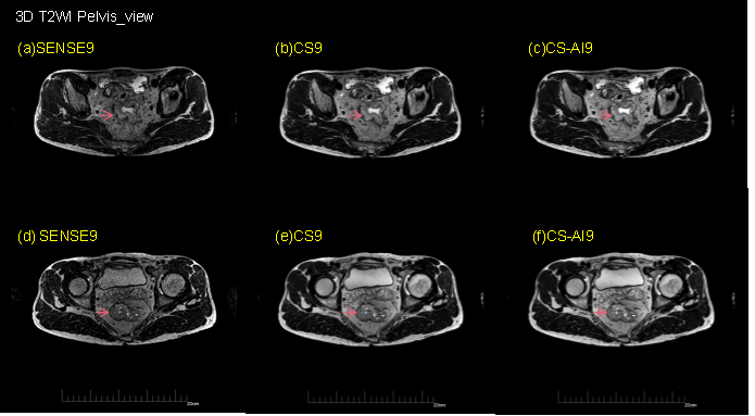
Figure 1. 3D T2WI Pelvis_view axial images for (a)(d) SENSE reference with acceleration factor9, (b) (e)Compressed SENSE and (c)(f) CS-AI with acceleration factor 9.
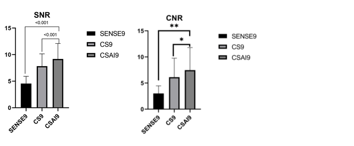
FIgure2.SNR and CNR measurements in SENSE、CS and CSAI.
DOI: https://doi.org/10.58530/2023/4277