4273
The value of MR perfusion and diffusion combined with tumor marker analysis in differentiating benign and malignant ovarian tumors
Jinsong Bai1, Haitao Zhang1, and shaoyu wang2
1Shanxi Hanzhong People's Hospital, Hanzhong, China, 2DI MR SMK, Siemens Healthineers, Shanghai, China
1Shanxi Hanzhong People's Hospital, Hanzhong, China, 2DI MR SMK, Siemens Healthineers, Shanghai, China
Synopsis
Keywords: Pelvis, Cancer, Ovarian cancer
The aim of our study was to compare the differential diagnostic efficacy of dynamic contrast enhanced magnetic resonance imaging (DCE-MRI), apparent diffusion coefficient (ADC), serum carbohydrate antigen 125 (CA125), human epididymal secretory protein 4 (HE4) and their combined application in benign and malignant ovarian tumors, and to explore the best method for the diagnosis of ovarian tumors.Introduction
The aim of this study was to evaluate the differential diagnostic efficacy of dynamic contrast enhanced magnetic resonance imaging (DCE-MRI), apparent diffusion coefficient (ADC), serum carbohydrate antigen 125 (CA125), human epididymal secretory protein 4 (HE4) and their combined application in benign and malignant ovarian tumors, and to explore the best method for the diagnosis of ovarian tumors.Methods
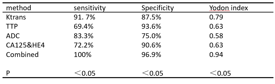
68 patients with ovarian tumors admitted to our hospital from January 2022 to September 2022 were enrolled in this retrospective analysis. 32 patients were confirmed as benign and 36 patients as malignant by pathology. All the MR scanning contained DCE-MRI and DWI sequences were performed on a 3T MR scanner (MAGNETOM Skyra, Siemens Healthineers, Erlangen, Germany). The DCE-MRI quantitative parameter Ktrans, semi quantitative parameter TTP were acquired after processing on the post-processing workstation(Syngo Via, Siemens Healthcare). The ADC map was generated after finished DWI scanning automatically. ADC values were obtained by sketching on the ADC maps, and the CA125 and HE4 indicators were retrieved through the LIS system for statistics. At the same time, the sensitivity and specificity of different examination methods and combined application to benign and malignant ovarian tumors were calculated based on the pathological diagnosis results as the gold standard.Results
The sensitivity of Ktrans, TTP, ADC, CA125 and HE4 in the diagnosis of ovarian benign and malignant tumors was 91.7%, 69.4%, 83.3% and 72.2% respectively, and the specificity was 87.5%, 93.6%, 75.0% and 90.6% respectively. The sensitivity and specificity of the combined application were 100% and 96.9% respectively in the diagnosis of ovarian malignant tumors; There was a statistical difference in the diagnostic efficacy of their respective and combined use for benign and malignant ovarian tumors (P<0.05).Conclusion
The sensitivity and specificity of DCE-MRI combined with ADC, CA125 and HE4 in differential diagnosis of benign and malignant ovarian tumors were higher than using single examination method alone, which has higher clinical application value.Acknowledgements
No acknowledgement found.References
No reference found.Figures
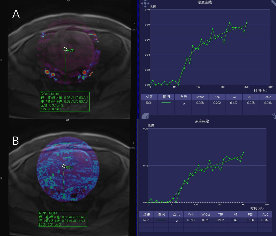
Figure1 A: Female, 65 years old, benign ovarian tumor (theca fibroma), Ktrans < 0.123 min-1, TTP > 0.85 min; B: Female, 49 years old, ovarian malignant tumor (stage III ovarian cancer), Ktrans > 0.123 min-1, TTP < 0.85 min
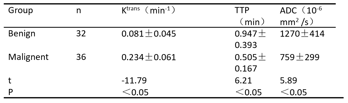
Table 1 Comparison of Ktrans, TTP and ADC between benign and malignant ovarian tumors (x ̅ ±s)
Comparison of effectiveness between individual inspection and joint inspection
DOI: https://doi.org/10.58530/2023/4273