4258
SNR Improvement with Ultrahigh Dielectric Constant (uHDC) Material for 7T 1H MRS Imaging1Pennsylvania State University College of Medicine, Hershey, PA, United States, 2Grodno State medical University, Grodno, Belarus
Synopsis
Keywords: High-Field MRI, Challenges
We investigated the effects of ultra-high dielectric constant (uHDC) materials for enhancing the B1 field at 300 MHz for potential MRS imaging 1H at 7.0 T. The uHDC material was able to increase the transmit efficiency. We calculated B1+ and B1+ efficiency for a phantom mouse brain with and without uHDC material and empirically obtained nominal flip angle maps and 1H metabolic spectra with and without uHDC. Our results show significant enhancement of Signal-to-Noise ratios (SNRs) of 1H spectra in the presence of the uHDC material.
Introduction
B1 field can be enhanced by introducing ultrahigh dielectric constant (uHDC) material into the RF coil setup1,2. This enhancement is attributed to displacement currents due to non-conservative E fields induced in the uHDC material. Conservative as well as non-conservative E fields generated by RF coils are known to increase noise levels, resulting in signal losses in samples being imaged. However, the uHDC material can partially shield conservative E-fields, reducing signal loses. In this study, we investigated the effects of uHDC materials for enhancing the B1 field at 300 MHz for MRS imaging 1H at 7.0 T.Materials and Methods
Figure 1 shows our experimental setup (for both the simulation and imaging) for electromagnetic field simulations using the CST Studio (2020) software. The scanner’s transmit birdcage volume coil was modeled as a high-pass birdcage with a phase increment of 45° between capacitor gaps. Coil polarization was set to quadrature. Simulations were run at a frequency of 300 MHz on a 0.67 mm isotropic grid with convergence levels set to -40 db. The resultant B1+ and electric field distributions were analyzed to calculate the transmit efficiency.Experiments were carried out on a 7T MRI scanner (Bruker BIOSPEC 70/20 USR) with a volume coil for transmission and receiver. This coil utilized two independent transmit channels. The coil polarization was set to quadrature for all experiments. The mouse brain phantom (diameter: 20 mm, length: 80 mm, conductivity: 0.05 S/m) was used. The diameter, thickness and length of the birdcage coil were 80 mm, 2 mm and 120 mm respectively. The diameter, thickness and length of the shield were 84 mm, 2 mm and 84 mm respectively.
A FLASH pulse sequence was used to acquire MRI data with variable flip angles (30° and 60°), TE/TR (Echo Time /Repetition Time) = 2.5/5000 ms, 64x20x20 mm Field of View (FOV) and 128x40x40 matrix size. Image volume slice thickness was 20.0 mm. Non-Selective RF hard pulse with duration of 0.5 ms was used. The data were acquired without uHDC and with uHDC material (thickness: 2 mm, inner diameter: 26 mm, length: 30 mm, permittivity: ~1000). Transmit efficiency maps and SNR maps were evaluated for the water phantom with and without uHDC material. Then we performed MRS Imaging for a dead mouse brain using the 2D-CSI pulse sequence with the following imaging parameters: TE/TR = 1.22/140 ms, Flip Angle = 65°, number of averages 128, Total Acquisition = 19 min, FOV = 24 mm by 24 mm, voxel size=3×3×3 mm3, slice thickness 3 mm
Results
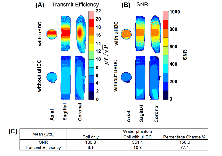
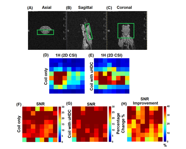
Figure 2 shows both the simulated B1+ and B1+ efficiency maps for the setup shown in Figure 1. They show an approximately 10-fold enhancement with uHDC material of 1000 permittivity. Figure 3 shows (A) estimated transmit efficiency and (B) SNR maps for the water phantom (axial, sagittal and coronal views) without and with uHDC material. SNR is increased by 156% in the water phantom with the uHDC (see Figure 3C). There was a 6-fold reduction in reference power when imaging with uHDC material compared to without uHDC material. B1+ efficiency in the water phantom with uHDC improved by 77%. Figure 4 shows (A) axial, (B) sagittal and (C) coronal views of the mouse brain images with the region of interest (green box) and 1H metabolic imaging maps (D) without and (E) with uHDC. The use of uHDC material produced higher SNR compared to the coil only SNR map of 1H imaging (Figure 4 D & E). The SNR is increased by 10% to 40% of 1H imaging within the mouse in the presence of uHDC.Discussion and Conclusion
Our results highlight the potential utility of uHDC material for enhancing SNR for 1H imaging at 7T. Conventionally, improving the overall SNR for in vivo MRI/MRS imaging is to increase the strength of the static magnetic field strength (B0). The SNR improvement, however, remains challenging for X-nuclear applications even with ultrahigh field systems. Utilizing uHDC materials may offer a novel approach to enhance SNR for high field low-gamma X-nuclear MRS imaging applications. Future studies should focus on the optimal HDC permittivity and geometry to further improve the ability of HDC material to shield conservative electric fields at a low frequency of 45 MHz potentially for HD0 imaging.Acknowledgements
We thank the Center for NMR Research and Center for Aging and Neurodegenerative Diseases of Penn State University College of Medicine for all the staffs' kind help and suggestion. This research is supported by the National Institute on Aging grants (1R01AG070088-01A1 and1R21AG064486)References
1. Yang, Q.X. et al., JMRI, 2013
2. Rupprecht, S., et al., Magnetic Resonance in Medicine, 2017
Figures
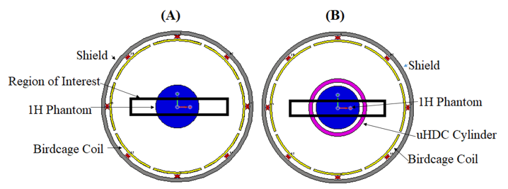
Figure 1. The experimental setup. (A) 1H phantom inside the birdcage coil (without uHDC material) and (B) 1H phantom placed inside the uHDC cylinder. The diameter, the thickness, and the length of the birdcage coil was 80 mm, 84 mm, and 120 mm respectively. The diameter, the thickness, and the length of the shield was 84 mm, 2 mm, and 84 mm. The uHDC cylinder had the following properties: permittivity/thickness/diameter/length = 1000/2.0 mm/26/30 mm. The 1H phantom had the following properties relative permittivity/conductivity/diameter= 60/0.05 S/m/20 mm.
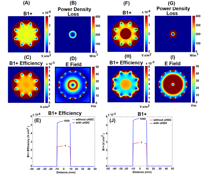
Figure 2. Simulated B1+ and B1+ efficiency maps (A, C) without uHDC and (F, H) with uHDC within the 1H phantom. B1+. Simulated images of power density loss and E field, without uHDC (B, D) and with uHDC (G, I), are also shown. The ID plots of the B1+ field (E) and B1+ Efficiency (J), without uHDC (red) and with uHDC (blue), are shown. Both the B1+ and B1+ efficiency show enhancement with uHDC material of 1000 permittivity.