4233
RF Field Enhancement with Ultrahigh Dielectric Constant Material for Micro Imaging at 7T1Pennsylvania State University College of Medicine, Hershey, PA, United States, 2Grodno State medical University, Grodno, Belarus
Synopsis
Keywords: High-Field MRI, High-Field MRI
In this study, we investigated the effects of ultra-high dielectric constant (uHDC) material for reducing noise and enhancing the B1 efficiency at 300 MHz for 1H nuclear applications at 7T. Using electromagnetic field simulations [CST Studio (2020) software] and imaging experiments, we estimated B1+ efficiency maps for a phantom mouse brain with and without uHDC material. Our results show significant enhancement of B1+ efficiency and Signal-to-Noise ratio (SNR) in the presence of the uHDC material.
Introduction
The effects of uHDC materials on RF coil field distributions are mainly due to induced displacement currents that can act as secondary RF field sources. These induced currents are proportional to the RF field frequency and permittivity of these uHDC materials, which can impact B1+ efficiency and SNR in nearby regions. Several approaches have been proposed to make uHDC materials, such as aqueous mixtures of powder or beads of dielectric materials1,2. Our lab uses BST ceramics to make these materials because it increases uHDC material density: 55% (slurry powder) and 74%–96% for beads. By increasing density, the permittivity values increase exponentially by factors of 2 to 103. These materials have significantly higher relative permittivity and lower loss that is one to three orders of magnitude lower than water-based mixtures. This study focused on the utility of uHDC material for microimaging. We combined a hollow uHDC cylinder with a murine phantom brain, then investigated RF field distributions through computer modeling and imaging experiments at 7T. We hypothesized that our uHDC material would alter RF field distributions and significantly improve the RF coil performance.Materials and Methods
We performed all electromagnetic field simulations using the CST Studio (2020) software. Figure 1 shows the EM field simulation setup for optimization. We used this setup to generate B1+ maps for different thicknesses of the uHDC hollow cylinder while keeping permittivity at 1000. We then investigate B1+ field changes along the diameter of the hollow cylinder with uHDC thickness. Figure 2 shows the simulation configuration for the phantom mouse brain. The scanner’s transmit birdcage volume coil was modeled as a 12 rod, high-pass birdcage. Simulations were performed at the 298.5 MHz frequency on a 0.67-mm isotropic grid and convergence levels set to -40 db with and without uHDC setups. Experiments used a Bruker 7T MRI scanner (BIOSPEC 70/20 USR). The phantom mouse (permittivity: 900) was made with distilled H2O, CuSO4 1 g/L, and NaCl 3.6 g/L. The Fast-low angle shot (FLASH) sequence, using a non-selective RF hard pulse, was used to collect data at 30° and 60° flip angles with the following parameters: TR/TE = 5000/2.5 ms; Voxel Size = 0.5 x 0.5 x 0.5 mm; FOV = 64 x 20 x 20 mm. The B1+ efficiency maps were computed using equation [1] below.>B1+, P, a , t , and g are RF field, dissipated power, flip angle, RF pulse duration, and gyromagnetic ratio respectively. The SNR Units method, implemented in MATLAB R2018b, was used to calculate SNR maps.
Results and Discussion
Figure 3 shows simulated B1+ maps at thicknesses of 2, 4, 6, and 6.5 mm of the uHDC hollow cylinder. The strongest B1+ map was obtained when uHDC thickness was 6 mm. Figure 4 shows simulated B1+ efficiency and SNR maps for the phantom mouse brain (axial views), without and with uHDC material. Quantitative comparisons for our experimental and simulated approaches are shown in Table 1. SNR is increased by 85% to 89% in the phantom mouse brain for both approaches, specifically near regions where uHDC material is located (see Table 1). Transmit power was reduced when imaging with uHDC material; there was a 7-fold reduction compared to without uHDC material. B1+ efficiency in the phantom brain with uHDC improved by 77% to 94% for both experimental and simulated approaches.Conclusion
Our results demonstrate that uHDC material can enhance the B1+ efficiency and improve SNR, which can be helpful for microimaging applications at ultra-high magnetic fields2. Future research should focus on optimizing uHDC permittivity and the thickness of hollow cylinders to further improve RF coil performance.Acknowledgements
We thank the Center for NMR Research and Center for Aging and Neurodegenerative Diseases of Penn State University College of Medicine for all the staffs' kind help and suggestion. This research is supported by the National Institute on Aging grants (1R01AG070088-01A1 and1R21AG064486)
References
[1]. Yang, Q.X. et al., JMRI, 2013. 38(2).
[2]. Rupprecht, S., et al., Magnetic Resonance in Medicine, 2017. 79(5).
[3]. Roemer, P. B., Edelstein, W. A., Hayes, C. E., Souza, S. P. and Mueller, O. M., The NMR phased array. Magnetic Resonance in Medicine, 1990.
Figures
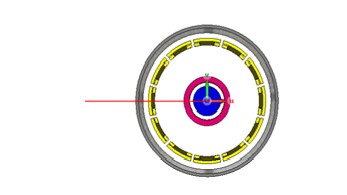
Figure 1. The simulation setup for 1H phantom placed inside the uHDC hollow cylinder (permittivity of 1000). Simulations were performed at 2, 4, 6, and 6.5 mm thickness of the uHDC hollow cylinder at a frequency of 298.5 MHz on a 0.67-mm isotropic grid and convergence levels set to -40 db in the CST Studio (2020). Field maps were calculated along the diameter shown in red.
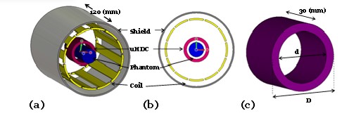
Figure 2. The simulation configuration of (a) phantom mouse brain inside the birdcage coil with 12 rods and (b) cross-sectional view of the simulation configuration of (a). The diameter, thickness and length of the coil was 80 mm, 2 mm and 120 mm respectively. The diameter, thickness and the length of the shield were 98 mm, 2 mm and 120 mm respectively. The outer and inner diameters, and length of the uHDC were D = 34 mm, d = 26 mm, and 30 mm (c). The 1H phantom had the following properties relative permittivity/conductivity/diameter= 58/0.05 S/m/20 mm)
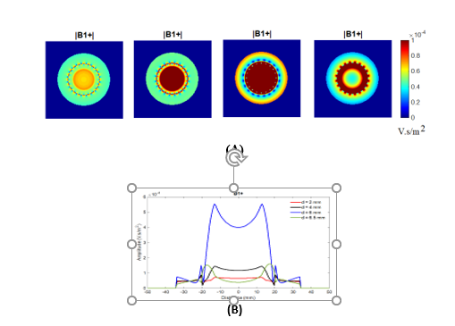
Figure 3. (A) B1+ maps for different thickness of the uHDC hollow cylinder (B) ID plots of the B1+ field for the thickness of 2 mm (red), 4 mm (black), 6 mm (blue) and 6.5 mm (green).
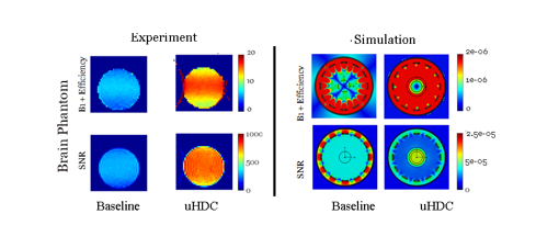
Figure 4. The measured and simulated B1+ efficiency and SNR maps (axial view) for the phantom mouse brain without uHDC material (baseline) and with uHDC material for experimental and simulated approaches: permittivity = 1000. Units of B1+ Efficiency is in V.s/m2
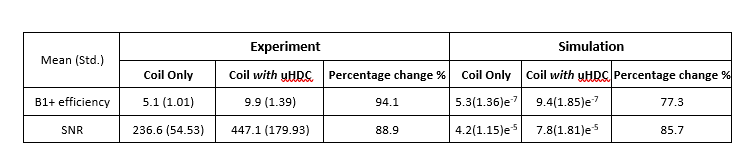
Table 1. Quantitative comparisons of B1+ efficiency and SNR for experimental and simulation results for the phantom mouse brain. Coil with uHDC and coil without uHDC setups are shown.