4229
Improved visualization of whole-brain structures and pathology at ultra-high field 7T MRI with parallel transmission universal pulses1Department of Radiology, Chinese PLA General Hospital, Beijing, China, 2MR Collaboration, Siemens Healthineers Ltd., Beijing, China, 3CEA NeuroSpin, Paris, France
Synopsis
Keywords: High-Field MRI, Parallel Transmit & Multiband
Ultra-high field 7T MRI provides a significant increase in signal-to-noise ratio and contrast for noninvasively visualizing the brain, but is challenging to use for whole-brain imaging. In this study, we investigated the clinical potential of parallel transmission universal pulses (UP) for improving visualization of whole-brain structures and pathology. Our results show that UP enables remarkable signal contrast improvement throughout the brain, which is promising to improve lesion detection (e.g., brain metastases) and structures visualization over the whole brain.Introduction
7T MRI allows better delineation of brain structures and subtle pathology due to the improved signal-to-noise ratio and tissue contrast at higher field strengths.1 Unfortunately, an increase in field strength also introduces non-uniformities in the radiofrequency transmit field that can lead to non-uniformly image contrast over the whole brain, especially in the cerebellum and the temporal lobes.2 Previous studies have demonstrated that parallel transmission UP technology is an effective way to resolve the challenge of whole-brain imaging at 7T MRI.3,4 However, the clinical potential of this technology has not been fully explored.Methods
This prospective study was approved by the Institutional Review Board and informed written consent was obtained from all participants. Non-enhanced T1-weighted magnetization-prepared rapid acquisition gradient-echo (MPRAGE) images with TrueForm and UP were obtained from 6 healthy participants, and contrast-enhanced T1-weighted MPRAGE images with TrueForm and UP were obtained from 6 patients. All imaging measurements were performed on a whole-body 7T scanner (MAGNETOM Terra, Siemens Healthcare, Erlangen, Germany) using an 8-channel transmitting and 32-channel receiving head coil (Nova Medical, Wilmington, MA, USA). The common set of parameters for both TrueForm and UPs were: repetition time = 2600 ms, echo time = 1.97 ms, inversion time = 1100 ms, field of view = 224 × 210 mm2, slice thickness = 0.7 mm, voxel size = 0.7 × 0.7 × 0.7 mm3, number of slices = 224. The acquisition time of the MPRAGE sequence with TrueForm and UP were 5 minutes 55 seconds and 5 minutes 28 seconds, respectively.Results
Figure 1 shows representative non-enhanced T1-weighted images obtained with UP (Figure 1A) and TrueForm (Figure 1B). The cerebellum and temporal lobes can be clearly depicted in the UP images, but is difficult to be observed in the TrueForm images.Representative contrast-enhanced T1-weighted images comparing the UP and TrueForm approaches in the presence of pathologic diseases are shown in Figures 2 and 3. The UP images are able to faithfully depict micrometastases (Figure 2A) and internal architecture (Figure 3A) of the brain metastases in the cerebellum. However, it is difficult to detect these lesions in the TrueForm images due to poor image contrast.
Discussion and conclusion
Image quality and signal contrast of the whole brain can be significantly improved with UP compared to TrueForm. UP is effective in enhancing signal homogeneity, especially in the cerebellum and temporal lobes, achieving high image quality for visualization of whole-brain structures and pathology. Broader application of UP may facilitate the clinical transformation of ultra-high field 7T MRI.Acknowledgements
This work is supported by the National Natural Science Foundation of China (81825012, 81730048 and 82151309). We would like to gratefully acknowledge Dr. Franck Mauconduit at CEA NeuroSpin for providing us the PASTEUR package used in this work.
References
[1] Rutland J W, Delman B N, Gill C M, et al. Emerging use of ultra-high-field 7T MRI in the study of intracranial vascularity: state of the field and future directions. AJNR Am J Neuroradiol. 2020;41:2–9.
[2] Oliveira Í A F, Roos T, Dumoulin S O, et al. Can 7T MPRAGE match MP2RAGE for gray-white matter contrast?. NeuroImage. 2021;240:118384.
[3] Gras V, Vignaud A, Amadon A, Bihan D, Boulant N. Universal pulses: A new concept for calibration-free parallel transmission. Magn Reson Med. 2017;77(2):635–643.
[4] Gras V, Mauconduit F, Vignaud A, et al. PASTeUR: Package of Anatomical Sequences using parallel Transmission UniveRsal kT-point pulses. In: Proceedings of the 27th Annual Meeting of ISMRM. Montréal, QC, Canada. 2019; p. 4626.
Figures
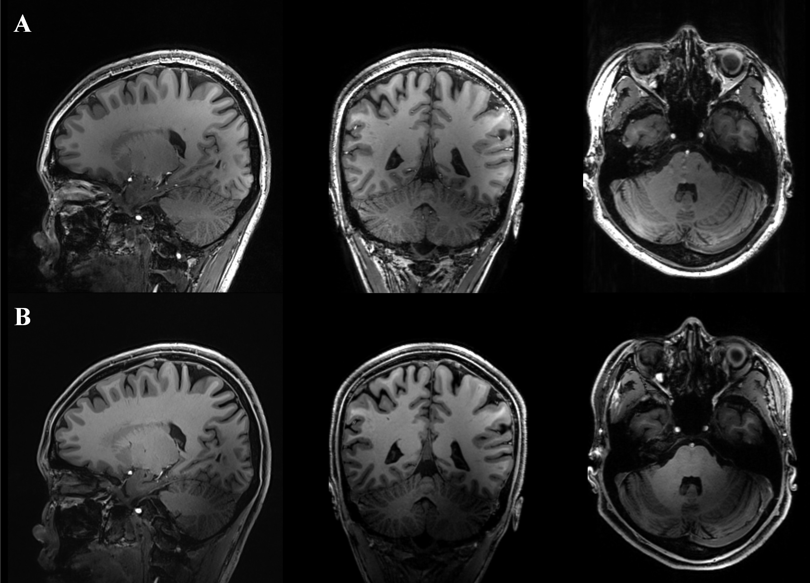
Figure 1. Representative T1-weighted images in a 22-year-old healthy participant comparing the UP (A) and TrueForm (B) approaches for visualization of whole-brain structures.
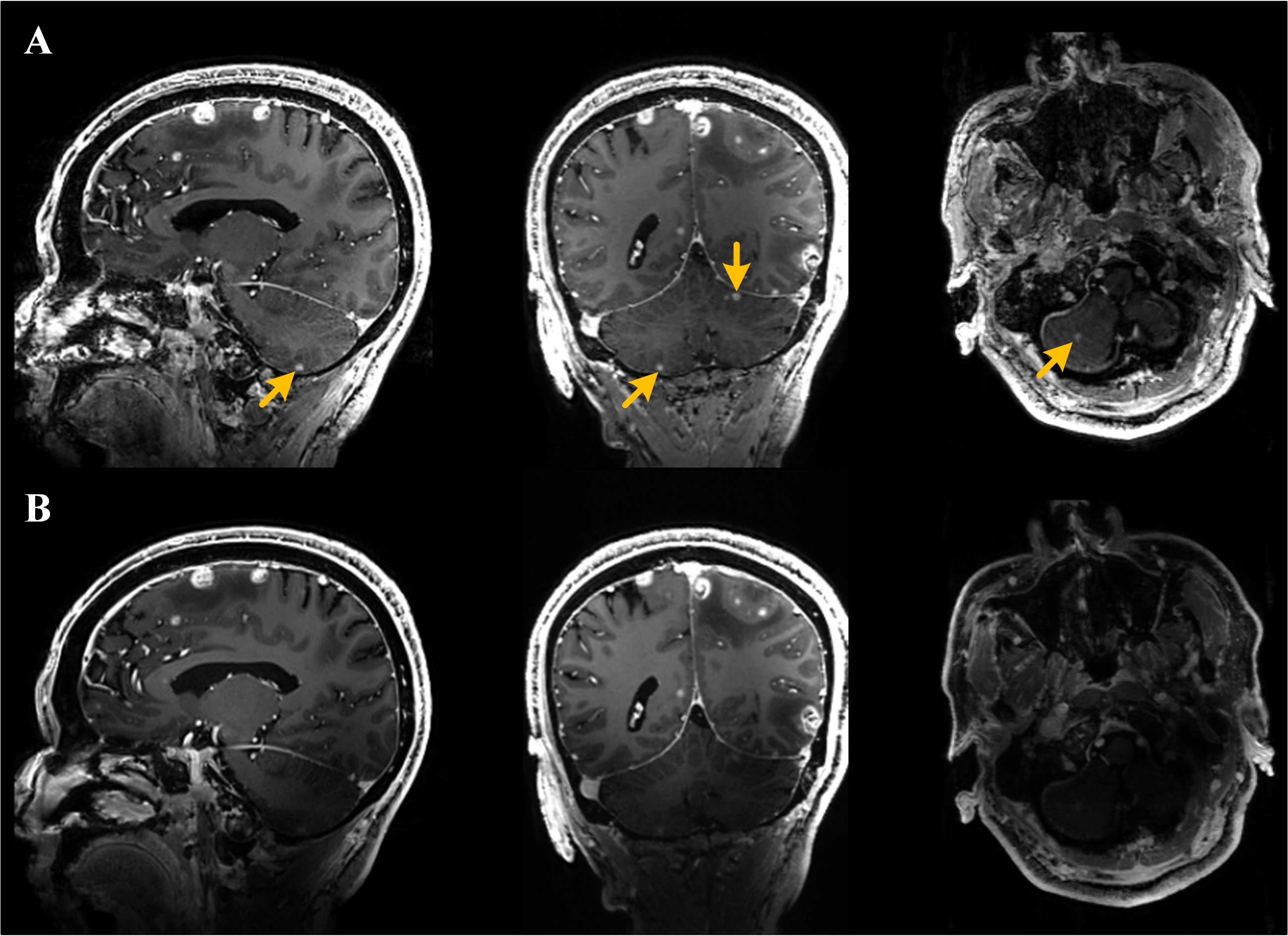
Figure 2. Contrast-enhanced T1-weighted images obtained using the UP (A) and TrueForm (B) approaches in a 75-year-old man with multiple brain metastases from lung cancer.
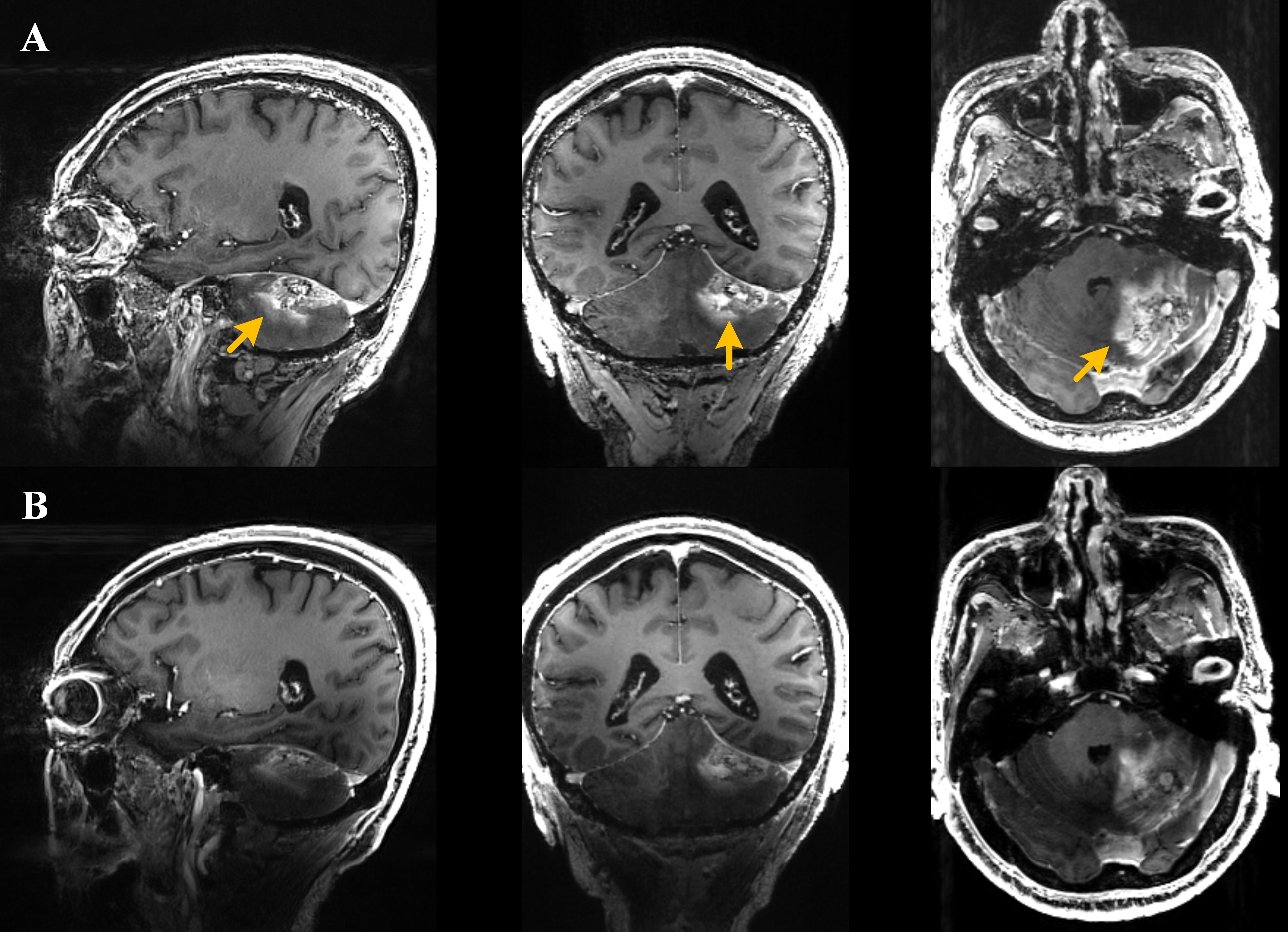
Figure 3. Contrast-enhanced T1-weighted images obtained using the UP (A) and TrueForm (B) approaches in a 67-year-old man with brain metastases from lung cancer after radiotherapy.