4227
A 48-channel head and neck neurovascular coil for MR vessel wall imaging at 5 T1Paul C. Lauterbur Imaging Research Center, Shenzhen Institutes of Advanced Technology, Chinese Academy of Sciences, Shenzhen, China, 2Key Laboratory for Magnetic Resonance and Multimodality Imaging of Guangdong Province, Shenzhen, China, 3Shanghai United Imaging Healthcare, Shanghai, China, 4Department of Biomedical Engineering, State University of New York at Buffalo, New York, NY, United States
Synopsis
Keywords: High-Field MRI, Head & Neck/ENT
Ultra-high field magnetic resonance imaging (MRI) has been increasing in use for human brain imaging. However, due to the absence of a head and neck neurovascular coil, joint imaging of intracranial and extracranial arterial vessel wall has not been applied in current ultra-high field MRI. In this study, a 48-channel head and neck receiver neurovascular coil for joint imaging of intracranial and extracranial arterial vessel wall at 5T was developed. Simultaneous high-resolution imaging of intracranial and extracranial arterial walls with an isotropic spatial resolution of 0.5 mm has been achieved, which further expands the application of ultra-high field imaging.Introduction
Ultra-high field MRI can provide high signal-to-noise ratio and has been used primarily in brain imaging1. Head and neck vessel wall imaging is critical in ultra-high field MRI. So far, no MRI system above 3T can perform simultaneous head and neck vessel wall imaging for the absence of a head and neck neurovascular coil. Most of the coils at ultra-high field MRI are only capable of head scanning2. In this study, a 48-channel head and neck neurovascular coil was developed for simultaneous high resolution imaging of intracranial and extracranial arterial walls in a 5T MRI system. Simultaneous high resolution imaging of intracranial and extracranial arterial walls with an isotropic spatial resolution of 0.5 mm were acquired.Methods
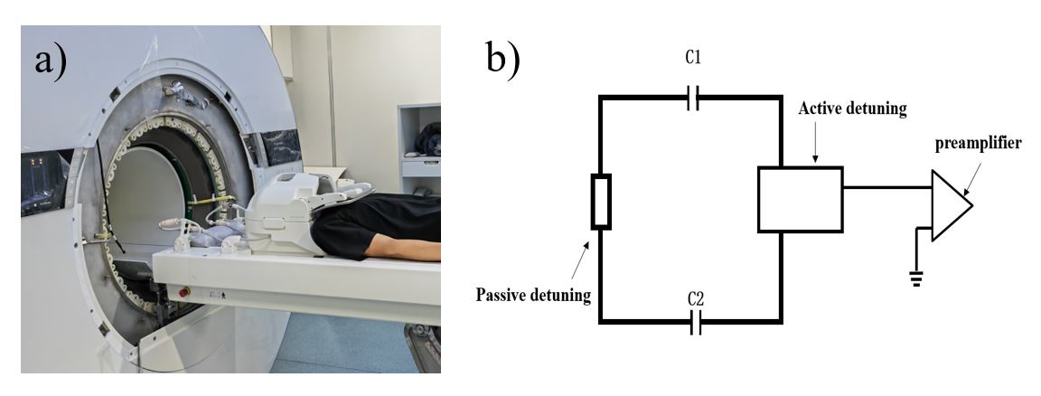
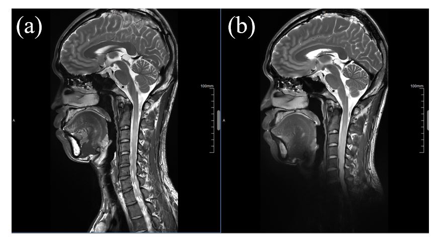
Figure 1 shows the photograph and the circuit schematic of the 48-channel head and neck neurovascular coil. To further reduce transmit field distortion that was caused by receive array, a passive detuning circuit was designed and placed at the opposite position of the active detuning circuit, as shown in Figure 1b. To diminish the common mode current on the cable shield, cable traps were added on the cables wired of the receive array. The coil was developed and tested on a 5T whole-body MRI scanner (Shanghai United Imaging Healthcare, Shanghai, China) with a volume transmit coil3.A T2-weighted fast spin echo (T2_FSE) sequence was applied for anatomy imaging of head and neckusing the 48-channel head and neck neurovascular coil and a quadrature birdcage/48-channel receiver coil assembly4. The parameters are shown as following: TR/TE=2000/107.8ms, flip angle=90o, FOV=350mm×200mm, matrix=532×304, bandwidth=200 Hz/pixel.
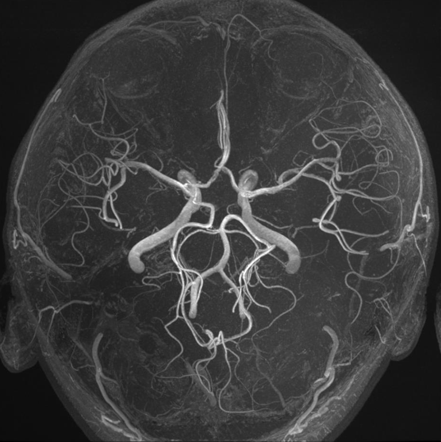
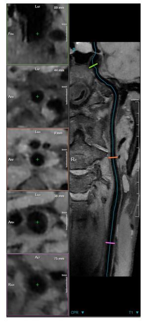
Magnetic resonance angiography (MRA) images were obtained using a time of flight (TOF) sequence: TE= 25, TR=4.4, Flip angle=15°,FOV=180mm×180mm, Matrix=448×448, reconstructed resolution=0.4×0.4×0.4mm, bandwidth=210Hz/pixel, uCS acceleration factor=3.5, total time=12:39. Vascular wall images were acquired using a T1-weighted 3D MATRIX sequence (T1_mx3d): TE=16.08/TR= 1245, flip angle=73o, FOV=192×232, matrix=384×464, reconstructed resolution=0.5mm×0.5mm×0.5mm, bandwidth=440Hz/pixel, uCS acceleration factor=6.0, total time=10:46.
Results
Figure 2 shows the T2_FSE images using the 48-channel head and neck neurovascular coil and the quadrature birdcage/48-channel receiver coil assembly, which demonstrate a larger imaging FOV by using the 48-channel head and neck neurovascular coil. Figure 3 shows the MRA images with a high resolution of 0.4 mm of the intracranial arteries from a healthy volunteer. Figure 4 shows head and neck vessel wall imaging acquired with a 0.5 mm resolution.Conclusions
The 48-channel head and neck neurovascular coil can provide a large imaging FOV that can coverage the head and neck. Simultaneous high-resolution imaging of intracranial and extracranial arterial walls with an isotropic spatial resolution of 0.5 mm has been achieved, which further expands the application of ultra-high field imaging.Acknowledgements
This work is supported by National Key Research and Development Program of China, 2021YFE0204400; the Strategic Priority Research Program of Chinese Academy of Sciences, XDB25000000; National Natural Science Foundation of China, U22A20344; Youth Innovation Promotion Association of CAS No. Y2021098; Key Laboratory Project of Guangdong Province, 2020B1212060051; Shenzhen city grant, RCYX20200714114735123.References
1. Okada T, Fujimoto K, Fushimi Y, Akasaka T, Thuy DHD, Shima A, Sawamoto N, Oishi N, Zhang Z, Funaki T, Nakamoto Y, Murai T, Miyamoto S, Takahashi R, Isa T. Neuroimaging at 7 Tesla: a pictorial narrative review. Quant Imaging Med Surg. 2022, 12(6):3406-3435.
2. Kraff O, Quick HH. Radiofrequency Coils for 7 Tesla MRI. Top Magn Reson Imaging. 2019, 28(3):145-158.
3. Che S, Fang F, Dong K, Han S, li Y. Whole Spine Imaging at 5.0T Using An 8-channel Volume Transmit Coil and 48-channel Receive Array. Proc. 30th Annual Meeting of ISMRM, London, 2022, p3224.
4. Wei Z, Chen Q, He Q, Zhang X, Liu X, Zheng H, Li Y. A quadrature birdcage/48-channel receiver coil assembly for human brain imaging at 5T. Proc. 30th Annual Meeting of ISMRM, London, 2022, p3223.
Figures